Tracking and mapping the health of damaged organs

Tissue engineering

Medical treatments for a variety of diseases have advanced dramatically in recent decades, but sometimes they come with a cost; namely damage to surrounding tissues and organs. That’s where stem cell research and regenerative medicine come in. Those fields seek to develop new ways of repairing the damage. But how do you see if those repairs are working? Researchers at Purdue say they have found a way to do just that.

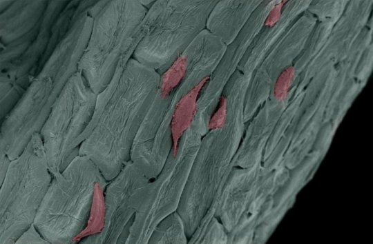
The researchers have developed a 3D technology that allows them to track, map and monitor what happens with cells and tissues that are being used to repair damage caused by disease or the treatment for the disease. By observing the cells and tissues they can see if they are staying where they are needed and if they are working.

The technology, published in the journal ACS Nano, uses tiny sensors placed on a flexible scaffold to monitor the new materials in the body. Ingeniously the scaffold is buoyant, so it can float and survive in the wet conditions found in many parts of the body.

In a news release, Chi Hwan Lee, the leader of the research team, says the device could help millions of people:

“Tissue engineering already provides new hope for hard-to-treat disorders, and our technology brings even more possibilities. This device offers an expanded set of potential options to monitor cell and tissue function after surgical transplants in diseased or damaged bodies. Our technology offers diverse options for sensing and works in moist internal body environments that are typically unfavorable for electronic instruments.”

Purdue created this video showing the device and explaining how it works.

Advancing stem cell research in many ways

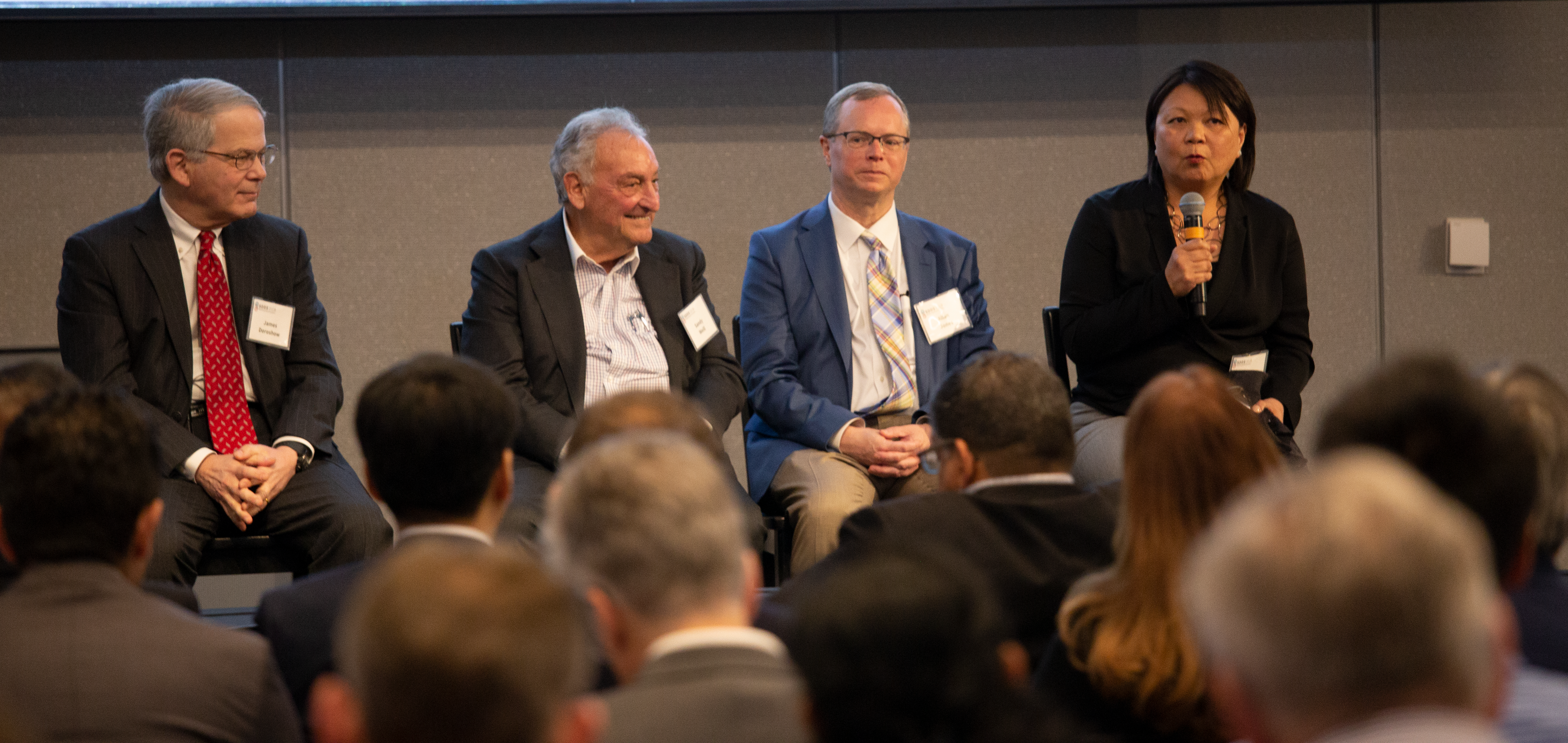
Speakers at the Alpha Stem Cell Clinics Network Symposium: Photo by Marco Sanchez

From Day One CIRM’s goal has been to advance stem cell research in California. We don’t do that just by funding the most promising research -though the 51 clinical trials we have funded to date clearly shows we do that rather well – but also by trying to bring the best minds in the field together to overcome problems.

Over the years we have held conferences, workshops and symposiums on everything from Parkinson’s disease, cerebral palsy and tissue engineering. Each one attracted the key players and stakeholders in the field, brainstorming ideas to get past obstacles and to explore new ways of developing therapies. It’s an attempt to get scientists, who would normally be rivals or competitors, to collaborate and partner together in finding the best way forward.

It’s not easy to do, and the results are not always obvious right away, but it is essential if we hope to live up to our mission of accelerating stem cell therapies to patients with unmet medical needs.

For example. This past week we helped organize two big events and were participants in another.

The first event we pulled together, in partnership with Cedars-Sinai Medical Center, was a workshop called “Brainstorm Neurodegeneration”. It brought together leaders in stem cell research, genomics, big data, patient advocacy and the Food and Drug Administration (FDA) to tackle some of the issues that have hampered progress in finding treatments for things like Parkinson’s, Alzheimer’s, ALS and Huntington’s disease.

We rather ambitiously subtitled the workshop “a cutting-edge meeting to disrupt the field” and while the two days of discussions didn’t resolve all the problems facing us it did produce some fascinating ideas and some tantalizing glimpses at ways to advance the field.

Alpha Stem Cell Clinics Network Symposium: Photo by Marco Sanchez

Two days later we partnered with UC San Francisco to host the Fourth Annual CIRM Alpha Stem Cell Clinics Network Symposium. This brought together the scientists who develop therapies, the doctors and nurses who deliver them, and the patients who are in need of them. The theme was “The Past, Present & Future of Regenerative Medicine” and included both a look at the initial discoveries in gene therapy that led us to where we are now as well as a look to the future when cellular therapies, we believe, will become a routine option for patients. 

Bringing these different groups together is important for us. We feel each has a key role to play in moving these projects and out of the lab and into clinical trials and that it is only by working together that they can succeed in producing the treatments and cures patients so desperately need.

Cierra Jackson: Photo by Marco Sanchez

As always it was the patients who surprised us. One, Cierra Danielle Jackson, talked about what it was like to be cured of her sickle cell disease. I think it’s fair to say that most in the audience expected Cierra to talk about her delight at no longer having the crippling and life-threatening condition. And she did. But she also talked about how hard it was adjusting to this new reality.

Cierra said sickle cell disease had been a part of her life for all her life, it shaped her daily life and her relationships with her family and many others. So, to suddenly have that no longer be a part of her caused a kind of identity crisis. Who was she now that she was no longer someone with sickle cell disease?

She talked about how people with most diseases were normal before they got sick, and will be normal after they are cured. But for people with sickle cell, being sick is all they have known. That was their normal. And now they have to adjust to a new normal.

It was a powerful reminder to everyone that in developing new treatments we have to consider the whole person, their psychological and emotional sides as well as the physical.

CIRM’s Dr. Maria Millan (right) at a panel presentation at the Stanford Drug Discovery Symposium. Panel from left to right are: James Doroshow, NCI; Sandy Weill, former CEO Citigroup; Allan Jones, CEO Allen Institute

And so on to the third event we were part of, the Stanford Drug Discovery Symposium. This was a high level, invitation-only scientific meeting that included some heavy hitters – such as Nobel Prize winners Paul Berg and  Randy Schekman, former FDA Commissioner Robert Califf. Over the course of two days they examined the role that philanthropy plays in advancing research, the increasingly important role of immunotherapy in battling diseases like cancer and how tools such as artificial intelligence and big data are shaping the future.

CIRM’s President and CEO, Dr. Maria Millan, was one of those invited to speak and she talked about how California’s investment in stem cell research is delivering Something Better than Hope – which by a happy coincidence is the title of our 2018 Annual Report. She highlighted some of the 51 clinical trials we have funded, and the lives that have been changed and saved by this research.

The presentations at these conferences and workshops are important, but so too are the conversations that happen outside the auditorium, over lunch or at coffee. Many great collaborations have happened when scientists get a chance to share ideas, or when researchers talk to patients about their ideas for a successful clinical trial.

It’s amazing what happens when you bring people together who might otherwise never have met. The ideas they come up with can change the world.

Attractive new regenerative medicine tool uses magnets to shape and stimulate stem cells

The ultimate goal of tissue engineers who work in the regenerative medicine field is to replace damaged or diseased organs with new ones built from stem cells. To accomplish the feat, these researchers are developing new tools and techniques to manipulate and specialize stem cells into three dimensional structures. Some popular methods – which we’ve blogged about often – include the use of bioscaffolds as well as 3D bioprinting . This week, a research team at the Laboratoire Matière et Systèmes Complexes in France has developed an attractive (pun intended!) new tool that uses magnetized stem cells to both manipulate and stimulate the cells into 3D shapes.

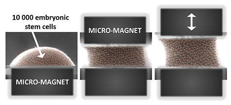
The magnetic stretcher: this all-in-one system can both form and mechanically stimulate an aggregate of magnetized embryonic stem cells. Image: © Claire Wilhelm / Laboratoire Matière et systèmes complexes (CNRS/Université Paris Diderot).

The study, reported on Monday in Nature Communications, used embryonic stem cells which were incubated with magnetic nanoparticles. The cells readily take up the nanoparticles which allowed the scientists to group the individual cells using magnets. But first the team needed to show that the nanoparticles had no negative effects on the cells. Comparing the iron nanoparticle-laden stem cells to iron-free cells showed no difference in the cells’ survival and their ability to divide.

It was also important to make sure the introduction of nanoparticles had no impact on the stem cell’s pluripotency; that is, its ability to maintain its unspecialized state. A visual check of the cells through a microscope showed that they grew together in rounded clumps, a hallmark of undifferentiated, pluripotent cells. In addition, the key genes that bestow pluripotency onto embryonic stem cells were still active after the addition of the nanoparticles.

The stem cells’ ability to mature into various cell types, like heart muscle or nerve, is key to any successful tissue engineering project. So, the next important assessment of these magnetized cells was to make sure their ability to differentiate, or specialize, was still intact. The typical first step to differentiating embryonic stem cells is to form so-called embryoid bodies (EBs), which are 3D groups of pluripotent stem cells which begin differentiating into the three fundamental tissues types: mesoderm (gives rise to muscle, bone, fat), ectoderm (gives rise to nerve, hair, eyes), endoderm (gives rise to intestines, liver). Using a popular technique, called the hanging drop method, the team showed that the presence of the nanoparticles did not negatively affect embryoid body formation.

In fact, the use of magnets to form embryoid bodies provided several advantages over the hanging drop method. The hanging drop technique requires multiple, time-consuming steps and the resulting embryoid bodies tend to be inconsistent in size and shape. Use of the magnets, on the other hand, instantaneously assembled the stem cells into consistently round aggregates. And by precisely adjusting the magnetic force used, the scientists could also vary the size of the embyroid body, which is an important variable to control since the embryoid size can impact its ability to differentiate.

While the magnet used to form the embryoid bodies was kept stable, the researchers included another magnet which they could move. With this setup, the team was able to stretch and shape the group of cells without the need of scaffolds or the need to physically contact the cells. Several previous studies, using flat, 2-dimensional petri dishes, have shown that the stiffness and flexibility of the dish can stimulate gene activity by affecting cell shape. In this study, the researchers found that when the magnet was moved in a cyclical pattern that imitates the rhythm of a heart beat, the embryoid bodies were, if you can believe it, nudged toward a heart muscle fate. A press release by France’s National Center of Scientific Research (CNRS), which funded the study, explained the big picture implications of this new technique:

“This “all-in-one” approach, which makes it possible to build and manipulate tissue within the same system, could thus prove to be a powerful tool both for biophysical studies and tissue engineering.”

Stem cell stories that caught our eye: spinal cord injury trial update, blood stem cells in lungs, and using parsley for stem cell therapies

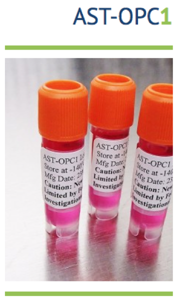
More good news on a CIRM-funded trial for spinal cord injury. The results are now in for Asterias Biotherapeutics’ Phase 1/2a clinical trial testing a stem cell-based therapy for patients with spinal cord injury. They reported earlier this week that six out of six patients treated with 10 million AST-OPC1 cells, which are a type of brain cell called oligodendrocyte progenitor cells, showed improvements in their motor function. Previously, they had announced that five of the six patients had shown improvement with the jury still out on the sixth because that patient was treated later in the trial.

 In a news release, Dr. Edward Wirth, the Chief Medical officer at Asterias, highlighted these new and exciting results:

 “We are excited to see the sixth and final patient in the AIS-A 10 million cell cohort show upper extremity motor function improvement at 3 months and further improvement at 6 months, especially because this particular patient’s hand and arm function had actually been deteriorating prior to receiving treatment with AST-OPC1. We are very encouraged by the meaningful improvements in the use of arms and hands seen in the SciStar study to date since such gains can increase a patient’s ability to function independently following complete cervical spinal cord injuries.”

Overall, the trial suggests that AST-OPC1 treatment has the potential to improve motor function in patients with severe spinal cord injury. So far, the therapy has proven to be safe and likely effective in improving some motor function in patients although control studies will be needed to confirm that the cells are responsible for this improvement. Asterias plans to test a higher dose of 20 million cells in AIS-A patients later this year and test the 10 million cell dose in AIS-B patients that a less severe form of spinal cord injury.

 Steve Cartt, CEO of Asterias commented on their future plans:

 “These results are quite encouraging, and suggest that there are meaningful improvements in the recovery of functional ability in patients treated with the 10 million cell dose of AST-OPC1 versus spontaneous recovery rates observed in a closely matched untreated patient population. We look forward to reporting additional efficacy and safety data for this cohort, as well as for the currently-enrolling AIS-A 20 million cell and AIS-B 10 million cell cohorts, later this year.”

Lungs aren’t just for respiration. Biology textbooks may be in need of some serious rewrites based on a UCSF study published this week in Nature. The research suggests that the lungs are a major source of blood stem cells and platelet production. The long prevailing view has been that the bone marrow was primarily responsible for those functions.

The new discovery was made possible by using special microscopy that allowed the scientists to view the activity of individual cells within the blood vessels of a living mouse lung (watch the fascinating UCSF video below). The mice used in the experiments were genetically engineered so that their platelet-producing cells glowed green under the microscope. Platelets – cell fragments that clump up and stop bleeding – were known to be produced to some extent by the lungs but the UCSF team was shocked by their observations: the lungs accounted for half of all platelet production in these mice.

Follow up experiments examined the movement of blood cells between the lung and bone marrow. In one experiment, the researchers transplanted healthy lungs from the green-glowing mice into a mouse strain that lacked adequate blood stem cell production in the bone marrow. After the transplant, microscopy showed that the green fluorescent cells from the donor lung traveled to the host’s bone marrow and gave rise to platelets and several other cells of the immune system. Senior author Mark Looney talked about the novelty of these results in a university press release:

Mark Looney, MD

“To our knowledge this is the first description of blood progenitors resident in the lung, and it raises a lot of questions with clinical relevance for the millions of people who suffer from thrombocytopenia [low platelet count].”

If this newfound role of the lung is shown to exist in humans, it may provide new therapeutic approaches to restoring platelet and blood stem cell production seen in various diseases. And it will give lung transplants surgeons pause to consider what effects immune cells inside the donor lung might have on organ rejection.

Add a little vanilla to this stem cell therapy. Typically, the only connection between plants and stem cell clinical trials are the flowers that are given to the patient by friends and family. But research published this week in the Advanced Healthcare Materials journal aims to use plant husks as part of the cell therapy itself.

Though we tend to focus on the poking and prodding of stem cells when discussing the development of new therapies, an equally important consideration is the use of three-dimensional scaffolds. Stem cells tend to grow better and stay healthier when grown on these structures compared to the flat two-dimensional surface of a petri dish. Various methods of building scaffolds are under development such as 3D printing and designing molds using materials that aren’t harmful to human tissue.

Human fibroblast cells growing on decellularized parsley.
Image: Gianluca Fontana/UW-Madison

But in the current study, scientists at the University of Wisconsin-Madison took a creative approach to building scaffolds: they used the husks of parsley, vanilla and orchid plants. The researchers figured that millions of years of evolution almost always leads to form and function that is much more stable and efficient than anything humans can create. Lead author Gianluca Fontana explained in a university press release how the characteristics of plants lend themselves well to this type of bioengineering:

Gianluca Fontana, PhD

“Nature provides us with a tremendous reservoir of structures in plants. You can pick the structure you want.”

The technique relies on removing all the cells of the plant, leaving behind its outer layer which is mostly made of cellulose, long chains of sugars that make up plant cell walls. The resulting hollow, tubular husks have similar shapes to those found in human intestines, lungs and the bladder.

The researchers showed that human stem cells not only attach and grow onto the plant scaffolds but also organize themselves in alignment with the structures’ patterns. The function of human tissues rely on an organized arrangement of cells so it’s possible these plant scaffolds could be part of a tissue replacement cell product. Senior author William Murphy also points out that the scaffolds are easily altered:

William Murphy, PhD

“They are quite pliable. They can be easily cut, fashioned, rolled or stacked to form a range of different sizes and shapes.”

And the fact these scaffolds are natural products that are cheap to manufacture makes this a project well worth watching.

Easier, Cheaper Stem Cell-Based Heart Muscle Sets Stage for Large-Scale Drug Development

The great inventions – like the automobile, the Internet or aviation – are marked as important turning points in human history. But it’s usually the additional tinkering that goes on in the ensuing years after the initial invention that makes the technology feasible in terms of cost, reproducibility and mass production.

The same holds true for the Nobel prize winning induced pluripotent stem cell (iPS) technique. The sight of human iPS-derived heart muscle cells, or cardiomyocytes, beating in a petri dish brought a lot of early excitement in the late 2000’s (and still does today) about the potential of using human cells rather than animal models to screen for novel heart disease therapies and to test for drug toxicity.

And since iPS cells can be created directly from the skin or blood of a heart disease patient, it opened new opportunities to better understand the cellular basis of heart disease.

The Earth isn’t flat and neither is the heart
But the human heart is more than a two dimensional layer of cardiomyocytes in a petri dish. As a result, more complex three dimensional miniature heart structures that better mimic cardiac function have been developed over time. Now a CIRM-funded research team at the Gladstone Institutes has gone a step further and devised a “Micro-Heart Muscle” (µHM) technique that is easy to make and uses much fewer cells. The method detailed in Scientific Reports yesterday is poised to make large-scale, high-throughput drug and toxicology testing for heart disease therapies a reality.

Prior to this current publication, the Engineered Heart Muscle (EHM) has been the gold standard for petri dish models of human heart function. To better reflect the cellular environment of heart tissue than a simple layer of heart cells, EHM is composed of a mixture of iPS-derived cardiomyocytes, fibroblasts, and extracellular matrix, a natural scaffold that supports the heart’s cellular structure. These components are grown into 3D molds in a lab dish and embedded into posts that give the muscle cells something to contract against. This setup provides a means to do detailed analysis of the impact of drugs on heart muscle function. While these miniature EHM structures successfully produce physiologically relevant heart tissue for testing in the lab, they nonetheless carry some practical limitations. The complexity of the molds and the need for millions of cells for each EHM tissue makes the cost of large-scale drug development experiments too high. And the use of extracellular matrix would muck up the miniaturized instrumentation that is used for these therapy development efforts.

Dog bones: the key to easier, cheaper muscle tissue
The Gladstone team, led by Bruce Conklin, has overcome these challenges with their Micro-Heart Muscle product. With some educated trial and error, they zeroed in on a simple dog bone shaped mold to grow the mixture of cells in. The shape of the mold encouraged the muscle cells to self organize and grow into contracting tissue without the need of extracellular matrix. And better yet, less than 10,000 cells were needed to form each tissue. Here’s a cool video, recorded by first author Nathaniel Huebsch, of the cells in action:

These technical improvements of the Micro-Heart Muscle could help make large-scale, systematic approaches to study heart disease and toxicology a reality. As you read Conklin’s summary of the results in a Gladstone press release, you can hear his excitement about the future applications of this method:

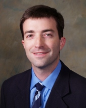
conklin-profile.jpg

Bruce Conklin, Gladstone Institutes Senior Investigator

“The beauty of this technique is that it is very easy and robust, but it still allows you to create three-dimensional miniature tissues that function like normal tissues. Our research shows that you can create these complex tissues with a simple template that exploits the inherent properties of these cells to self-organize. We think that the micro heart muscle will provide a superior resource for conducting research and developing therapies for heart disease.”


Micro-Heart Muscle: the Model T of iPS innovations?
Karl Benz is generally credited as the inventor of the automobile, but a few years later it was Henry Ford’s efficient assembly line and manufacturing process that helped make mass production and affordability of the car possible. In a somewhat similar way, Shinya Yamanaka’s iPS technique will no doubt go down as one of the greatest inventions in the 21st century and maybe Conklin’s team’s Micro-Heart Muscle will also feature prominently in the history books as a follow up innovation that made the development of heart disease therapies possible.

Bruce-&-Nate-'web

Senior author Bruce Conklin and first author Nathaniel Huebsch [Photo: Chris Goodfellow]

Meet ITOP: A One Stop Shop for 3D Printing Body Parts

“They have managed to create what appears to be the goose that really does lay golden eggs!”

That was how UK surgeon Martin Birchall described it to BBC News. The goose in this case is a 3D bioprinter, and the golden eggs are the human sized tissues that the bioprinter successfully constructed. This breakthrough for the field of tissue engineering was reported on Monday in Nature Biotechnology by a research team led by Anthony Atala, director of Wake Forest Institute for Regenerative Medicine.

Bioprinting: yes, it’s actually a thing

earbioprinting copy

The 3D bioprinting process. Image: Wake Forest, Nature Biotechnology

To some, printing human body parts may sound like a far-fetched story torn from a science fiction novel but, in reality, development of bioprinters has been underway for a number of years. The bioprinting process isn’t all that different from an inkjet printer except a mixture of gelatin, or hydrogel, and living cells is used as the “ink” to incrementally build a defined  biological structure.

As amazing as this technology is, it has met some limitations. Printing cells into 2D shapes and small 3D structures is doable but a lack of structural stability limits building more complex, human-scale tissues. Also, because oxygen and nutrients can only diffuse about 0.004 inches through living tissue, the cells located inside a large bioprinted structure have trouble with long-term survival (check out yesterday’s blog for a mind-blowning story about one lab’s use of cotton candy to deal with this diffusion issue). These challenges have to be overcome before 3D bioprinting can be used for the repair and replacement of human-sized tissues and organs.

Enter ITOP
That’s where the Atala team’s Integrated Tissue-Organ Printer (ITOP), developed over ten years, comes into the picture. For better structural stability, the printer is configured to deliver the cell/hydrogel “bio-ink” within a stronger type of gel, with the fancy name Pluronic F127, that helps the printed cells maintain their shape during the printing process. Afterward, the Pluronic F127 scaffolding mold is simply washed away from the bioprinted tissue.

ITOPcloseup

Close-up view of ITOP: a 3D bioprinter. Image: Wake Forest, Nature Biotechnology.

To ensure adequate oxygen diffusion into the bioprinted tissue, a biodegradable polymer, PCL, is dispensed from the printer at regular intervals; this creates microchannels as stand-ins for blood vessels to help oxygen and nutrients readily reach the interior areas of the tissues.  As a bonus, PCL takes about 2 years to biodegrade, providing long term stability.

To prove these innovations of the ITOP actually work, the team built three different tissues: a jawbone fragment, an ear, and muscle.

The making of a jawbone
For reconstruction of the jawbone, a 3D computer model was generated from actual CT scan data of a human jaw with a missing piece of bone – something that might be seen in a traumatic injury to a combat soldier (the work is funded in part by the Armed Forces Institute for Regenerative Medicine). That data was fed into the ITOP with coordinates of the precise printing pattern necessary to rebuild the shape of the jaw fragment. In this case, printing was carried out using human amniotic fluid stem cells (AFSC). With the right cues, these stem cells readily specialize into osteogenic, or bone-forming, cells.

Sure enough, 28 days after being cultured in liquid nutrients containing bone-promoting factors, the surface of the bioprinted human jaw showed calcium deposits, the tell-tale signs of bone formation. How would bioprinted bone fare in a living mammal? To find out, the researchers transplanted small discs of AFSC fabricated bone into a bone defect in mice. After 5 months, the transplanted bone was thriving with plenty of blood vessels and no necrosis, or cell death, inside the bone.

The implications of this bone study are pretty cool. In an interview with BBC news, Atala envisions a not so distant future clinical scenario:

“We’d bring the patient in, do the imaging and then we would take the imaging data and transfer it through our software to drive the printer to create a piece of jawbone that would fit precisely in the patient.”

Vincent van Gogh could have used this technology
But the possibilities don’t end with bone. Next, the team tested the ITOP’s talents at building the complex shapes of the outer human ear. In this case, the printer was loaded up with cartilage-producing cells called chondrocytes. The authors posted a fascinating video of the ear bioprinting process in their online publication.

ear_wakeforest

A human-sized bioprinted ear. Image: Wake Forest, Nature Biotechnology

Image: Wake Forest, Nature Biotechnology.

After five weeks in liquid nutrients, a matrix of cartilage had grown throughout the ear. And to look at tissue growth in an animal, the ear was implanted under the skin of the mice. A couple of months after implantation, even more cartilage had formed and the shape of the ear was intact.

But wait there’s more: printing skeletal muscle
Since both the jaw bone and ear cartilage represent hard tissues, the team sought to reconstruct muscle, a soft tissue, with the ITOP. Muscle-forming cells, or myoblasts, were printed to mimic the muscle fiber bundles seen in native skeletal muscle. After growing a week in the lab under conditions that stimulate muscle cell formation, the muscle-like fibers were implanted into rats. Two week after implantation, the bioprinted muscle had not only grown into well-organized muscle fibers, they also were functional in that they were responsive to electrical stimulation.

3D bioprinting: getting closer to reality
Atala is the first to admit that a lot more testing is needed to safely bring this technology into a clinical setting for human use. But as he states in a Wake Forest press release, the ITOP brings 3D bioprinting a step closer to reality:

“This novel tissue and organ printer is an important advance in our quest to make replacement tissue for patients. It can fabricate stable, human-scale tissue of any shape. With further development, this technology could potentially be used to print living tissue and organ structures for surgical implantation.”


3D Printing Cells with DNA Velcro

Print

The complex, 3D micro-anatomy of the human liver. (Image source: WikiMedia Commons)

One of the Holy Grails of stem cell research is growing body parts to replace those damaged by disease or injury. Enormous strides have been made in a key first step: mastering recipes for maturing stem cells into various specialized cell types. But a lawn of, say, liver cells in a petri dish is not a functioning liver. Organs have complex, three-dimensional structures with intricate communication between multiple cell types.

Scientists are actively devising methods to overcome this challenge. For instance, cultivating cells onto biological scaffolds help mold the cells into the shape of a particular organ or tissue. And retooled 3D printers using “bio ink” can seed layers of different cells onto these scaffolds to create specified structures.

This week, a UCSF team added an ingenious new tool to this tissue engineering tool kit.  As reported on Monday in Nature Methods, the lab of Zev Gartner took advantage of DNA’s Velcro-like chemistry to build layers of different cell types in a specified pattern.

DNA – it’s not just for genetics anymore

DNAbasepairing

A DNA fragment is made of two complimentary strands that bind together with high specificity. (Image source: Visionlearning)

DNA is a molecule made of two thin strands. Each strand is specifically attracted to the other based on a unique sequence of genetic information. So if two strands of a short DNA fragment are peeled apart, they will only rejoin to each other and not some other fragment with a different sequence.  While DNA usually resides in the nucleus of a cell, the team worked out a method to temporarily attach copies of a strand of DNA on the outside of, let’s call it, “cell A”. The opposite strand of that DNA fragment was attached to “cell B”. When mixed together the two cells became attached to each other via the matching DNA sequences. Other cells with different DNA fragments floated on by.

The screen shot below from a really neat time-lapse video, which accompanies the research publication, shows how a rudimentary 3D cell structure could be built with a series of different cell-DNA fragment combinations. In this case, the team first attached DNA fragments onto a petri dish in a specific pattern. At the thirty-second mark in the video, you can see that cells with matching DNA fragments have attached to the DNA on the dish.

Screen Shot 2015-09-02 at 8.48.16 AM

This video demonstrates the assembly of 3D cell structures with the help of DNA “Velcro” (image source: Todhunter et al. Nature Methods 2015 Aug 31st)

The new technique, dubbed DNA programmed assembly of cells (DPAC), opens up a lot possibilities according to Gartner in a UCSF press release:

 “We can take any cell type we want and program just where it goes. We can precisely control who’s talking to whom and who’s touching whom at the earliest stages. The cells then follow these initially programmed spatial cues to interact, move around, and develop into tissues over time.”

The Quest still continues with possible victories along the way

 Of course, this advance is still a far cry from the quest for whole organs derived from stem cells. The cell assemblies using DPAC can only be grown up to about 100 microns, the thickness of a human hair. Beyond that size, the innermost cells get starved of oxygen and nutrients. Gartner says that obstacle is a current focus in the lab:

“We’re working on building functional blood vessels into these tissues. We can get the right cells in the right positions but haven’t figured out how to perfuse them with blood or a substitute efficiently yet.”

In the meantime, building these small 3D “organoids” from stem cells certainly could be put to good use as a means to test drug toxicity on human tissue or as a way to study human disease.

Related Links:


CIRM-Funded Scientists Make New Progress Toward Engineering a Human Esophagus

Creating tissues and organs from stem cells—often referred to as ‘tissue engineering’—is hard. But new research has discovered that the process may in fact be a little easier than we once thought, at least in some situations.

Engineered human esophageal tissue [Credit: The Saban Research Institute].

Engineered human esophageal tissue [Credit: The Saban Research Institute].

Last week, scientists at The Saban Research Institute of Children’s Hospital Los Angeles announced that the esophagus—the tube that transports food, liquid and saliva between the mouth and the stomach—can be grown inside animal models after injecting the right mix of early-stage, or ‘progenitor,’ esophageal cells.

These findings, published in the journal Tissue Engineering Part A, are an important step towards generating tissues and organs that have been damaged due to disease or—in some cases—never existed in the first place.

According to stem cell researcher Tracy Grikscheit, who led the CIRM-funded study, the researchers first implanted a biodegradable ‘scaffold’ into laboratory mice. They then injected human progenitor cells into the mice and watched as they first traveled to the correct location—and then began to grow. The ability to both migrate to the right location and differentiate into the right cell type, without the need for any external coaxing, is crucial if scientists are to successfully engineer such a critical type of tissue.

“Different progenitor cells can find the right ‘partner’ in order to grow into specific esophageal cell types—and without the need for [outside] growth factors,” explained Grikscheit in a news release. “This means that successful tissue engineering of the esophagus is simpler than we previously thought.”

Grikscheit, who is also a pediatric surgeon as Children’s Hospital Los Angeles, was particularly hopeful with how their findings might one day be used to treat children born with portions of the esophagus missing—as well as adults suffering from esophageal cancer, the fastest-growing cancer in the U.S.

“We have demonstrated that a simple and versatile, biodegradable polymer is sufficient for the growth of a tissue-engineered esophagus from human cells. This not only serves as a potential source of tissue, but also a source of knowledge—as there are no other robust models available for studying esophageal stem cell dynamics.”

Want to learn more about tissue engineering? Check out these video highlights from a recent CIRM Workshop on the field.

ISSCR 2014: Tony Atala, Jason Burdick and the Power of Tissue Engineering

The progress in tissue engineering in just the past two decades has been like the construction industry moving from simple lean-to structures to homes with plumbing, heating and cooling systems. We are not yet ready to build a high-rise—think of a beating functioning heart—but we are making major strides toward that goal.

One of the founders of the field, Wake Forest’s Dr. Tony Atala, led off this morning plenary session at the annual meeting of the International Society for Stem Cell Research. He started trying to build simple organs in 1990. His talk nicely mapped his progress through four levels of complexity of structure.

Tony Atala speaks about tissue engineering in a 2011 TED talk (credit: Wikipedia)

Tony Atala speaks about tissue engineering in a 2011 TED talk (credit: Wikipedia)

The first level, accomplished by a few teams, was our largest organ, skin, which is relatively simple because it is flat. Next, came simple hollow organs like blood vessels and the urethra that carries urine from the bladder. He followed that with more complex hollow organs, first the bladder and more recently the vagina. Last up were complex solid organs: the heart and the penis. He expects to begin clinical trials with the latter soon, which is eagerly anticipated by our military dealing with the aftermath IED explosive injuries from the wars in Iraq and Afghanistan.

He noted that researchers in the field quickly learned that just throwing cells on scaffolds and hoping they knew what to do was not enough in most cases. They need to grow blood vessels so they can get nourishment and communicate with their surroundings and they often have to make multiple cell types. His own work here benefited from a bit of geographic serendipity. His lab at the time was on the same floor as Judah Folkman’s at Harvard affiliated Children’s Hospital. Folkman is the father of the field of angiogenesis, the art of growing blood vessels.

Atala showed slides comparing injecting cells where you need new muscle, to cells plus scaffold, and finally to the two combined with a vessel growth factor. The three-way combo far outperformed the others. He published his first study using this technique for a hollow simple organ, the urethra, in 2011. At that point his patients had been living with the functional new organ for six years. They work and last.

Researchers almost always place a cell-scaffold complex in a soup of nutrients and growth factors called a bioreactor before implanting it. But at the time of implant, the organ is not mature. Atala said the body acts like a “finishing bioreactor” to fill out and strengthen the organ, which becomes fully mature around six months after implant. He showed images of this in-body growth in his first patients who had been born without a complete vagina and were given a fully functioning organ. He just published that study two months ago, eight years after the implants in order to make sure they stayed functional over time.

He then showed his animal model work creating a penis in rabbits. Being a highly vascular organ it required much more structure. He used a donor organ that had all its cells chemically washed away to leave just the intracellular scaffold. This structure helped guide the blood vessel growth and the rabbits succeeded in mating and having offspring.

His lab has begun early stage work for both liver and heart. They have created miniature livers about the size of a half dollar that are able to produce the appropriate proteins and metabolize drugs. They have used a 3-D printer to build two chambers of a heart that are able to beat in a dish, but their structure has not been stable. So, he noted much more work lies ahead for complex organs.

The second speaker, Jason Burdick from the University of Pennsylvania, concentrated on making better scaffolds for the stem cells, which can have three enhanced properties:

  1. they can be instructive, they can tell cells what to do;
  2. they can be dynamic, they can react to their environment and the cells around them;
  3. they can lead to heterogeneity, they can provide varied instructions so you get the different cell types that you need for a complex tissue.

He discussed two examples, the first was growing better cartilage (as he joked, for injured World Cup soccer players). One problem with early gels used as scaffold was they held the cells individually apart from each other limiting their ability to communicate with each other. This cell-to-cell cross talk is key to tissue maturation. He showed how you could chemically alter the gel to enhance this communication. He also showed how you could implant the gels with microspheres loaded with growth factors to deliver instructions to the cells.

Burdick’s second example focused on minimizing injury after an induced heart attack in rodents. But instead of loading the gel with cells, they loaded it with microspheres that release chemicals that summons the stem cells waiting quietly in reservoirs in all of us. They saw sustained release of the chemicals for 21 days and significant improvement in heart function.

But he closed with a fun twist. The first heart experiment used a strict time-release formulation. He said it would be much better if the chemicals were released at the points the heart needs it the most. So, he is working on a system that releases the chemical based on the levels of an enzyme the heart makes when it is injured. He is hoping this right-amount-at-the-right-time formula will be even better.

We have a short video of the highlights of a workshop we held on tissue engineering that you can watch to get a better feel for where the field is going.

Don Gibbons