CCSF’s CIRM Bridges scholars: the future of stem cell research is in good hands

In need of an extra dose of inspiration? You might read a great book or listen to that podcast your friend recommended. You might even take a stroll along the beach. But I can do you one better: go to a conference poster session where young stem cell scientists describe their research.

That’s what I did last week at the City College of San Francisco’s (CCSF) Bioscience Symposium held at UC San Francisco’s Genentech Hall. It’s a day-long conference that showcases the work of CCSF Bioscience interns and gives them a chance to present the results of their research projects, network with their peers and researchers, hear panelists talk about careers in biotechnology and participate in practice job interviews.

Bridges_CCSF_2018b

CCSF’s CIRM Bridges Scholars (clockwise from top left): Vanessa Lynn Herrara, Viktoriia Volobuieva, Christopher Nosworthy and Sofiana E. Hamama.

Bridges_CCSF_2018

CCSF’s CIRM Bridges Scholars (clockwise from top left): Seema Niddapu, Mark Koontz, Karolina Kaminska and Iris Avellano

Eight of the dozens of students in attendance at the Symposium are part of the CIRM-funded Bridges Stem Cell Internship program at CCSF. It’s one of 14 CIRM Bridges programs throughout the state that provides paid stem cell research internships to students at universities and colleges that don’t have major stem cell research programs. Each Bridges internship includes thorough hands-on training and education in stem cell research, and direct patient engagement and outreach activities that engage California’s diverse communities.

In the CCSF Bridges Program, directed by Dr. Carin Zimmerman, the students do a 9-month paid internship in top notch labs at UCSF, the Gladstone Institutes and Blood System Research Institute. As I walked from poster to poster and chatted with each Bridges scholar, their excitement and enthusiasm for carrying out stem cell research was plain to see. It left me with the feeling that the future of stem cell research is in good hands and, as I walked into the CIRM office the next day, I felt re-energized to tackle the Agency’s mission to accelerate stem cell treatment for patients with unmet medical needs. But don’t take my word for it, listen to the enthusiastic perspectives of Bridges scholars Mark Koontz and Iris Avellano in this short video.

Making stem cell-derived liver cells to study fatty liver disease

Non-alcoholic fatty liver disease (NAFLD) affects approximately 30% of the population, with that number increasing to 75% in obese individuals. Shockingly, the number of cases is expected to increase 21% by the year 2030 in the United States alone.

liver_fattyliverNAFLD refers to a broad range of liver conditions, which are all characterized by abnormally high levels of fat deposits in the livers of people who do not drink excessive amounts of alcohol. While not always fatal, NAFLD can lead to liver cirrhosis, or extensive scaring of the liver tissue. Cirrhosis, in turn, can cause life-threatening conditions such as liver cancer or liver failure. Whether or not N

AFLD will lead to extensive liver damage is not well understood and the primary therapeutic option is weight loss with no FDA-approved drug options. The projected increase in NALD cases combined with the poor treatment options makes this disease a significant public health burden.

Studying NALD can be quite complicated because the liver is complex organ made up of multiple different cell types. Investigators at the University of Edinburgh have simplified some of this complexity by figuring out a way to generate liver cells in a dish.

In studies published in the Philosophical Transactions of the Royal Society B, these scientists used human embryonic stem cells to generate hepatocyte-like cells (HLCs), or cells that are highly similar to liver cells isolated from humans. When exposed to fatty acids, they saw that the HLCs exhibited hallmarks of NAFLD, such as fat accumulation in liver cells, and changes in gene expression that are indicative of NAFLD.

In a press release, Dr. David Hay, one of the two senior investigators of this study, states:

david hay

Dr. David Hay

“Our ability to generate human hepatocytes from stem cells, using semi-automated procedures, allows us to study the mechanisms of human liver disease in a dish and at scale.”


This approach is particularly valuable because it would replace the need to use cancer cell lines for this type of work. While valuable for many reasons, research done in cancer cells lines can be difficult to draw therapeutic conclusions from, because cell lines have significant genetic alternations from normal cells. Generating liver cells from human stem cells provides an important tool for high throughput screening of medically relevant therapies for NALD.


Can stem cells help people recover from a stroke? Join us for a Facebook Live event this Thursday, May 31 for the answers

AskExpertsMAY2018[1]

Stroke is one of the leading causes of death in the US and the leading cause of serious, long-term disability. But could stem cell therapies change that and help people who’ve had a brain attack?  Could stem cells help repair the damage caused by a stroke and restore a person’s ability to speak normally, to be able to walk without a limp or regain strength in their hands and arms?

To find out the answers to these and other questions joins us for “Ask the Expert”, a special Facebook Live event this Thursday, May 31, from noon till 1pm PDT

 The event will feature Dr. Gary Steinberg, the Chair of Neurosurgery at Stanford University. Dr. Steinberg is currently running a CIRM-funded clinical trial targeting stroke.

We will also be joined by CIRM Senior Science Officer Lila Collins, PhD who can talk about the broad range of other projects using stem cells to help people recover from a stroke.

We are also delighted to welcome Sonia Coontz, who suffered a devastating stroke several years ago and made a remarkable recovery after getting a stem cell therapy.

To join us for the event, all you have to do is go to our Facebook page on Thursday at noon (PDT) and you should see a video playing, which you can watch on mobile or desktop. Click the video to enter viewing mode.

Also, make sure to “like” our page before the event to receive a notification that we’ve gone live.

And we want to hear from you, so you will be able to post questions for the experts to answer or, you can email them directly to us at info@cirm.ca.gov

We look forward to seeing you there.


Stem Cell Roundup: Jake Javier’s amazing spirit; TV report highlights clinic offering unproven stem cell therapies

JakeJavier_A_0107_20161207142726_JakeJavier_SeesTheDay

Jake Javier: Photo Michael Clemens, Sees the Day

In the Roundup we usually focus on studies that highlight advances in stem cell research but today we’re going to do something a little different. Instead of relying on print for our stories, we’re turning to video.

We begin with a piece about Jake Javier. Regular readers of our blog will remember that Jake is the young man who broke his neck the day before he graduated high school, leaving him paralyzed from the upper chest down.

After enrolling in the CIRM-funded Asterias clinical trial, and receiving a transplant of 10 million stem cells, Jake regained enough use of his arms and hands to be able to go to Cal Poly and start his life over.

This video highlights the struggles and challenges he faced in his first year, and his extraordinary spirit in overcoming them.

(thanks to Matt Yoon and his Creative Services team at Cal Poly for this video)

Going Undercover

The second video is from the NBC7 TV station in San Diego and highlights one of the big problems in regenerative medicine today, clinics offering unproven therapies. The investigative team at NBC7 went undercover at a stem cell clinic seminar where presenters talked about “the most significant breakthrough in natural medicine” for improving mobility and reducing pain. As the reporter discovered, the reality didn’t live up to the promise.

NBC7 Investigative Report


CIRM invests in stem cell clinical trial targeting lung cancer and promising research into osteoporosis and incontinence

Lung cancer

Lung cancer: Photo courtesy Verywell

The five-year survival rate for people diagnosed with the most advanced stage of non-small cell lung cancer (NSCLC) is pretty grim, only between one and 10 percent. To address this devastating condition, the Board of the California Institute for Regenerative Medicine (CIRM) today voted to invest almost $12 million in a team from UCLA that is pioneering a combination therapy for NSCLC.

The team is using the patient’s own immune system where their dendritic cells – key cells in our immune system – are genetically modified to boost their ability to stimulate their native T cells – a type of white blood cell – to destroy cancer cells.  The investigators will combine this cell therapy with the FDA-approved therapy pembrolizumab (better known as Keytruda) a therapeutic that renders cancer cells more susceptible to clearance by the immune system.

“Lung cancer is a leading cause of cancer death for men and women, leading to 150,000 deaths each year and there is clearly a need for new and more effective treatments,” says Maria T. Millan, M.D., the President and CEO of CIRM. “We are pleased to support this program that is exploring a combination immunotherapy with gene modified cell and antibody for one of the most extreme forms of lung cancer.”

Translation Awards

The CIRM Board also approved investing $14.15 million in four projects under its Translation Research Program. The goal of these awards is to support promising stem cell research and help it move out of the laboratory and into clinical trials in people.

Researchers at Stanford were awarded almost $6 million to help develop a treatment for urinary incontinence (UI). Despite being one of the most common indications for surgery in women, one third of elderly women continue to suffer from debilitating urinary incontinence because they are not candidates for surgery or because surgery fails to address their condition.

The Stanford team is developing an approach using the patient’s own cells to create smooth muscle cells that can replace those lost in UI. If this approach is successful, it provides a proof of concept for replacement of smooth muscle cells that could potentially address other conditions in the urinary tract and in the digestive tract.

Max BioPharma Inc. was awarded almost $1.7 million to test a therapy that targets stem cells in the skeleton, creating new bone forming cells and blocking the destruction of bone cells caused by osteoporosis.

In its application the company stressed the benefit this could have for California’s diverse population stating: “Our program has the potential to have a significant positive impact on the lives of patients with osteoporosis, especially in California where its unique demographics make it particularly vulnerable. Latinos are 31% more likely to have osteoporosis than Caucasians, and California has the largest Latino population in the US, accounting for 39% of its population.”

Application Title Institution CIRM funding
TRAN1-10958 Autologous iPSC-derived smooth muscle cell therapy for treatment of urinary incontinence


Stanford University


$5,977,155


TRAN2-10990 Development of a noninvasive prenatal test for beta-hemoglobinopathies for earlier stem cell therapeutic interventions


Children’s Hospital Oakland Research Institute


$1,721,606


TRAN1-10937 Therapeutic development of an oxysterol with bone anabolic and anti-resorptive properties for intervention in osteoporosis  

MAX BioPharma Inc.


$1,689,855


TRAN1-10995 Morphological and functional integration of stem cell derived retina organoid sheets into degenerating retina models


UC Irvine


$4,769,039


Boosting immune system cells could offer a new approach to treating Lou Gehrig’s disease

ALS

Amyotrophic lateral sclerosis (ALS), also known as Lou Gehrig’s disease, is one of those conditions that a lot of people know about but don’t know a lot about. If they are fortunate it will stay that way. ALS is a nasty neurodegenerative disease that attacks motor neurons, the cells in the brain and spinal cord that control muscle movement. As the disease progresses the individual loses their ability to walk, talk, eat, move and eventually to breathe. There are no effective treatments and no cure. But now research out of Texas is offering at least a glimmer of hope.

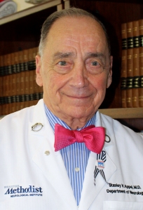
Dr. Stanley Appel, a neurologist at the Houston Methodist Neurological Institute noticed that many of the ALS patients he was treating had low levels of regulatory T cells, also known as Tregs. Tregs play a key role in our immune system, suppressing the action of molecules that cause inflammation and also helping prevent autoimmune disease.

In an article on Health News Digest Appel said:

Stanley Appel

Dr. Stanley Appel: Photo courtesy Australasian MND Symposium

“We found that many of our ALS patients not only had low levels of Tregs, but also that their Tregs were not functioning properly. We believed that improving the number and function of Tregs in these patients would affect how their disease progressed.”

And so that’s what he and his team did. They worked with M.D. Anderson Cancer Center’s Stem Cell Transplantation and Cellular Therapy program on a first-in-human clinical trial. They took blood from three people with different stages of ALS, separated the red and white blood cells, and returned the red blood cells to the patient. They then separated the Tregs from the white blood cells, increased their number in the lab, and then reinfused them into the patients, in a series of eight injections over the course of several months.

Their study, which appears in the journal Neurology,® Neuroimmunology & Neuroinflammation, found that the therapy appears to be safe without any serious side effects.

Jason Thonhoff, the lead author of the study, says the therapy also appeared to help slow the progression of the disease a little.

“A person has approximately 150 million Tregs circulating in their blood at any given time. Each dose of Tregs given to the patients in this study resulted in about a 30 to 40 percent increase over normal levels. Slowing of disease progression was observed during each round of four Treg infusions.”

Once the infusions stopped the disease progression resumed so clearly this is not a cure, but it does at least suggest that keeping Tregs at a healthy, high-functioning level may help slow down ALS.

CIRM is funding two clinical trials targeting ALS. One is a Phase 1 clinical trial with Clive Svendsen’s team at Cedars-Sinai Medical Center, the other is a Phase 3 project with Brainstorm Cell Therapeutics.

The Mother of Modern Medicine: Henrietta Lacks’ Portrait Unveiled at National Portrait Gallery

Back during my research scientist days, using HeLa cells for my experiments was as commonplace as a carpenter reaching for his hammer at a construction site. What makes these cells so handy is their robustness: they are easy to maintain in the lab where they divide indefinitely in petri dishes.

HeLaV_M

Scanning electron micrograph of just-divided HeLa cells.
Credit: National Center for Microscopy and Imaging Research

Henrietta Lacks and the Story of HeLa Cells
The reason they grow so readily is because they originally came from a patient’s tumor. For the longest time I had been under the impression that “HeLa” stood for Helen Lang, supposedly the patient’s name. It wasn’t until Rebecca Skloot’s award-winning book, “The Immortal Life of Henrietta Lacks”, was published in 2010, that I learned their true identity.

Only 31 years old, Henrietta Lacks died of cervical cancer in 1951. Before she died, cells from her cancer were collected without her permission or knowledge. Noticed for their remarkable ability to continually divide in cell culture, these cells, labelled as “HeLa”, became the first human cell line. Though Henrietta Lacks is long gone, her cells still live on in research labs all over the world and have been instrumental to many important discoveries and over 10,000 patents.

The Mother of Modern Medicine: The Portrait

lacks1

Henrietta Lacks (HeLa): The Mother of Modern Medicine by Kadir Nelson (see full portrait here)

The story of Lacks’ contribution to science can now be appreciated not only in the form of words on a page but also paint on canvas. Last week, the Smithsonian’s National Portrait Gallery, in collaboration with the National Museum of African American History and Culture, installed Kadir Nelson’s 2017 portrait of Lacks on the museum’s first-floor presentation wall.

In a Smithsonian.com article, painting and sculpture curator, Dorothy Moss, explained that Lacks’ portrait will be installed next to portraits of more recognizable Americans like Barack Obama and Susan B. Anthony where she hopes it acts as a, “signal to the kinds of history we want to tell. We want to make sure that people who have not been written into traditional narratives of history are visible immediately when our visitors enter. It will spark a conversation about people who have made a significant impact on science yet have been left out of history.”

When you look at the painting, be sure to notice some subtle details that help tell Lacks’ story, like the two missing buttons in her dress that symbolize her cells that were taken without her permission and the “Flower of Life” wall paper pattern meant to represent immortality.

CIRM’s Commitment to the Patient
It’s the learning from the unethical treatment of patients like Henrietta Lacks that in recent years has driven more focus on protecting patients and given them a voice when it comes to their care and their participation in medical research.

This commitment to patients is at the forefront of everything we do at CIRM. For instance, our 29-member Governing Board is composed of ten patient advocates, our CIRM-funded clinical trials are supported by Clinical Advisory Panels (CAPs) that include a patient advocate at the table and our mission itself is wholly focused on accelerating stem cell treatments to patients with unmet medical needs.

I think it’s very appropriate that Henrietta Lack’s portrait is titled, “The Mother of Modern Medicine” because of her legacy of empowering patients to advocate for the development of life-saving therapies.

New findings about muscle stem cells reveals the potential for growing replacement organs

Chrissa Kioussi’s group at Oregon State University has made exciting advances in further unraveling the scientific mysteries of stem cells. In work detailed in Scientific Reports, this group found that muscle-specific stem cells actually have the ability to make multiple different cell types.

muscle_bicep_FaceBook_shutterstock_162592241

Pumping up our knowledge about muscle stem cells

Initially, this group was interested in understanding how gene expression changes during embryonic development of skeletal muscle. To understand this process, they labeled muscle stem cells with a kind of fluorescent dye, called GFP, which allowed them to isolate these cells at different stages of development.  Once isolated, they determined what genes were being expressed by RNA sequencing. Surprisingly, they found that in addition to genes involved in muscle formation, they also identified activation of genes involved in the blood, nervous, immune and skeletal systems.

This work is particularly exciting, because it suggests the existence of stem cell “pockets,” or stem cells that are capable of not only making a specific cell type, but an entire organ system.

In a press release, Dr. Kioussi said:

chrissa_kioussi

Chrissa Kioussi, PhD

“That cell populations can give rise to so many different cell types, we can use it at the development stage and allow it to become something else over time… We can identify these cells and be able to generate not one but four different organs from them — this is a prelude to making body parts in a lab.” 

This study is particularly exciting because it gives more credence to the idea that entire limbs can be reconstructed from a small group of stem cells. Such advances could have enormous meaning for individuals who have lost body parts due to amputation or disease.

Stem Cell Roundup: Protein shows promise in treating deadliest form of breast cancer: mosquito spit primes our body for disease

Triple negative breast cancerTriple negative breast cancer is more aggressive and difficult to treat than other forms of the disease and, as a result, is more likely to spread throughout the body and to recur after treatment. Now a team at the University of Southern California have identified a protein that could help change that.

The research, published in the journal Nature Communications, showed that a protein called TAK1 allows cancer cells from the tumor to migrate to the lungs and then form new tumors which can spread throughout the body. There is already an FDA-approved drug called OXO that has been shown to block TAK1, but this does not survive in the blood so it’s hard to deliver to the lungs.

The USC team found a way of using nanoparticles, essentially a tiny delivery system, to take OXO and carry it to the lungs to attack the cancer cells and stop them spreading.

triple_negative_breast_cancer_particle_graphic-768x651In a news release Min Yu, the principal investigator on the team, said that although this has only been tested in mice the results are encouraging:

“For patients with triple-negative breast cancer, systemic chemotherapies are largely ineffective and highly toxic. So, nanoparticles are a promising approach for delivering more targeted treatments, such as OXO, to stop the deadly process of metastasis.”

Mosquito spit and your immune system

Mosquito

Mosquito bite: Photo courtesy National Academy of Sciences

Anyone who has ever been bitten by a mosquito knows that it can be itchy and irritable for hours afterwards. But now scientists say the impact of that bite can last for much longer, days in fact, and even help prime your body for disease.

The scientists say that every time a mosquito bites you they inject saliva into the bite to keep the blood flowing freely. But that saliva also has an impact on your immune system, leaving it more vulnerable to diseases like malaria.

OK, so that’s fascinating, and really quite disgusting, but what does it have to do with stem cells? Well, researchers at the National Institute of Health’s (NIH) Malaria and Vector Research Laboratory in Phnom Penh, Cambodia engrafted human stem cells into mice to study the problem.

They found that mice with the human stem cells developed more severe symptoms of dengue fever if they were bitten by a mosquito than if they were just injected with dengue fever.

In an article in Popular Science Jessica Manning, an infectious disease expert at the NIH, said previously we had no idea that mosquito spit had such a big impact on us:

“The virus present in that mosquito’s saliva, it’s like a Trojan horse. Your body is distracted by the saliva [and] having an allergic reaction when really it should be having an antiviral reaction and fighting against the virus. Your body is unwittingly helping the virus establish infection because your immune system is sending in new waves of cells that this virus is able to infect.”

The good news is that if we can develop a vaccine against the saliva we may be able to protect people against malaria, dengue fever, Zika and other mosquito-borne diseases.

A scalable, clinic-friendly recipe for converting skin cells to muscle cells

Way back in 1987, about two decades before Shinya Yamanaka would go on to identify four proteins that can reprogram skin cells into induced pluripotent stem cells (iPSCs), Harold Weintraub’s lab identified the first “master control” protein, MyoD, which can directly convert a skin cell into a muscle cell. Though MyoD opened up new approaches for teasing out the molecular mechanisms of a cell’s identity, it did not produce therapeutic paths for replacing muscle damaged by disease and injury.

That’s because MyoD-generated muscle cells are not amenable to a clinical setting. For a cell therapy to be viable, you need to manufacture large amounts of your product to treat many people. But these MyoD cells do not grow well enough to be effective to serve as a cell replacement therapy. Generating iPSC-derived muscle cells provides the potential of overcoming this limitation but the capacity of the embryonic stem cell-like iPSC for unlimited growth carries a risk of forming tumors after the transplanting iPSC-derived cell therapies into the muscle.

169572_web

This image shows iMPCs stained for markers of muscle stem, progenitor and differentiated cells. iMPCs recapitulate muscle differentiation in a dish. Credit: Ori Bar-Nur and Mattia Gerli

A recent study in Stem Cell Reports, by Konrad Hochedlinger’s lab at Massachusetts General Hospital and the Harvard Stem Cell Institute, may provide a work around. The team came up with a recipe that calls for the temporary activation of MyoD in mouse skin cells, along with the addition of three molecules that boost cell reprogramming. The result? Cells they dubbed induced myogenic progenitor cells, or iMPCs, that can make self-sustaining copies of themselves and can be scaled up for manufacturing purposes. On top of that, they show that these iMPCs carry the hallmarks of muscle stem cells and generate muscle fibers when transplanted into mice with leg injuries without signs of tumor formation.

A lot of work still remains to be done, like confirming that these iMPCs truly have the same characteristics as muscle stem cells. But if everything pans out, the potential applications for people suffering from various muscle disorders and injuries is very exciting, as co-first author Mattia FM Gerli, PhD points out in a press release:

in7czFjH_400x400

Mattia FM Gerli, PhD

“Patient-specific iMPCs could be used for personalized medicine by treating patients with their own genetically matched cells. If disease-causing mutations are known, as is the case in many muscular dystrophies, one could in principle repair the mutation in iMPCs prior to transplantation of the corrected cells back into the patient.”