Stem cell stories that caught our eye: watching tumors grow, faster creation of stem cells, reducing spinal cord damage, mini organs

Here are some stem cell stories that caught our eye this past week. Some are groundbreaking science, others are of personal interest to us, and still others are just fun.

Video shows tumors growing. A team at the University of Iowa used video to capture breast cancer cells recruiting normal cells to the dark side where they help tumors grow.

Led by David Soll, the team reports that cancer cells secrete a cable that can reach out and actively grab other cells. Once the cable reaches another cell, it pulls it in forming a larger tumor.

 “There’s nothing but tumorigenic cells in the bridge (between cells),” Soll said in a story in SciCasts, “and that’s the discovery. The tumorigenic cells know what they’re doing. They make tumors.”

They published their work in the American Journal of Cancer Research, and in a press release they suggested the results could provide an alternative to the theory that cancer stem cells are the engine of tumor growth.  I would guess that before too long, someone will find a way to merge the two theories into one, more cohesive story of how cancer grows.


3-D home creates stem cells quicker. Using a 3-D gel to grow the cells, a Swiss team reprogrammed skin cells into iPS-type stem cells in half the time that it takes in a flat petri dish. Since these induced Pluripotent Stem cells have tremendous value now in research and potentially in the future treating of patients, this major improvement in a process that has been notoriously slow and inefficient is great news.

The senior researcher Matthias Lutoff from Polytechnique Federale explained that the 3-D environment gave the cells a home closer to the environment where they would grow in someone’s body. In an article in Healthline, he described the common method used today:

 “What we currently have available is this two dimensional plastic surface that many, many stem cells really don’t like at all.”

At CIRM our goal is to get this research done as quickly as possible and to find ways to scale up any therapy so that it becomes practical to make it available to all patients who need it. Healthline quoted our CIRM scientist colleague Kevin Whittlesey on how the work would be a boon for stem cells scientists with its ability to shave months off the process of creating iPS cells.


Help for recent spinal cord injury.  A team at Case Western Reserve University in Cleveland used the offspring of stem cells that they are calling multi-potent adult progenitor cells (MAPCs) to modulate the immune response after spinal cord injury. They wanted to preserve some of the role of the immune system in clearing debris after an injury but prevent any overly rambunctious activity that would result in additional damage to healthy tissue and scarring.

a6353-spinalcord

They published their work in Scientific Reports and at the web portal MD the senior researcher Jerry Silver described the project as targeting a specific immune cell, the macrophage, in the early days following stroke in mice:


 “These were kinder, gentler macrophages. They do the job, but they pick and choose what they consume. The end result is spared tissue.”

The team injected the MAPCs into the mice one day after injury. Those cells were observed to go mostly to the spleen, which is know to be a reservoir for macrophages, and from their the MAPCs seemed to modulate the immune response.

 “There was this remarkable neuroprotection with the friendlier macrophages,” Silver explained. “The spinal cord was just bigger, healthier, with much less tissue damage.”


Rundown on all the mini-organs.  Regular readers of The Stem Cellar know researchers have made tremendous strides toward growing replacement organs from stem cells. You also know that with a few exceptions, like bladders and the esophagus, these are not ready for transplant into people.

Live Science web site does a fun rundown of progress with 11 different organs. They hit the more advanced esophagus and cover the early work on the reproductive tract, with items on fallopian tubes, vaginas and the penis. But most of the piece covers the early stage research that results in mini-organs, or as some have dubbed them, organoids. The author includes brain, heart, kidney, lung, stomach and liver. They also throw it the recent full ear grown on a scaffold.

Each short item comes with a photograph, mostly beautiful fluorescent microscopic images of cells forming the complex structures that become rudimentary organs.

3D printed human ear.

3D printed human ear.

Mini-stomachs.

Mini-stomachs.

This past summer we wrote about an article on work at the University of Wisconsin on the many hurdles that have to be leapt to get actual replacement organs. Progress is happening faster that most of us expected, but we still have a quite a way to go.

From Science Fiction to Science Fact: Gene Editing May Make Personalized Therapies for Blindness

Have you seen the movie Elysium? It’s a 2013 futuristic science fiction film starring one of my favorite actors Matt Damon. The plot centers on the economic, social and political disparities between two very different worlds: one, an overpopulated earth where people are poor, starving, and have little access to technology or medical care, the other, a terraformed paradise in earth’s orbit that harbors the rich, the beautiful, and advanced technologies.

Med-Bays.

Med-Bays.

The movie is entertaining (I give it 4 stars, Rotten Tomatoes says 67%), but as a scientist, one of the details that stuck out most was the Med-Bays. They’re magical, medical machines that can diagnose and cure any disease, regrow body parts, and even make people young again.

Wouldn’t it be wonderful if Med-Bays actually existed? Unfortunately, we currently lack the capabilities to bring this technology out of the realm of science fiction. However, recent efforts in the areas of personalized stem cell therapies and precision medicine are putting paths for creating potential cures for a wide range of diseases on the map.

One such study, published in Scientific Reports, is using precision medicine to help cure patients with a rare eye disease. Scientists from the University of Iowa and Columbia University Medical Center used CRISPR gene editing technology to fix induced pluripotent stem cells (iPS cells) derived from patients with an inherited form of blindness called X-linked retinitis pigmentosa (XLRP). The disease is caused by a single genetic mutation in the RPGR gene, which causes the retina of the eye to break down, leaving the patient blind or with very little vision. (For more on RP and other diseases of blindness, check out our Stem Cells in your Face video.)

CRISPR is a hot new tool that allows scientists to target and change specific sequences of DNA in the genome with higher accuracy and efficiency than other gene editing tools. In this study, researchers were concerned that it would be hard for CRISPR to correct the RPGR gene mutation because it’s located in a repetitive section of DNA that can be hard to accurately edit. After treating patient stem cells with the CRISPR modifying cocktail, the scientists found that the RPGR mutation had a 13% correction rate, which is comparable to other iPS cell based CRISPR editing studies.

Skin cells from a patient with X-linked Retinitis Pigmentosa were transformed into induced pluripotent stem cells and the blindness-causing point mutation in the RPGR gene was corrected using CRISPR/Cas9. Image by Vinit Mahajan.

Stem cells derived from a patient with X-linked Retinitis Pigmentosa. (Image by Vinit Mahajan)

The authors claim that this is the first study to successfully correct a genetic mutation in human stem cells derived from patients with degenerative retinal disease. The study is important because it indicates that XLRP patients can benefit from personalized stem cell therapy where scientists make individual patient iPS cell lines, use precision medicine to genetically correct the RPGR mutation, and then transplant healthy retinal cells derived from the corrected stem cells back into the same patients to hopefully give them back their sight.

Senior author on the study, Vinit Mahajan explained in a University of Iowa news release:

Vinit Mahajan

Vinit Mahajan

“With CRISPR gene editing of human stem cells, we can theoretically transplant healthy new cells that come from the patient after having fixed their specific gene mutation. And retinal diseases are a perfect model for stem cell therapy, because we have the advanced surgical techniques to implant cells exactly where they are needed.”

It’s important to note that this study is still in its early stages. Stephen Tsang, a co-author on the study, commented:

“There is still work to do. Before we go into patients, we want to make sure we are only changing that particular, single mutation and we are not making other alterations to the genome.”


Related Links:

New drug kicks the cancer stem cell addiction

Did you know that cancer stem cells have an addiction problem? This might sound bizarre, but the science checks out.

Cancer stem cells are found in many different types of cancer tumors. They have the uncanny ability to survive even the most aggressive forms of treatment. After weathering the storm, cancer stem cells are able to divide and repopulate an entire tumor and even take road trips to create tumors in other areas of the body.

How cancer stem cells are able to survive and thrive is a question that is being actively pursued by scientists who aim to develop new strategies that target these cells.

Cancer stem cells have a Wnt addiction

To understand why a cancer stem cell is so good at staying alive and creating new tumors, you need to get down to the protein signaling level, which is basically a cascade of protein interactions that begin at the cell surface and instruct certain activities inside the cell. During embryonic development, one of the signaling pathways that’s activated is the Wnt pathway. It’s responsible for keeping embryonic stem cells in a pluripotent state where they maintain the ability to become any cell type.

As embryonic stem cells mature into adult cells, Wnt signaling plays different roles. It helps stem cells differentiate or change into cells of various tissues and helps maintain the health and integrity of those tissues. Because Wnt signaling has varying functions depending on the developmental stage of the cells, it’s important for cells to properly regulate this pathway.

It turns out that cancer stem cells don’t do this. Typically cells need to receive certain biochemical signals to activate the Wnt pathway, but cancer stem cells acquire genetic mutations and evolve such that this pathway is constantly activated. They ramp up their Wnt signaling and never turn it off. This “Wnt addiction” allows them to stay alive and flourish in a cancerous stem cell state.

Kicking the Wnt Addiction

A team at the Max Delbruck Center (MDC) in Germany decided to kick this Wnt addiction and make cancer stem cells go cold turkey. They published their results in the journal Cancer Research this week.

Their strategy involved targeting proteins called transcription factors, the activators of genes, that are turned on during aberrant Wnt signaling in cancer stem cells. The transcription factor they focused on is called TCF4. In normal cells, biochemical signals are required to activate the Wnt cascade and a protein called beta-catenin, which transmits signals to transcription factors like TCF4 that then turn on genes. In cancer stem cells, this signal isn’t required because the Wnt pathway is permanently switched on leaving TCF4 free to activate genes that promote tumor cell survival and growth.

The researchers thought that if they could break up the partnership between beta-catenin and TCF4, that they might be able to block Wnt signaling and kill the life-line of the cancer stem cells. They screened a library of drugs and identified a small molecule called LF3 that was able to block the interaction between beta-catenin and TCF4.

A new drug kills that cancer stem cells. The image on the left shows beta catenin (red) in cell nuclei indicating that these are cancer stem cells. The image on the right shows that the new substance sucessfully removed beta catenin from the nuclei. Picture by Liang Fang for the MDC

Cancer stem cells express beta-catenin shown in red on the left. On the right, drug treatment blocks Wnt signaling and removes beta-catenin from the cancer stem cells. (Image: Liang Fang for the MDC)

The scientists tested the LF3 molecule in mice with tumors derived from human colon cancer stem cells. Senior author on the study, Walter Birchmeier, explained in an MDC press release:

Walter

Walter Birchmeier

“We observed a strong reduction of tumor growth. What remained of the tumors seemed to be devoid of cancer stem cells – LF3 seemed to be powerfully triggering these cells to differentiate into benign tissue. At the same time, no signaling systems other than Wnt were disturbed. All of these factors make LF3 very promising to further develop as a lead compound, aiming for therapies that target human tumors whose growth and survival depend on Wnt signaling.”

Upon further analysis, they found that LF3 prevented cancer stem cells from dividing into more stem cells and migrating to other tissues. Instead, they differentiated into non-cancerous tissues. Importantly, the drug did not negatively affect the function of healthy cells nearby. This is a logical concern as Wnt signaling is activated in healthy adult tissue, just in a different way than in stem cells.

This study offers a new angle for cancer treatment. Not only does LF3 force cancer stem cells to kick their “Wnt addiction”, it also spares healthy cells and tissues. This drug sounds like a promising option for patients who suffer from aggressive, recurring tumors caused by cancer stem cells.


Related Links:


Super stem cell exhibit opens in San Diego

Stem cell exhibit

The best science museums are like playgrounds. They allow you to wander around, reading, watching and learning and being amazed as you go. It’s not just a feast for the mind; it’s also fun for the hands.  You get to interact with and experience science, pushing buttons, pulling levers, watching balls drop and electricity spark.

The best science museums bring out the kid in all of us.

This Saturday a really great science museum is going to be host to a really great exhibition. The Reuben H. Fleet Science Center in San Diego is the first stop on a California tour for “Super Cells: The Power of Stem Cells”. The exhibit is coming here fresh from a successful tour of Canada and the UK.

The exhibit is a “hands-on” educational display that demonstrates the importance and the power of stem cells, calling them “our body’s master cells.” It uses animations, touch-screen displays, videos and stunning images to engage the eyes and delight the brain.

stem cell exhibit 2Each of the four sections focuses on a different aspect of stem cell research, from basic explanations about what a stem cell is, to how they change and become all the different cells in our body. It has a mini laboratory so visitors can see how research is done; it even has a “treatment” game where you get to implant and grow cells in the eye, to see if you can restore sight to someone who is blind.


In a news release the Fleet Science Center celebrated the role that stem cells play in our lives:

“Stem cells are important because each of us is the result of only a handful of tiny stem cells that multiply to produce the 200 different types of specialized cells that exist in our body. Our stem cells continue to be active our whole lives to keep us healthy. Without them we couldn’t survive for more than three hours!”

It is, in short, really fun and really cool.

Of course we might be a tad biased here as we helped produce and develop the exhibit in collaboration with the Sherbrooke Museum of Science and Nature in Canada, the Canadian Stem Cell Network, the Centre for Commercialization of Regenerative Medicine in Canada; the Cell Therapy Catapult in the UK, and EuroStemCell.

stem cell exhibit 3

The exhibit is tri-lingual (English, Spanish and French) because our goal was to create a multi-lingual global public education program. San Diego was an obvious choice for the first stop on the California tour (with LA and the Bay Area to follow) because it is one of the leading stem cell research hubs in the U.S., and a region where CIRM has invested almost $380 million over the last ten years.

As our CIRM Board Chair, Jonathan Thomas, said:

“One of our goals at CIRM is to help spread awareness for the importance of stem cell research. San Diego is an epicenter of stem cell science and having this exhibition displayed at the Reuben H. Fleet Science Center is a wonderful opportunity to engage curious science learners of all ages.”

The Super Cells exhibit runs from January 23 to May 1, 2016, in the Main Gallery of the Reuben H. Fleet Science Center. The exhibition is included with the cost of Fleet admission.

For more information, visit the Reuben H. Fleet Science Center website.

Protective cell therapy could mean insulin independence for diabetic patients

This has already been a productive year for diabetes research. Earlier this month, scientists from UCSF and the Gladstone Institutes successfully made functional human pancreatic beta cells from skin, providing a new and robust method for generating large quantities of cells to replace those lost in patients suffering from type 1 diabetes.

Today marks another breakthrough in the development of stem cell therapies for diabetes. Scientists from MIT and the Harvard Stem Cell Institute published a new method in Nature Medicine that encapsulates and protects stem cell-derived pancreatic beta cells in a way that prevents them from being attacked by the immune system after transplantation.

Protecting transplanted cells from the immune system

Stem cell therapy holds promise for diabetes for a number of reasons. First, scientists now have the ability to generate large numbers of insulin producing pancreatic beta cells from human skin and stem cells. This obviates the need for donor beta cells, which are always in short supply and high demand. Second, there’s the issue of the immune system. Transplanting beta cells from a donor into a patient will trigger an immunological reaction, which can only be abated by a lifetime regimen of immunosuppressive drugs.

One way that scientists have addressed the issue of immune rejection is to transplant stem cell-derived beta cells in a protected capsule. A CIRM-funded company called ViaCyte has developed a medical device that acts like a replacement pancreas but is surgically implanted under the skin. It contains human beta cells derived from embryonic stem cells and has a membrane barrier that allows only certain molecules to pass in and out of the device. This way, the foreign pancreatic cells are shielded from the immune system, but they can still respond to changing blood sugar levels in the patient by secreting insulin into the blood stream.

Another way that scientists trick the immune system in diabetes patients uses a similar strategy but instead of a medical device that protects a large population of cells, they encapsulate individual islets (clusters of beta cells) using biomaterials.

However, previous attempts using a biomaterial called alginate to encapsulate islets caused an immune response in the form of fibrosis, or scar tissue, and cell death. Additionally, transplanted alginate microspheres were only able to achieve glycemic control, or control of blood sugar levels, temporarily in animal models.

In the Nature Medicine study, the scientists developed a new method for beta cell encapsulation where they used a chemically modified version of the alginate microspheres – triazole-thiomorpholine dioxide (TMTD) – that didn’t cause an immune reaction and was able to maintain glycemic control in mice that had diabetes.

New protective method makes diabetic mice insulin independent

The scientists tested the conventional alginate microspheres and the modified TMTD-alginate microspheres containing embryonic stem cell-derived human beta islets in diabetic mice.

Encapsulated beta islets were transplanted into diabetic mice. (Nature Medicine)

Encapsulated beta islets were transplanted into diabetic mice. (Nature Medicine)

They found that the conventional smaller alginate microspheres caused fibrosis while larger TMTD-alginate microspheres did not. They observed that the modified TMTD-alginate microspheres were able to achieve glycemic control for over 70 days after transplantation while conventional microspheres didn’t perform as well.

The scientists also looked at the immune response to both types of alginate spheres. They saw lower numbers of immune cells and less fibrosis surrounding the transplanted TMTD microspheres compared to the conventional microspheres.

The final studies were the icing on the cake. The asked whether the modified TMTD microspheres were able to maintain long-term glycemic control or insulin independence, which would mean sustaining blood glucose levels in diabetic mice for over 100 days. They studied diabetic mice that received TMTD microspheres for 174 days. At 150 days, they performed a glucose test and saw that the diabetic mice were just as good at regulating glucose levels as normal mice. Furthermore, after 6 months, these mice showed no build up of fibrotic tissue, indicating that the modified microspheres weren’t causing an immune response and these mice didn’t need immunosuppressive drugs.

What the experts had to say…

This study was picked up by STATnews, which also mentioned another related study published in Nature Biotechnology that tested various alginate derivatives in rodent and monkey models of diabetes.

Julia Greenstein, vice president of discovery research at JDRF, discussed the implications of both studies with STATnews:

“This is really the first demonstration of the ability of these novel materials in combination with a stem-cell derived beta cell to reverse diabetes in an animal model. Our goal is to bring that kind of biological cure across the spectrum of type 1 diabetes.”

First author on both studies, Arturo Vegas, also gave his thoughts and discussed future applications:

Arturo Vegas

Arturo Vegas

“From very early on, we were getting great success. Everything kind of fell into place. You saw less foreign body response. The human beta cells survived exquisitely well. I think we’ve advanced the ball pretty far, almost as far you could get in an academic environment. The talk is shifting toward doing something clinically.”

According to STATnews, Vegas and his team are working on tests now in monkey models. “Vegas said that if the primate studies are successful, the next step will be developing a therapy to be used in people.”


Related Links:

Stem cell stories that caught our eye: colon cancer relapse and using age, electricity and a “mattress” to grow better hearts

Here are some stem cell stories that caught our eye this past week. Some are groundbreaking science, others are of personal interest to us, and still others are just fun.

Stem cells yield markers for relapse in colon cancer. Some colon cancer patients do fine after surgery without any chemotherapy, but it has been hard to predict which ones. A CIRM-funded team at the University of California, San Diego, with collaborators at Stanford and Columbia Universities, found a predictor for the need for chemotherapy by looking at the patients’ cancer stem cells.

colon cancer stem cells

Patients whose colon cancer stem cells tested positive for CDX2 (brown) had a better prognosis.

Previously researchers have looked for markers in the tumors themselves for differences between those who require chemotherapy and those who don’t. Those efforts generally come up empty handed. The current team instead looked for differences in the patient’s cancer stem cells. They found that patients whose stem cells lacked one protein marker called CDX2 did poorer with surgery alone and were candidates for follow-up chemotherapy.

The team published its work in this week’s New England Journal of Medicine and it got wide pickup by online news outlets, but that coverage varied somewhat depending on which group the reporters called. Medical News Today provides the Columbia angle. Newswise distributed a press release with the San Diego voice and BlackDoctor.org used quotes from Stanford as well as the American Cancer Society. The latter lets Stanford’s Michael Clark remind readers that this was a retrospective look back at prior cancer patients and the conclusions need confirmatory studies.

 “The data is extremely strong, but you need a prospective analysis to be 100 percent sure. It should be validated in a prospective trial.”


Three studies aim for better heart cells. While researchers have been turning stem cells into heart muscle in lab dishes for several years, getting them to function like normal heart cells either in the dish or when transplanted into animals has been tough. Three research groups published studies this week showing different approaches to making better heart muscle.

normal heart cells

Normal heart muscle cells, courtesy Kyoto University

Age matters

 Biologists at Japan’s Kyoto University found a sweet spot in the age of new muscle cells when they were most likely to engraft and survive when transplanted in animals. They first created reprogrammed iPS-type stem cells and then matured them toward becoming heart muscle for four, eight, 20 and 30 days. The 20-day cells proved the most able to engraft in the mouse hearts and improve their function as seen by echocardiography.

The Kyoto team published its results in Scientific Reports and BiotechDaily wrote an article on the work.

Give them a jolt. 

A group of physician engineers at Columbia University found that exposing lab grown heart muscle cells to electrical stimulation that mimicked the signals the cells would receive in a fetus resulted in stronger, more synchronized heart muscle. They started by engineering the heart muscle cells to grow in three dimensions and then added the electrical signals.

 “We applied electrical stimulation to mature these cells, regulate their contractile function, and improve their ability to connect with each other. In fact, we trained the cell to adopt the beating pattern of the heart, improved the organization of important cardiac proteins, and helped the cells to become more adult-like,” said Gordana Vunjak-Novakovic, the lead author on the paper published in Nature Communications.

 NewsMedical picked up the university’s press release.

Give them a mattress. 

 A team at Vanderbilt University in Tennessee found that growing the heart muscle cells on a commonly used lab gel called Matrigel resulted in cells with a shape and contractile function that matched normal heart tissue. The Matrigel formed a cushiony substrate that one team member referred to as a “mattress” for the cells to grow on that is more like the living environment in an animal than the usual lab dish.

ScienceDaily ran the university’s press release about the study published in Circulation Research. In the release, the team speculated that the matrigel worked through a combination of the flexibility of the gel and unknown growth factors released by the gel itself.

With heart disease still a leading cause of death, learning how to make better repair tissue could lead to major improvements in quality and length of lives. Of the 600-plus stem cell clinical trails currently active around the world, at least 70 target heart disease, but very few are striving to provide new tissue to repair damaged heart muscle. Generally, they are using stem cells that secrete various factors that help the heart heal itself. CIRM funds one of those trials being conducted by Capricor.

Computer “Magic” Helps Scientists Morph One Cell’s Identity Into Another

Mogrify. Sounds like one of Harry Potter’s spells, doesn’t it? In reality, it’s something cooler than that. As reported on Tuesday in Nature Genetics, Mogrify is a new research tool that uses the magic of mathematics and computer programming to help stem cell scientists determine the necessary ingredients to convert one human cell type into another.

mogrifyharrypotter

It may sound like a magical spell but Mogrify is based on real science to help researchers predict what factors are needed to convert a given cell into another. Image credit: Warner Bros.

Now, make no mistake, the stem cell field already has the knowhow to manipulate the identity of cells and stem cells in order to study human disease and work toward cell therapies. Got a human embryonic stem cell? Scientists can specialize, or differentiate, that into an insulin-producing pancreatic cell or a beating heart muscle cell to name just two examples. Got a skin cell from an autistic patient? Using the induced pluripotent stem cell (iPS) technique, researchers have worked out the steps to transform that skin cell into an embryonic stem cell-like state and then differentiate it to a nerve cell – providing new insights into the disorder. This iPS technique can even be skipped altogether to directly convert a skin cell into, say, a liver cell through a technique called transdifferentiation.

But these methods require trial and error to pinpoint the right combination of genetic on/off switches to “flip” in the cells. These switches are called transcription factors, proteins that bind to DNA and activate or repress genes. The interaction between transcription factors and genes that give a cell it’s specific identity is extremely complex. To mimic these interactions in a lab dish, scientists use their expert knowledge and make educated guesses about which combinations of genes to modulate to generate certain cell types. Still, trial and error is a necessary part of the workflow which can require months and even years of work. And with about 2000 transcription factors and 400 cell types in humans, there’s an enormous number of possible combinations to potentially test.

Meet Mogrify
This is where Mogrify, a computational algorithm developed by a collaboration between scientists at the University of Bristol in the UK and Monash University in Australia, comes into the picture. Without lifting a pipette, Mogrify appears to be able to determine the most likely combination of transcription factors to transdifferentiate a given cell type into another without forcing the cell back to an embryonic stem cell state.

Mogrify was applied to FANTOM5, a dataset created by a large international effort to describe gene activity networks in all the cell types of the human body. With Mogrify and FANTOM5 in hand, the team first validated their algorithm by making predictions for transdifferentiation recipes that have already been established in scientific publications. For example, Mogrify correctly predicted that the transcription factor, MYOD1, could directly convert a skin cell to a muscle cell, one of the early examples of transdifferentiated cells described back in the 1980’s by the lab of Harold Weintraub. Altogether these “in silico” validation experiments recovered the correct published transcription factors at a rate of 84% compared to 31% and 51% for two other computer algorithms published by independent groups. And in 6 out of the 10 conversion experiments, Mogrify predicted 100% of the required transcription factors. As the team points out in their research article, had Mogrify been available to these scientists, they would have saved a lot of time:

“If Mogrify had been used in the original studies, the experiments could have been a success the first time.”

In addition to these validation tests, the team also tried out Mogrify in lab experiments without the help of previous publications. In one of the experiments they asked Mogrify to suggest transdifferentiation factors for converting adult fibroblasts, which are collagen-producing cells, into keratinocytes, the cells that make up the outer layer of our skin.  The algorithm predicted a set of five transcription factors which were then introduced into the fibroblasts in the lab. Within three weeks, most of the fibroblasts had converted into cells resembling keratinocytes – they had the appropriate protein markers on their surface and had taken on the typical shape seen in keratinocytes.

mogrify

The image shows the results of converting fibroblasts (collagen producing cells) to keratinocytes (skin cells) using the Mogrify algorithm. In the image it can be seen that the converted keratinocytes, which are stained green, have a ‘cobble-stone’ pattern while fibroblasts have a long thin morphology. Credit: Nature Genetics & Rackham et al.

Insights and Questions
I think Mogrify is a fascinating example of how machines and human brain power together can push the envelope of biological discoveries. Through laboratory research, scientists gradually build mental models of various cellular processes. These mental models are sources of thought experiments that they test in the lab. Yet, the countless interactions between genes, proteins and cells is so complex that the intuition of even the greatest scientific minds breaks down at some point. That’s where researchers can leverage the insight of tools like Mogrify.

Will Mogrify be a breakthrough game-changer in the world of stem cell science? Only time will tell as more scientists around the world put it to use. And thanks to the team, one can start using it right now because it’s available to anyone online. Just select your starting and finishing cell types from a pull down menu to begin.

mogrify_screenshot2

Screenshot from Mogrify.net. Just select your desired starting and finishing cell types and Mogrify recommends which transcription factors to use for your cell conversion. 

Will Mogrify completely eliminate the need to do some trial and error? Not likely, as the authors knowledge, but it’s a great starting point. If scientists can dramatically shorten the time needed to generate the cells related to their particular disease of interest, then they can more quickly move on to the hard work ahead: gaining a deeper understanding of the disease and developing cures. Julian Gough, professor of bioinformatics at the University of Bristol and one of the senior researchers on the report, spoke of the potential impact of Mogrify in a university press release:

“The ability to produce numerous types of human cells will lead directly to tissue therapies of all kinds, to treat conditions from arthritis to macular degeneration, to heart disease. The fuller understanding, at the molecular level of cell production leading on from this, may allow us to grow whole organs from somebody’s own cells.”


Training the Next Generation of Stem Cell Scientists

Nobel prize winners don’t come out of thin air, they were all young, impressionable kids at one point in time.  If you ask any award-winning scientists how they got into science research, many of them would likely tell you about an inspiring teacher, an encouraging parent, or a hands-on research opportunity that inspired or helped them to pursue a scientific career.

Not every student is lucky enough to have one of these experiences, and many students, especially those from low income families, might never be exposed to good science or have the opportunity to pursue a career as a scientist.

CIRM is changing this for students in California by committing a significant portion of its funds to educating and training future stem cells scientists.

Yesterday, the Board approved over $42 million to fund two of CIRM’s educational programs, the Bridges to Stem Cell Research and Therapy Awards (Bridges) and the Summer Program to Accelerate Regenerative Medicine Knowledge (SPARK).

Bridging the Stem Cell Gap

The Bridges program supports undergraduate and master’s level students by providing paid research internships at California universities or colleges that don’t have a major stem cell research program. This program has evolved over the past seven years since it began, and now includes training and education courses in stem cell research, and direct patient engagement and outreach activities within California’s diverse communities.

CIRM’s president, Randy Mills explained in a press release:

Randy Mills, Stem Cell Agency President & CEO

Randy Mills, CIRM President & CEO

“The goal of the Bridges program is to prepare undergraduate and Master’s level students in California for a successful career in stem cell research. That’s not just a matter of giving them money, but also of giving them good mentors who can help train and guide them, of giving them meaningful engagement with patients and patient advocates, so they have a clear vision of the impact the work they are doing can have on people’s lives.”

Chairman of the CIRM Board, Jonathan Thomas, added:

Jonathan Thomas

Jonathan Thomas, Chairman of the CIRM Board

“The Bridges program has been incredibly effective in giving young people, often from disadvantaged backgrounds, a shot at a career in science. Of the 700 students who have completed the program, 95 percent are either working in a lab, enrolled in school or applying to graduate school. Without the Bridges program this kind of career might have been out of reach for many of these students.”

The CIRM Board voted to approve $40.13 million for the Bridges program, which will fund 14 programs at California state universities and city colleges. Each program will be able to support ten students for five years.

SPARKing Interest in Stem Cells

The SPARK program supports summer research internships for high school students that represent the diversity of the state’s population. It evolved from an earlier educational program called Creativity, and now emphasizes community outreach, direct patient engagement activities, and social media training along with training in stem cell research techniques.

“SPARK is all about helping cultivate high school students who are interested in science, and showing them it’s possible to have a career doing something they love,” said Randy Mills.

The Board approved $2.31 million for the SPARK program, which will provide California institutions funding support for five to ten students each year. Seven programs received funding including the Children’s Hospital Oakland Research Institute, UC San Francisco, UC Davis, Cedars-Sinai, City of Hope, USC and Stanford.

2015 Creativity Program students (now called SPARK).

2015 Creativity Program students (now called SPARK).

Training the Next Generation

For years, national leaders, including President Obama, have warned that without skilled, experienced researchers, the U.S. is in danger of losing its global competitiveness in science. But cuts in federal funding for research mean this is a particularly challenging time to begin a scientific career.

Our goal with the Bridges and SPARK programs is to address both these issues and support young scientists as they get the experience they need to launch their careers.


Related Links:

Patients beware: warnings about shady clinics and suspect treatments

stem-cells therapy?

Every day we get a call from someone seeking help. Some are battling a life-threatening or life-changing disease. Others call on behalf of a friend or loved one. All are looking for the same thing; a treatment, better still a cure, to ease their suffering.

Almost every day we have to tell them the same thing; that the science is advancing but it’s not there yet. You can almost feel the disappointment, the sense of despair, on the other end of the line.

If it’s hard for us to share that news, imagine how much harder it is for them to hear it. Usually by the time they call us they have exhausted all the conventional therapies. In some cases they are not just running out of options, they are also running out of time.

Chasing hope

Sometimes people mention that they went to the website of a clinic that was offering treatments for their condition, claiming they had successfully treated people with that disease or disorder. This week I had three people mention the same clinic, here in the US, that was offering them “treatments” for multiple sclerosis, traumatic brain injury and chronic obstructive pulmonary disease (COPD). Three very different problems, but the same approach was used for each one.

It’s easy to see why people would be persuaded that clinics like this could help them. Their websites are slick and well produced. They promise to take excellent care of patients, often helping take care of travel plans and accommodation.

There’s just one problem. They never offer any scientific evidence on their website that the treatments they offer work. They have testimonials, quotes from happy, satisfied patients, but no clinical studies, no results from FDA-approved clinical trials. In fact, if you explore their sites you’ll usually find an FAQ section that says something to the effect of they are “not offering stem cell therapy as a cure for any condition, disease, or injury. No statements or implied treatments on this website have been evaluated or approved by the FDA. This website contains no medical advice.”

What a damning but revealing phrase that is.

Now, it may be that the therapies they are offering won’t physically endanger patients – though without a clinical trial it’s impossible to know that – but they can harm in other ways. Financially it can make a huge dent in someone’s wallet with many treatments costing $10,000 or more. And there is also the emotional impact of giving someone false hope, knowing that there was little, if any, chance the treatment would work.

Shining a light in shady areas

U.C. Davis stem cell researcher, CIRM grantee, and avid blogger Paul Knoepfler, highlighted this in a recent post for his blog “The Niche” when he wrote:

Paul Knoepfler

Paul Knoepfler

“Patients are increasingly being used as guinea pigs in the stem cell for-profit clinic world via what I call stem cell shot-in-the-dark procedures. The clinics have no logical basis for claiming that these treatments work and are safe.

As the number of stem cell clinics continues to grow in the US and more physicians add on unproven stem cell injections into their practices as a la carte options, far more patients are being subjected to risky, even reckless physician conduct.”

As if to prove how real the problem is, within hours of posting that blog Paul posted another one, this time highlighting how the FDA had sent a Warning Letter to the Irvine Stem Cell Treatment Center saying it had serious concerns about the way it operates and the treatments it offers.

Paul has written about these practices many times in the past, sometimes incurring the wrath of the clinic owners (and very pointed letters from their lawyers). It’s to his credit that he refuses to be intimidated and keeps highlighting the potential risks that unapproved therapies pose to patients.

Making progress

As stem cell science advances we are now able to tell some patients that yes, there are promising therapies, based on good scientific research, that are being tested in clinical trials.

There are not as many as we would like and none have yet been approved by the FDA for wider use. But those will come in time.

For now we have to continue to work hard to raise awareness about the need for solid scientific evidence before more people risk undergoing an unproven stem cell therapy.

And we have to continue taking calls from people desperate for help, and tell them they have to be patient, just a little longer.

***

If you are considering a stem cell treatment, the International Society for Stem Cell Research had a terrific online resource, A Closer Look at Stem Cells. In particular, check out the Nine Things to Know about Stem Cell Treatments page.


Stem cell stories that caught our eye: Both parents’ diets impact health of offspring, also lab grown fallopian tubes and testicles

Here are some stem cell stories that caught our eye this past week. Some are groundbreaking science, others are of personal interest to us, and still others are just fun.

A special post on stem cells and reproductive health

 Mom needs to balance her Omegas. To produce a healthy baby with a robust brain you need young vigorous nerve stem cells. But when mothers-to-be eat a diet with too much of one omega fat and not enough of another, the nerve stem cells in the fetus age too quickly, and produce offspring with smaller brains and abnormal behavior—at least in a mouse study.

salmonA similar diet can occur in human populations where more oils come from nuts and other seeds that have mostly Omega 6 fats rather than fish that have oils with Omega 3 fats. Researchers at Japan’s Tohoku University published the study on the impact of a diet with an omega fat imbalance in the journal Stem Cells and the university posted a press release on EurekAlert.
 

Dad’s diet can impact offspring, too.  Several groups published studies recently showing a man’s diet can impact his fertility and the health of any offspring. One of those studies linked a low protein diet to changes in genes responsible for the healthy development of stem cells in the fetus.

That mouse study was published this week in Science along with a second study on male mice that consumed a high fat diet and produced offspring with a reduced ability to process sugars. A story on Yahoo News briefly describes the two studies along with a couple others on father’s diets from the past two years.


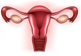
Lab-grown fallopian tubes—or bits of them. A woman’s fallopian tubes, those tiny shafts that transport eggs from the ovary to the uterus, present a major challenge to researchers trying to improve fertility. The cause of infertility for many couples may reside in those tiny tubes but they are almost impossible to study in the developing fetus or adults.

fallopian tubesA German research team made major strides in overcoming this barrier by growing bits of the inner portion of fallopian tubes in the lab. They used cells from the lining of donor fallopian tubes that have stem cell-like qualities and grew them in conditions that mimicked the environment of that portion of the growing embryo. Like many other teams who have grown mini organs, or organoids, they found that if you choose the right cells they have an incredible ability to self-form multilayered complex tissues. In this case the epithelial cells they used formed hollow spheres that have the characteristics of natural fallopian tubes.

 “That happened without any additional instruction whatsoever,” one of the researchers, Mirjana Kessler, told Science alert. “The entire blueprint of the fallopian tube must therefore be stored in the epithelial cells.”

The work is far from being able to offer a woman with damaged fallopian tubes a new chance for fertility. But it does offer researchers a great new tool for studying how the tiny organs form and potentially how to repair them.


And for the men, potential lab grown testicles. The Wake Forest team led by Anthony Atala that has pioneered growing simple organs such as urinary tract bladders from stem cells, has now grown tiny human testicles in the lab. However, none of their miniature organs grown so far could never produce enough product to fully do its job.

 “The future plans are to grow the testicular tissue, expand the cells and put it back into the patient,” Atala, told Motherboard in a story quoted in LatinosHealth. “But for a whole testicle, there is a very rich blood-vessel supply and that’s the challenge. We can make them small, but we’re working hard to make them larger.”

The U.S. Defense Department funds the work because of the number of soldiers who have had their reproductive ability damaged by war injuries. However, everyone on the research team predicts it will be many years before they can make fully functional organs to help out these war heroes.