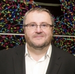
UC Davis Stem Cell Director Jan Nolta Shares Her Thoughts on the Importance of Mentoring Young Scientists

Dr. Jan Nolta (UC Davis Health)

Jan Nolta is a scientific rockstar. She is a Professor at UC Davis and the Director of the Stem Cell Program at the UC Davis School of Medicine. Her lab’s research is dedicated to developing stem cell-based treatments for Huntington’s disease (HD). Jan is a tireless advocate for both stem cell and HD research and you’ll often see her tweeting away about the latest discoveries in the field to her followers.

What I admire most about Dr. Nolta is her dedication to educating and mentoring young students. Dr. Nolta helped write the grant that funded the CIRM Bridges master’s program at Sacramento State in 2009. Over the years, she has mentored many Bridges students (we blogged about one student earlier this year) and also high school students participating in CIRM’s SPARK high school internship program. Many of her young trainees have been accepted to prestigious colleges and universities and gone on to pursue exciting careers in STEM.

I reached out to Dr. Nolta and asked her to share her thoughts on the importance of mentoring young scientists and supporting their career ambitions. Below is a summary of our conversation. I hope her passion and devotion will inspire you to think about how you can get involved with student mentorship in your own career.


Describe your career path from student to professor.

I was an undergraduate student at Sacramento State University. I was a nerdy student and did research on sharks. I was planning to pursue a medical degree, but my mentor, Dr. Laurel Heffernan, encouraged me to consider science. I was flabbergasted at the suggestion and asked, “people pay you to do this stuff??” I didn’t know that you could be paid to do lab research. My life changed that day.

I got my PhD at the University of Southern California. I studied stem cell gene therapy under Don Kohn, who was a fabulous mentor. After that, I worked in LA for 15 years and then went back home to UC Davis in 2007 to direct their Stem Cell Program.

It was shortly after I got to Davis that I reconnected with my first mentor, Dr. Heffernan, and we wrote the CIRM Bridges grant. Davis has a large shared translational lab with seven principle investigators including myself and many of the Bridges students work there. Being a scientist can be stressful with grant deadlines and securing funding. Mentoring students is the best part of the job for me.

Why is it important to fund educational programs like Bridges and SPARK?

There is a serious shortage of well-trained specialists in regenerative medicine in all areas of the workforce. The field of regenerative medicine is still relatively new and there aren’t enough people with the required skills to develop and manufacture stem cell treatments. The CIRM Bridges program is critical because it trains students who will fill those key manufacturing and lab manager jobs. Our Bridges program at Sacramento State is a two-year master’s program in stem cell research and lab management. They are trained at the UC Davis Good Manufacturing Practice (GMP) training facility and learn how to make induced pluripotent stem cells (iPSCs) and other stem cell products. There aren’t that many programs like ours in the country and all of our students get competitive job offers after they complete our program.

We are equally passionate about our high school SPARK program. It’s important to capture students’ interests early whether they want to be a scientist or not. It’s important they get exposed to science as early as possible and even if they aren’t going to be a scientist or healthcare professional, it’s important that they know what it’s about. It’s inspiring how many of these students stay in STEM (Science, Technology, Engineering and Math) because of this unique SPARK experience.

Jan Nolta with the 2016 UC Davis SPARK students.

Can you share a student success story?

I’m so proud of Ranya Odeh. She was a student in our 2016 SPARK program who worked in my lab. Ranya received a prestigious scholarship to Stanford largely due to her participation in the CIRM SPARK program. I got to watch her open the letter on Instagram, and it was a really incredible experience to share that part of her life.

I’m also very proud of our former Bridges student Jasmine Carter. She was a mentor to one of our SPARK students Yasmine this past summer. She was an excellent role model and her passion for teaching and research was an inspiration to all of us. Jasmine was hoping to get into graduate school at UC Davis this fall. She not only was accepted into the Neuroscience Graduate Program, but she also received a prestigious first year program fellowship!

UC Davis Professors Jan Nolta and Kyle Fink with CIRM Bridges student Jasmine Carter

[Side note: We’ve featured Ranya and Jasmine previously on the Stem Cellar and you can read about their experiences here and here.]

Why is mentoring important for young students?

I can definitely relate to the importance of having a mentor. I was raised by a single mom, and without scholarships and great mentors, there’s no way I would be where I am today. I’m always happy to help other students who think maybe they can’t do science because of money, or because they think that other people know more than they do or are better trained. Everybody who wants to work hard and has a passion for science deserves a chance to shine. I think these CIRM educational programs really help the students see that they can be what they dream they can be.

What are your favorite things about being a mentor?

Everyday our lab is full of students, science, laughter and fun. I love coming in to the lab. Our young people bring new ideas, energy and great spirit to our team. I think every team should have young trainees and high school kids working with them because they see things in a different way.

Do you have advice for mentoring young scientists?

You can sum it up in one word: Listen. Ask them right away what their dreams are, where do they imagine themselves in the future, and how can you help them get there. Encourage them to always ask questions and let them know that they aren’t bothering you when they do. I also let my students know that I’m happy to be helping them and that the experience is rewarding for me as well.

So many students are shy when they first start in the lab and don’t get all that they can out of the experience. I always tell my students of any age: what you really want to do is try in life. Follow your tennis ball. Like when a golden retriever sees a tennis ball going by, everything else becomes secondary and they follow that ball. You need to find what that tennis ball is for you and then just try to follow it.

What advice can you give to students who want to be scientific professors or researchers?

Find somebody who is a good mentor and cares about you. Don’t go into a lab where the Principle Investigator (PI) is not there most of the time. You will get a lot more out of the experience if you can get input from the PI.

A good mentor is more present in the lab and will take you to meetings and introduce you to people. I find that often students read papers from well-established scientists, and they think that their positions are unattainable. But if they can meet them in person at a conference or a lecture, they will realize that all of the established scientists are people too. I want young students to know that they can do it too and these careers are attainable for anybody.

Meet the high school student who moonlights as a neuroscientist

As part of our CIRM scholar blog series, we’re featuring the research and career accomplishments of CIRM funded students. Today, you’ll read about one of our former SPARK high school students.


Emma Friedenberg and former CIRM SPARK Director Karen Ring at the 2017 SPARK Conference.

Emma Friedenberg is a high school senior at Campbell Hall in North Hollywood, California. She’s also an up-and-coming neuroscientist who has her sights set on unraveling the complexities of the brain and discovering cures for degenerative brain diseases. Emma spent the summer of 2017 studying Huntington’s disease in the lab of Dr. Virginia Mattis at the Cedars-Sinai Medical Center. Her internship was possible because of the CIRM SPARK high school educational program which gives California students the opportunity to do stem cell research for a summer.

Below is an interview with Emma about her SPARK experience and how the program is helping her pursue her passions for research and medicine.

Q: How did you learn about the CIRM SPARK program and why did you want to apply?

I’ve been a clinical volunteer at Cedars-Sinai Medical Center for two years in the Intensive Care Unit and the Neurology and Spine Unit. I was submitting my application to return as a volunteer when I explored Cedars-Sinai’s Outreach website page and found the CIRM SPARK program. I knew immediately it was a perfect fit. I plan on studying neuroscience in college with an intention of obtaining my medical degree and becoming a surgeon. The CIRM SPARK program at Cedars within the Board of Governor’s Regenerative Medicine Institute had an option to be involved specifically in the Brain Program. In Dr. Virginia Mattis’ lab, I studied translational stem cell therapies for neurodegenerative diseases, in particular Huntington’s Disease. As Cedars-Sinai calls it, a “bench to bedside” approach is an unparalleled and invaluable experience and huge advantage in science.

Q: What was your SPARK research project?

At Cedars-Sinai, I was mentored by Dr. Virginia Mattis in her stem cell lab. The Mattis Lab researches stem cell therapies for Huntington’s disease (HD), a neurodegenerative brain disease. HD is caused by a loss of neurons, specifically medium spiny neurons in the striatum of Huntington’s patients. We used induced pluripotent stem cells to model HD in a petri dish to study the development of the disease and to create medium spiny neurons that could one day be transplanted into Huntington’s patients to replace lost and damaged cells.

Medium spiny neurons made from Huntington’s disease patient induced pluripotent stem cells. (Image credit: Mattis Lab, Cedars Sinai)

My primary research in the Mattis Lab was experimenting on our cell line to find the most time and cost-effective procedure to produce large populations of medium spiny neurons, because current methods are expensive and largely inefficient. However, my internship was not limited to the laboratory. I spent a significant amount of time shadowing doctors in the ALS Clinic.

Q: What was your experience in the CIRM SPARK program like?

In one word, the CIRM SPARK program was incredible –a one of a kind opportunity. The sciences are my personal passion and the cornerstone of my academic pursuits. The CIRM SPARK program has bolstered my scientific knowledge and provided practical experience in a real-world laboratory environment. A career in medicine is a significant commitment, and I’m confident the CIRM SPARK program was a beneficial start to obtaining my goals.

Cedars-Sinai SPARK students celebrating the completion of their 2017 internships.

Q: What do you value most about your SPARK experience?

It was wonderful to be part of a program which understood collaboration and offered a plethora of learning opportunities outside of the wet lab. What I will keep with me is not only techniques of immunocytochemistry and microscopy, but also the advice and encouragement from accomplished scientists like Clive Svendsen and my mentor Virginia Mattis.

Q: What are your future goals?

I plan on studying neuroscience in college with an intention of obtaining my medical degree and becoming a surgeon.

Q: Who is your scientific idol and why?

I recently read Dr. Eric Kandel’s book, The Age of Insight: The Quest to Understand the Unconscious in Art, Mind, and Brain, from Vienna 1900 to the Present. Dr. Kandel is a neuroscientist and a Professor at Columbia University. He received the Nobel Prize for his work in memory storage using Aplysia, a type of sea slug. His book examines how the human brain responds to art. What I find so inspiring about his book is his interdisciplinary approach to science, a combination of neuroscience, psychoanalysis, biology, and art. The human brain is so complicated that it can be studied from numerous perspectives, from biology to chemistry to electrophysiology. It is not until we can begin to merge these understandings that we will begin to unlock the secrets of the brain. Dr. Kandel is not only a scientist, but an intellectual.

Q: What is your favorite thing about being a scientist?

For centuries, the human brain was an anomaly, unexplainable by science. With 100 billion neurons and 100 trillion connections, the brain is the most complex network in the universe. How the brain functions as an information-processing organ and regulates emotion, behavior, and cognition as well as basic body functions like breathing remains a mystery. In recent years, there has been significant progress in brain research. Scientists are on the brink of major breakthroughs, but there is significant work to do particularly on neurological brain disorders. Being a scientist means living on the cutting-edge of human innovation. I enjoy being able to both ask and answer questions that will benefit humankind.


Related Links:

The Story of a South African Bubble Boy and a Gene Therapy That Gave Him His Life Back

Ayaan Isaacs, health24

Ayaan Isaacs was born in South Africa on March 4th, 2016 as a seemingly healthy baby. But only a few days in to life, he contracted a life-threatening liver infection. He thankfully survived, only to have the doctors discover a few weeks later that he had something much more troubling – a rare disease that left him without a functioning immune system.

Ayaan was diagnosed with X-linked severe combined immunodeficiency (SCID), which is often referred to as ‘bubble baby’ disease because patients are extremely susceptible to infection and must live in sterile environments. SCID patients can be cured with a blood stem cell transplant if they have a genetically matched donor. Unfortunately for Ayaan, only a partially matched donor was available, which doesn’t guarantee a positive outcome.

Ayaan’s parents were desperate for an alternative treatment to save Ayaan’s life. It was at this point that they learned about a clinical trial at St. Jude Children’s Research hospital in Memphis, Tennessee. The trial is treating SCID patients with a stem cell gene therapy that aims to give them a new functioning immune system. The therapy involves extracting the patient’s blood-forming stem cells and genetically correcting the mutation that causes SCID. The corrected blood stem cells are then transplanted back into the patient where they rebuild a healthy immune system.

Ayaan was able to enroll in the trial, and he was the first child in Africa to receive this life-saving gene therapy treatment. Ayaan’s journey with bubble boy disease was featured by South Africa’s health24 earlier this year. In the article, his mom Shamma Sheik talked about the hope that this gene therapy treatment brought to their family.

“No child should have to die just because they are unable to find a donor. Gene therapy offered Ayaan a chance at life that he ordinarily would not have had. I was fortunate to have found an alternative therapy that is working and already showing remarkable results. We are mindful that this is still an experimental treatment and there are complications that can arise; however, I am very optimistic that he will return to South Africa with a functioning immune system.”

Carte Blanche, an investigative journalism program in South Africa, did a feature video of Ayaan in February. Although the video is no longer available on their website, it did reveal that four months after Ayaan’s treatment, his condition started to improve suggesting that the treatment was potentially working.

We’ve written previously about another young boy named Ronnie who was diagnosed with X-linked SCID days after he was born. Ronnie also received the St. Jude stem cell gene therapy in a CIRM-funded clinical trial at the UCSF Benioff Children’s Hospital. Ronnie was treated when he was six months old and just celebrated his first birthday as a healthy, vibrant kid thanks to this trial. You can hear more about Ronnie’s moving story from his dad, Pawash Priyank, in the video below.

Our hope is that powerful stories like Ayaan’s and Ronnie’s will raise awareness about SCID and the promising potential of stem cell gene therapies to cure patients of this life-threatening immune disease.

Ronnie and his parents celebrating his 1st birthday. (Photo courtesy of Pawash Priyank)


Related Links:

Celebrating Exciting CIRM-Funded Discovery Research on World Parkinson’s Day

April 11th is World Parkinson’s Disease Awareness Day. To mark the occasion, we’re featuring the work of CIRM-funded researchers who are pursuing new, promising ideas to treat patients with this debilitating neurodegenerative disease.


Birgitt Schuele, Parkinson’s Institute

CIRM Grant: Quest Award – Discovery Stage Research

Research: Birgitt and her team at the Parkinson’s Institute in Sunnyvale, California, are using CRISPR gene editing technology to reduce the levels of a toxic protein called alpha synuclein, which builds up in the dopaminergic brain cells affected by Parkinson’s disease.

Birgitt Schuele

“My hope is that I can contribute to stopping disease progression in Parkinson’s. If we can develop a drug that can get rid of accumulated protein in someone’s brain that should stop the cells from dying. If someone has early onset PD and a slight tremor and minor walking problems, stopping the disease and having a low dose of dopamine therapy to control symptoms is almost a cure.”

Parkinson’s disease in a dish. Dopaminergic neurons made from Parkinson’s patient induced pluripotent stem cells. (Image credit: Birgitt Schuele)


Jeanne Loring, Scripps Research Institute

CIRM Grant: Quest Award – Discovery Stage Research

Research: Jeanne Loring and her team at the Scripps Research Institute in La Jolla, California, are deriving dopaminergic neurons from the iPSCs of Parkinson’s patients. Their goal is to develop a personalized, stem cell-based therapy for PD.

Jeanne Loring

“We are working toward a patient-specific neuron replacement therapy for Parkinson’s disease.  By the time PD is diagnosed, people have lost more than half of their dopamine neurons in a specific part of the brain, and loss continues over time.  No drug can stop the loss or restore the neurons’ function, so the best possible option for long term relief of symptoms is to replace the dopamine neurons that have died.  We do this by making induced pluripotent stem cells from individual PD patients and turning them into the exact type of dopamine neuron that has been lost.  By transplanting a patient’s own cells, we will not need to use potentially dangerous immunosuppressive drugs.  We plan to begin treating patients in a year to two years, after we are granted FDA approval for the clinical therapy.”

Skin cells from a Parkinson’s patient (left) were reprogrammed into induced pluripotent stem cells (center) that were matured into dopaminergic neurons (right) to model Parkinson’s disease. (Image credit: Jeanne Loring)


Justin Cooper-White, Scaled BioLabs Inc.

CIRM Grant: Quest Award – Discovery Stage Research

Research: Justin Cooper-White and his team at Scaled Biolabs in San Francisco are developing a tool that will make clinical-grade dopaminergic neurons from the iPSCs of PD patients in a rapid and cost-effective manner.

Justin Cooper-White

“Treating Parkinson’s disease with iPSC-derived dopaminergic neuron transplantation has a strong scientific and clinical rationale. Even the best protocols are long and complex and generally have highly variable quality and yield of dopaminergic neurons. Scaled Biolabs has developed a technology platform based on high throughput microfluidics, automation, and deep data which can optimize and simplify the road from iPSC to dopaminergic neuron, making it more efficient and allowing a rapid transition to GMP-grade derivation of these cells.  In our first 6 months of CIRM-funded work, we believe we have already accelerated and simplified the production of a key intermediate progenitor population, increasing the purity from the currently reported 40-60% to more than 90%. The ultimate goal of this work is to get dopaminergic neurons to the clinic in a robust and economical manner and accelerate treatment for Parkinson’s patients.”

High throughput differentiation of dopaminergic neuron progenitors in  microbioreactor chambers in Scaled Biolabs’ cell optimization platform. Different chambers receive different differentiation factors, so that optimal treatments for conversion to dual-positive cells can be determined (blue: nuclei, red: FOXA2, green: LMX1A).


Xinnan Wang, Stanford University

CIRM Grant: Basic Biology V

Research: Xinnan Wang and her team at Stanford University are studying the role of mitochondrial dysfunction in the brain cells affected in Parkinson’s disease.

Xinnan Wang

“Mitochondria are a cell’s power plants that provide almost all the energy a cell needs. When these cellular power plants are damaged by stressful factors present in aging neurons, they release toxins (reactive oxygen species) to the rest of the neuron that can cause neuronal cell death (neurodegeneration).  We hypothesized that in Parkinson’s mutant neurons, mitochondrial quality control is impaired thereby leading to neurodegeneration. We aimed to test this hypothesis using neurons directly derived from Parkinson’s patients (induced pluripotent stem cell-derived neurons).”

Dopaminergic neurons derived from human iPSCs shown in green, yellow and red. (Image credit: Atossa Shaltouki, Stanford)


Related Blogs:

Stem Cell Roundup: watching brain cells in real time, building better heart cells, and the plot thickens on the adult neurogenesis debate

Here are the stem cell stories that caught our eye this week.

Watching brain cells in real time

This illustration depicts a new method that enables scientists to see an astrocyte (green) physically interacting with a neuronal synapse (red) in real time, and producing an optical signal (yellow). (Khakh Lab, UCLA Health)

Our stem cell photo of the week is brought to you by the Khakh lab at UCLA Health. The lab developed a new method that allows scientists to watch brain cells interact in real time. Using a technique called fluorescence resonance energy-transfer (FRET) microscopy, the team can visualize how astrocytes (key support cells in our central nervous system) and brain cells called neurons form connections in the mouse brain and how these connections are affected by diseases like Alzheimer’s and ALS.

Baljit Khakh, the study’s first author, explained the importance of their findings in a news release:

“This new tool makes possible experiments that we have been wanting to perform for many years. For example, we can now observe how brain damage alters the way that astrocytes interact with neurons and develop strategies to address these changes.”

The study was published this week in the journal Neuron.


Turn up the power: How to build a better heart cell (Todd Dubnicoff)

For years now, researchers have had the know-how to reprogram a donor’s skin cells into induced pluripotent stem cells (iPSCs) and then specialize them into heart muscle cells called cardiomyocytes. The intervening years have focused on optimizing this method to accurately model the biology of the adult human heart as a means to test drug toxicity and ultimately develop therapies for heart disease. Reporting this week in Nature, scientists at Columbia University report an important step toward those goals.

The muscle contractions of a beating heart occur through natural electrical impulses generated by pacemaker cells. In the case of lab-grown cardiomyocytes, introducing mechanical and electrical stimulation is required to reliably generate these cells. In the current study, the research team showed that the timing and amount of stimulation is a critical aspect to the procedure.

The iPS-derived cardiomyocytes have formed heart tissue that closely mimics human heart functionality at over four weeks of maturation. Credit: Gordana Vunjak-Novakovic/Columbia University.

The team tested three scenarios on iPSC-derived cardiomyocytes (iPSC-CMs): no electrical stimulation for 3 weeks, constant stimulation for 3 weeks, and finally, two weeks of increasingly higher stimulation followed by a week of constant stimulation. This third setup mimics the changes that occur in a baby’s heart just before and just after birth.

These scenarios were tested in 12 day-old and 28 day-old iPSC-CMs. The results show that only the 12 day-old cells subjected to the increasing amounts of stimulation gave rise to fully mature heart muscle cells. On top of that, it only took four weeks to make those cells. Seila Selimovic, Ph.D., an expert at the National Institutes of Health who was not involved in the study, explained the importance of these findings in a press release:

“The resulting engineered tissue is truly unprecedented in its similarity to functioning human tissue. The ability to develop mature cardiac tissue in such a short time is an important step in moving us closer to having reliable human tissue models for drug testing.”

Read more at: https://phys.org/news/2018-04-early-bioengineered-human-heart-cells.html#jCp


Yes we do, no we don’t. More confusion over growing new brain cells as we grow older (Kevin McCormack)

First we didn’t, then we did, then we didn’t again, now we do again. Or maybe we do again.

The debate over whether we are able to continue making new neurons as we get older took another twist this week. Scientists at Columbia University said their research shows we do make new neurons in our brain, even as we age.

This image shows what scientists say is a new neuron in the brain of an older human. A new study suggests that humans continue to make new neurons throughout their lives. (Columbia University Irving Medical Center)

In the study, published in the journal Cell Stem Cell, the researchers examined the brains of 28 deceased donors aged 14 to 79. They found similar numbers of precursor and immature neurons in all the brains, suggesting we continue to develop new brain cells as we age.

This contrasts with a UCSF study published just last month which came to the opposite conclusion, that there was no evidence we make new brain cells as we age.

In an interview in the LA Times, Dr. Maura Boldrini, the lead author on the new study, says they looked at a whole section of the brain rather than the thin tissues slices the UCSF team used:

“In science, the absence of evidence is not evidence of absence. If you can’t find something it doesn’t mean that it is not there 100%.”

Well, that resolves that debate. At least until the next study.

CIRM’s Industry Alliance Program: Facilitating Partnerships to Advance Stem Cell Therapies

Some things are better together. Take for instance macaroni and cheese, eggs and bacon, cookies and ice cream. Each of these things are fine on their own, but together, they become something more powerful and delicious.

The right partnerships can bring out the best in things. At CIRM, we fully embrace this concept. That’s why we’re launching the Industry Alliance Program (IAP). It’s a new partnering opportunity to bring the most promising stem cell, gene therapy, and regenerative medicine programs to market where they can help people with unmet medical needs.

CIRM is the world’s largest stem cell research funding institution dedicated to helping patients by accelerating the development of quality stem cell treatments. We’re currently funding 244 active stem cell research programs including 39 ongoing clinical trials.

The CIRM IAP is designed to give pharma, biotech and VC firms direct access to CIRM’s growing stem cell portfolio. These partners work in the stem cell and regenerative medicine field and will be connected to CIRM-funded scientists working on projects relevant to their interests.

In a news release, CIRM’s President and CEO, Dr. Maria T. Millan, explained:

Maria T. Millan

“The goal of the IAP is to secure industry partnerships and funding for CIRM’s translational and clinical-stage projects. Our Agency provides researchers the initial funding to advance promising projects towards the clinic. Now, we’re going a step further by offering a program that facilitates connections between industry partners and our grantees. These companies can offer support or additional funding needed to give these promising projects the best chance for success and the best chance of helping patients.”

The first two companies to join the IAP are BlueRock Therapeutics and Vivo Capital. BlueRock is a Cambridge, Massachusetts-based company that is pioneering cell therapies for degenerative diseases while Vivo Capital is a global venture capital firm that invests in life sciences and healthcare companies.

CIRM will continue to selectively recruit new partners to the IAP with the goal of building a collaborative network to support the development and commercialization of CIRM-funded programs.

Neil Littman, CIRM’s Director of Business Development, concluded:

Neil Littman

“The IAP is essentially a built-in concierge service for the stem cell space. Our unique vantage-point both inside and outside of California – spanning discovery, translation, and clinical trials – allows us to effectively match CIRM-funded programs with the strategic objectives of our IAP partners.  We’re excited to work with partners such as BlueRock and Vivo who have a demonstrated commitment to advance stem cell-based therapies to the market.”

For more information about CIRM’s new IAP program, visit our website.

Stem Cell Roundup: Crafty Cancer, Fighting Viruses, and Brainstorm ALS Trial Expands to Canada

TGIF! Here is your weekly dose of stem cell news…

Shapeshifting cancer cells

This week’s awesome stem cell photo comes with a bizarre story and bonus video footage.

New research from Duke has found that some lung cancer cells with errors in transcription factors begin to resemble their nearest relatives – the cells of the stomach and gut. (Credit – Tata Lab, Duke University)

Researchers at Duke University were studying lung tumor samples and discovered something that didn’t quite belong. Inside the lung tumors were miniature parts of the digestive system including the stomach, duodenum and small intestine. It turns out that the lung cancer cells (and cancer cells in general) are super crafty and had turned off the expression of a gene called NKX2-1. This gene is a master switch that tells developing cells to turn into lung cells. Without this command, cells switch their identity and mature into gut tissue instead. By manipulating these master switches, cancer cells are able to develop resistance to chemotherapy and other cancer treatments.

So, what does this bizarre finding mean for cancer research? Purushothama Rao Tata, first author on the Developmental Cell study, provided an answer in a news release:

“Cancer biologists have long suspected that cancer cells could shape shift in order to evade chemotherapy and acquire resistance, but they didn’t know the mechanisms behind such plasticity. Now that we know what we are dealing with in these tumors – we can think ahead to the possible paths these cells might take and design therapies to block them.”

For more cool photos and insights into this study, watch the Duke Univeristy video below.


Secrets to the viral-fighting ability of stem cells uncovered (Todd Dubnicoff)

I’ve been writing about stem cells for many years and thought I knew most of the basic info about these amazing cells. But up until this week, I had no idea that stem cells are known to fight off viral infections much better than other cells. It does makes sense though. Stem cells give rise to and help maintain all the organs and tissues of the body. So, it would be bad news if, let’s say, a muscle stem cell multiplied to repair damaged tissue while carrying a dangerous virus.

How exactly stem cells fend off attacking viruses is a question that has eluded researchers for decades. But this week, results published in Cell by Rockefeller University scientists may provide an answer.

Stem cells lacking their protective genes are susceptible to infection by the dengue virus, in red. (Rockefeller University)

The researchers found that liver cells and stem cells defend themselves against viruses differently. In the presence of a virus, liver cells and most other cells react by releasing large amounts of interferon, a protein that acts as a distress signal to other cells in the vicinity. That signal activates hundreds of genes responsible for attracting protective immune cells to the site of infection.

Stem cells, however, are always in this state of emergency. Even in the absence of interferon, the antiviral genes were activated in stem cells. And when the stem cells were genetically engineering to lack some of the antiviral genes, the cells no longer could stop viral infection.

In a press release, senior author Charles Rice explained the importance of this work:

“By understanding more about this biology in stem cells, we may learn more about antiviral mechanisms in general.”


CIRM-funded clinical trial for ALS now available next door – in Canada (Kevin McCormack)

In kindergarten we are taught that it’s good to share. So, we are delighted that a Phase 3 clinical trial for ALS – also known as Lou Gehrig’s disease – that CIRM is helping fund is now expanding its reach across the border from the U.S. into Canada.

Brainstorm Cell Therapeutics, the company behind the therapy, says it is going to open a clinical trial site in Canada because so many Canadians have asked for it.

The therapy, as we described in a recent blog post, takes mesenchymal stem cells from the patient’s own bone marrow. Those cells are then modified in the lab to be able to churn out specific proteins that can help protect the brain cells attacked by ALS. The cells are then transplanted back into the patient and the hope is they will slow down, maybe even stop the progression of the disease.

Earlier studies showed the therapy was safe and seemed to benefit some patients. Now people with ALS across our northern border will get a chance to see if it really works.

Chaim Lebovits, the president and chief executive officer of BrainStorm, said in a press release:

“Although there are thousands of patients worldwide with ALS, we initially designed the Phase 3 trial to enroll U.S.-based patients only, primarily to make it easier for patient follow-up visits at the six U.S. clinical sites. However, due to an outpouring of inquiry and support from Canadian patients wanting to enroll in the trial, we filed an amendment with the FDA [the U.S. Food and Drug Administration] to allow Canada-based ALS patients to participate.”

We are happy to share.

Say Hello to CIRM’s New Active Awards Portfolio Dashboard (Video Included!)

It takes a lot of time, money and effort to develop a promising stem cell research idea into an effective treatment that can help patients. Oftentimes, you don’t hear about the early-stage research that goes into developing a particular treatment until it reaches the clinic.

CIRM recognizes the importance of investing in all stages of stem cell research and has an impressive portfolio of over 160 active projects spanning discovery, translation, and clinical-stage research.

To help you understand the breadth of our funding efforts, and to highlight our expanding research pipeline, we’ve created the Active Awards Portfolio Dashboard on our website. This interactive tool makes it easy to search through the active research projects that we’re currently funding, and filter these projects by disease focus, technology type or stage of research.

Watch the short video below to learn more about our new Dashboard and how to use it.

The Active Awards Dashboard reflects our Agency’s commitment to investing in the full range of stem cell research and to helping the most promising research projects advance to the next level.

For those of you interested in learning more about the 45 active clinical trials we’re funding, be sure to check out the companion Clinical Trials Dashboard on our website, featured previously on the Stem Cellar blog.

Tiny blood vessels in the brain can spur the growth of spinal motor neurons

Last week, researchers from Cedars-Sinai Medical Center added a new piece to the complex puzzle of what causes neurodegenerative disorders like amyotrophic lateral sclerosis (ALS). The team discovered that the tiny blood vessels in our brains do more than provide nutrients to and remove waste products from our brain tissue. It turns out that these blood vessels can stimulate the growth of new nerve cells called spinal motor neurons, which directly connect to the muscles in our body and control how they move. The study, which was funded in part by a CIRM Discovery research-stage Inception award, was published in the journal Stem Cell Reports.

The Cedars team used a combination of human induced pluripotent stem cells (iPSCs) and organ-on-a-chip technology to model the cellular microenvironment of the spinal cord. They matured the iPSCs into both spinal motor progenitor cells and brain endothelial cells (which line the insides of blood vessels). These cells were transferred to an organ-chip where they were able to make direct contact and interact with each other.

Layers of spinal motor neuron cells (top, in blue) and capillary cells (bottom, in red) converge inside an Organ-Chip. Neurons and capillary cells interact together along the length of the chip. (Cedars-Sinai Board of Governors Regenerative Medicine Institute).

The researchers discovered that exposing the spinal motor progenitor cells to the blood vessel endothelial cells in these organ-chips activated the expression of genes that directed these progenitor cells to mature into spinal cord motor neurons.

Hundreds of spinal motor neurons spontaneously communicate through electrical signals inside an Organ-Chip. Neurons fire individually (flashing dots) and in synchronized bursts (bright waves). (Cedars-Sinai)

First author on the study, Samuel Sances, explained their findings in a news release:

“Until now, people thought these blood vessels just delivered nutrients and oxygen, removed waste and adjusted blood flow. We showed that beyond plumbing, they are genetically communicating with the neurons.”

The team also showed the power of stem cell-based organ-chip platforms for modeling diseases like ALS and answering key questions about why these diseases occur.

“What may go wrong in the spinal neurons that causes the motor neurons to die?” Sances asked. “If we can model an individual ALS patient’s tissues, we may be able to answer that question and one day rescue ALS patients’ neurons through new therapies.”

Clive Svendsen, a CIRM grantee and the senior author on the study, said that his team will conduct additional studies using organ-chip technology to study the interactions between iPSC-derived neurons and blood vessels of healthy individuals and ALS patients. Differences in these cellular interactions in diseased patient cells could offer new targets for developing ALS therapies.

The current study is a collaboration between Cedars and a Boston company called Emulate, Inc. Emulate developed the organ-chip technology and is collaborating with Svendsen at Cedars to not only model neurodegenerative diseases, but also model other organ systems. Be sure to check out our recent blog about their efforts to create a stem cell-based gut-on-a-chip, which they hope will pave the way for personalized treatments for patients with gastrointestinal diseases like Chrohn’s and inflammatory bowel disease.

Stem Cell Patch Restores Vision in Patients with Age-Related Macular Degeneration

Stem cell-derived retinal pigmented epithelial cells. Cell borders are green and nuclei are red. (Photo Credit: Dennis Clegg, UCSB Center for Stem Cell Biology and Engineering)

Two UK patients suffering from vision loss caused by age-related macular degeneration (AMD) have regained their sight thanks to a stem cell-based retinal patch developed by researchers from UC Santa Barbara (UCSB). The preliminary results of this promising Phase 1 clinical study were published yesterday in the journal Nature Biotechnology.

AMD is one of the leading causes of blindness and affects over six million people around the world. The disease causes the blurring or complete loss of central vision because of damage to an area of the retina called the macula. There are different stages (early, intermediate, late) and forms of AMD (wet and dry). The most common form is dry AMD which occurs in 90% of patients and is characterized by a slow progression of the disease.

Patching Up Vision Loss

In the current study, UCSB researchers engineered a retinal patch from human embryonic stem cells. These stem cells were matured into a layer of cells at the back of the eye, called the retinal pigment epithelium (RPE), that are damaged in AMD patients. The RPE layer was placed on a synthetic patch that is implanted under the patient’s retina to replace the damaged cells and hopefully improve the patient’s vision.

The stem cell-based eyepatches are being implanted in patients with severe vision loss caused by the wet form of AMD in a Phase 1 clinical trial at the Moorfields Eye Hospital NHS Foundation Trust in London, England. The trial was initiated by the London Project to Cure Blindness, which was born from a collaboration between UCSB Professor Peter Coffey and Moorsfields retinal surgeon Lyndon da Cruz. Coffey is a CIRM grantee and credited a CIRM Research Leadership award as one of the grants that supported this current study.

The trial treated a total of 10 patients with the engineered patches and reported 12-month data for two of these patients (a woman in her 60s and a man in his 80s) in the Nature Biotech study. All patients were given local immunosuppression to prevent the rejection of the implanted retinal patches. The study reported “three serious adverse events” that required patients to be readmitted to the hospital, but all were successfully treated. 12-months after treatment, the two patients experienced a significant improvement in their vision and went from not being able to read at all to reading 60-80 words per minute using normal reading glasses.

Successfully Restoring Sight

Douglas Waters, the male patient reported on, was diagnosed with wet AMD in July 2015 and received the treatment in his right eye a few months later. He spoke about the remarkable improvement in his vision following the trial in a news release:

“In the months before the operation my sight was really poor, and I couldn’t see anything out of my right eye. I was struggling to see things clearly, even when up-close. After the surgery my eyesight improved to the point where I can now read the newspaper and help my wife out with the gardening. It’s brilliant what the team have done, and I feel so lucky to have been given my sight back.”

This treatment is “the first description of a complete engineered tissue that has been successfully used in this way.” It’s exciting not only that both patients had a dramatic improvement in their vision, but also that the engineered patches were successful at treating an advanced stage of AMD.

The team will continue to monitor the patients in this trial for the next five years to make sure that the treatment is safe and doesn’t cause tumors or other adverse effects. Peter Coffey highlighted the significance of this study and what it means for patients suffering from AMD in a UCSB news release:

Peter Coffey

“This study represents real progress in regenerative medicine and opens the door to new treatment options for people with age-related macular degeneration. We hope this will lead to an affordable ‘off-the-shelf’ therapy that could be made available to NHS patients within the next five years.”