Novartis acquisition validates CIRM’s impact on cystinosis treatment

Dr. Stephanie Cherqui

The future looks brighter for cystinosis patients and their families.  

As one of 7,000 rare diseases, cystinosis causes an abnormal build-up of the amino acid cystine that can lead to organ failure and premature death. With little to no treatment options, children and young adults primarily affected by the condition often have a poor prognosis – until now.  

Thanks to $17 million in early funding from the California Institute for Regenerative Medicine (CIRM) and the unwavering dedication of researchers like Dr. Stephanie Cherqui at UC San Diego (UCSD), a gene therapy to address the genetic defects of cystinosis is making its way through the field.

Cherqui’s stem cell gene therapy offers a potential breakthrough by modifying a patient’s own blood stem cells with a functional version of the defective CTNS gene to address the underlying cause of the disease.  

Last month, Novartis, a pharmaceutical company with extensive expertise in clinical-stage gene therapy and access to infrastructure, purchased the gene therapy for $87.5 million from AVROBIO. CIRM funded the preclinical work for this study, which involved completing the testing needed to apply to the Food and Drug Administration (FDA) as well as a subsequent clinical trial.  

“CIRM played a significant role in advancing the stem cell gene therapy program for cystinosis from bed-to-bedside. Through its funding and support, CIRM has allowed me to perform the pre-clinical studies required for an investigational new drug application, and then to conduct the first phase of the clinical trial in an academic setting,” said Dr. Cherqui.  

Not only will the Novartis acquisition expedite the global availability of this therapy for people with cystinosis, but it also affirms CIRM’s pivotal role in the development of early-stage research that can turn into tangible treatments. 

“The Novartis acquisition has the potential to accelerate clinical development of novel hematopoietic gene therapy for cystinosis patients. We’re excited for the cystinosis patient community as well as the UCSD, UCLA and AVROBIO teams that led this advancement,” said Dr. Shyam Patel, Senior Director of Business Development and Alliance Management at CIRM. “Furthermore, the Novartis acquisition validates CIRM’s Clinical funding model, which has supported this program since 2016 through preclinical studies as well as through the ongoing first-in-human clinical trial that was coordinated through the UCSD CIRM Alpha Clinic.” 

The impact of this acquisition goes far beyond the walls of research laboratories. For those with cystinosis, it brings a renewed sense of hope and possibility.  

Making transplants easier for kids, and charting a new approach to fighting solid tumors.

Every year California performs around 100 kidney transplants in children but, on average, around 50 of these patients will have their body reject the transplant. These children then have to undergo regular dialysis while waiting for a new organ. Even the successful transplants require a lifetime of immunosuppression medications. These medications can prevent rejection but they also increase the risk of infection, gastrointestinal disease, pancreatitis and cancer.

Dr. Alice Bertaina and her team at Stanford University were awarded $11,998,188 to test an approach that uses combined blood stem cell (HSC) and kidney transplantation with the goal to improve outcomes with kidney transplantation in children. This approach seeks to improve on the blood stem cell preparation through an immune-based purification process.

In this approach, the donor HSC are transplanted into the patient in order to prepare for the acceptance of the donor kidney once transplanted. Donor HSC give rise to cells and conditions that re-train the immune system to accept the kidney. This creates a “tolerance” to the transplanted kidney providing the opportunity to avoid long-term need for medications that suppress the immune system.

Pre-clinical data support the idea that this approach could enable the patient to stop taking any immunosuppression medications within 90 days of the surgery.

Dr. Maria T. Millan, President and CEO of CIRM, a former pediatric transplant surgeon and tolerance researcher states that “developing a way to ensure long-term success of organ transplantation by averting immune rejection while avoiding the side-effects of life-long immunosuppression medications would greatly benefit these children.”

The CIRM Board also awarded $7,141,843 to Dr. Ivan King and Tachyon Therapeutics, Inc to test a drug showing promise in blocking the proliferation of cancer stem cells in solid tumors such as colorectal and gastrointestinal cancer.

Patients with late-stage colorectal cancer are typically given chemotherapy to help stop or slow down the progression of the disease. However, even with this intervention survival rates are low, usually not more than two years.

Tachyon’s medication, called TACH101, is intended to target colorectal cancer (CRC) stem cells as well as the bulk tumor by blocking an enzyme called KDM4, which cancer stem cells need to grow and proliferate.

In the first phase of this trial Dr. King and his team will recruit patients with advanced or metastatic solid tumors to assess the safety of TACH101, and determine what is the safest maximum dose. In the second phase of the trial, patients with gastrointestinal tumors and colorectal cancer will be treated using the dose determined in the first phase, to determine how well the tumors respond to treatment.  

The CIRM Board also awarded $5,999,919 to Dr. Natalia Gomez-Ospina and her team at Stanford University for a late-stage preclinical program targeting Severe Mucopolysaccharidosis type 1, also known as Hurler syndrome. This is an inherited condition caused by a faulty gene. Children with Hurler syndrome lack an enzyme that the body needs to digest sugar. As a result, undigested sugar molecules build up in the body, causing progressive damage to the brain, heart, and other organs. There is no effective treatment and life expectancy for many of these children is only around ten years.

Dr. Gomez-Ospina will use the patient’s own blood stem cells that have been genetically edited to restore the missing enzyme. The goal of this preclinical program is to show the team can manufacture the needed cells, to complete safety studies and to apply to the US Food and Drug Administration for an Investigational New Drug (IND), the authorization needed to begin a clinical trial in people.

Finally the Board awarded $20,401,260 to five programs as part of its Translational program. The goal of the Translational program is to support promising stem cell-based or gene projects that accelerate completion of translational stage activities necessary for advancement to clinical study or broad end use. Those can include therapeutic candidates, diagnostic methods  or devices and novel tools that address critical bottlenecks in research.

The successful applicants are:

APPLICATIONTITLEPRINCIPAL INVESTIGATOR – INSTITUTIONAMOUNT  
TRAN4-14124Cell Villages and Clinical Trial in a Dish with Pooled iPSC-CMs for Drug DiscoveryNikesh Kotecha — Greenstone Biosciences  $1,350,000
TRAN1-14003Specific Targeting Hypoxia Metastatic Breast Tumor with Allogeneic Off-the-Shelf Anti-EGFR CAR NK Cells Expressing an ODD domain of HIF-1αJianhua Yu — Beckman Research Institute of City of Hope  $6,036,002  
TRAN1-13983CRISPR/Cas9-mediated gene editing of Hematopoietic
stem and progenitor cells for Friedreich’s ataxia
Stephanie Cherqui — University of California, San Diego  $4,846,579
TRAN1-13997Development of a Gene Therapy for the Treatment of
Pitt Hopkins Syndrome (PHS) – Translating from Animal Proof of Concept to Support Pre-IND Meeting
Allyson Berent — Mahzi Therapeutics  $4,000,000
TRAN1-13996Overcoming resistance to standard CD19-targeted CAR
T using a novel triple antigen targeted vector
William J Murphy — University of California, Davis  $4,168,679

Reasons to be thankful this Thanksgiving: creative nerds

We at the California Institute for Regenerative Medicine have a lot to be thankful for this Thanksgiving. We get to work with some extraordinary colleagues, we get to know some remarkable patient advocates who are pioneers in volunteering for stem cell and gene therapies, and we have a front row seat in a movement that is changing the face of medicine.

We also get to work with some brilliant scientists and help support their research. As if we needed any reminders of how important that funding is, we thought we would share this video with you. It’s from the talented post docs and researchers at the University of California San Diego. It’s a delightful parody of the Cyndi Lauper classic “Girls Just Wanna Have Fun”. Only in this case it’s “Nerds Just Wanna Have Funds.”

Enjoy, and Happy Thanksgiving.

How the Tooth Fairy is helping unlock the secrets of autism

Our 2021-22 Annual Report is now online. It’s filled with information about the work we have done over the last year (we are on a fiscal calendar year from July 1 – June 30), the people who have helped us do that work, and some of the people who have benefited from that work. One of those is Dr. Alysson Muotri, a professor in the Departments of Pediatrics and Cellular & Molecular Medicine at the University of California, San Diego.

Dr. Alysson Muotri, in his lab at UCSD

For Dr. Alysson Muotri, trying to unlock the secrets of the brain isn’t just a matter of scientific curiosity, it’s personal. He has a son with autism and Dr. Muotri is looking for ways to help him, and millions of others like him around the world.

He created the Tooth Fairy project where parents donated more than 3,000 baby teeth from  children with autism and children who are developing normally. Dr. Muotri then turned cells from those teeth into neurons, the kind of brain cell affected by autism. He is using those cells to try and identify how the brain of a child with autism differs from a child who is developing normally.

“We’ve been using cells from this population to see what are the alterations (in the gene) and if we can revert them back to a normal state. If you know the gene that is affected, and autism has a strong genetic component, by genome sequencing you can actually find what are the genes that are affected and in some cases there are good candidates for gene therapy. So, you just put the gene back. And we can see that in the lab where we are correcting the gene that is mutated, the networks start to function in a way that is more neurotypical or normal. We see that as highly promising, there’s a huge potential here to help those individuals.”

He is also creating brain organoids, three-dimensional structures created from stem cells that mimic some of the actions and activities of the brain. Because these are made from human cells, not mice or other animals, they may be better at indicating if new therapies have any potential risks for people.

“We can test drugs in the brain organoids of the person and see if it works, see if there’s any toxicity before you actually give the drug to a person, and it will save us time and money and will increase our knowledge about the human brain.”

He says he still gets excited seeing how these cells work. “It’s amazing, it’s a miracle. Every time I see it, it’s like seeing dolphins in the sea because it’s so beautiful.”

Dr. Muotri is also a big proponent of diversity, equity and inclusion in scientific research. He says in the past it was very much a top-down model with scientists deciding what was important. He says we need to change that and give patients and communities a bigger role in shaping the direction of research.

“I think this is something we scientists have to learn, how to incorporate patients in our research. These communities are the ones we are studying, and we need to know what they want and not assume that what we want is what they want. They should be consulted on our grants, and they should participate in the design of our experiments. That is the future.”

An experimental gene therapy with a hairy twist

In October 2019, 20-year-old Jordan Janz became the first person in the world to receive an experimental therapy for cystinosis. Cystinosis is a rare genetic disorder characterized by the accumulation of an amino acid called cystine in different tissues and organs of the body including the kidneys, eyes, muscles, liver, pancreas, and brain. This accumulation of cystine ultimately leads to multi-organ failure, eventually causing premature death in early adulthood. On average, cystinosis patients live to 28.5 years old. By that calculation, Janz didn’t have a lot of time.

The treatment was grueling but worth it. The experimental gene therapy funded by the California Institute for Regenerative Medicine seemed to work and Janz began to feel better. There was, however, an unexpected change. Janz’s almost white, blonde hair had settled into a darker tone. Of all the things the gene therapy was expected to alter such as the severity of his cystinosis symptoms hair color was not one of them. Eventually, the same phenomenon played out in other people: So far in the gene-therapy trial, four of the five patients all of whom are white have gotten darker hair.

The outcome, while surprising to researchers, didn’t seem to be a sign of something going awry, instead they determined that it might be a very visible sign of the gene therapy working.

The sudden hair-color changes were surprising to Stephanie Cherqui, a stem-cell scientist at UC San Diego and the principal investigator of the gene-therapy trial. However, it didn’t seem to be a sign of something going awry, instead Cherqui and her colleagues determined that it might be a very visible sign of the gene therapy working.

But exactly how did genetically modifying Janz’s (and other participants’) blood cells change his hair color? In this instance, scientists chose to genetically tweak blood stem cells because they have a special ability: Some eventually become white blood cells, which then travel to all different parts of the body.

Janz’s new white blood cells were genetically modified to express the gene that is mutated in cystinosis, called CTNS. Once they traveled to his eyes, skin, and gut, the white blood cells began pumping out the missing protein encoded by the gene. Cells in the area began taking up the protein and clearing away long accumulated cystine crystals. In Janz, the anti-cystine proteins from his modified blood cells must have reached the hair follicles in his skin. There, they cleared out the excess cystine that was blocking normal melanin production, and his hair got darker.

Hair color is one way in which patients in the clinical trial are teaching scientists about the full scope of the CTNS gene. The investigators have since added hair biopsies to the trial in order to track the color changes in a more systematic fashion.

Read the full article on The Atlantic.

Meet the man who is unlocking the secrets of autism and sending mini-brains into space

THIS BLOG IS ALSO AVAILABLE AS AN AUDIO CAST

Dr. Alysson Muotri, UC San Diego

Normally if you meet someone who has a mini-fridge filled with brains, your first thought is to call the police. But when that someone is Dr. Alysson Muotri, a professor at U.C. San Diego, your second thought is “do tell me more.”

Alysson is a researcher who is fascinated by the human brain. He is working on many levels to try and unlock its secrets and give us a deeper understanding of how our brains evolved and how they work.

One of the main focuses of his work is autism (he has a son on the autism spectrum) and he has found a way to see what is happening inside the cells affected by autism—work that is already leading to the possibility of new treatments.

As for those mini-brains in his lab? Those are brain organoids, clumps of neurons and other cells that resemble—on a rudimentary level—our brains. They are ideal tools for seeing how our brains are organized, how the different cells signal and interact with each other. He’s already sent some of these brain organoids into space.

Brain in space

Alysson talks about all of this, plus how our brains compare to those of Neanderthals, on the latest episode of our podcast, Talking ‘Bout (re)Generation.

It’s a fascinating conversation. Enjoy.

Stem Cell Agency Board Invests in Therapy Targeting Deadly Blood Cancers

THIS BLOG IS ALSO AVAILABLE AS AN AUDIO CAST

Dr. Ezra Cohen, photo courtesy UCSD

Hematologic malignancies are cancers that affect the blood, bone marrow and lymph nodes and include different forms of leukemia and lymphoma. Current treatments can be effective, but in those patients that do not respond, there are few treatment options. Today, the governing Board of the California Institute for Regenerative Medicine (CIRM) approved investing $4.1 million in a therapy aimed at helping patients who have failed standard therapy.

Dr. Ezra Cohen, at the University of California San Diego, and Oncternal Therapeutics are targeting a protein called ROR1 that is found in B cell malignancies, such as leukemias and lymphomas, and solid tumors such as breast, lung and colon. They are using a molecule called a chimeric antigen receptor (CAR) that can enable a patient’s own T cells, an important part of the immune system, to target and kill their cancer cells. These cells are derived from a related approach with an antibody therapy that targets ROR1-binding medication called Cirmtuzumab, also created with CIRM support. This CAR-T product is designed to recognize and kill cancer stem cells that express ROR1.

This is a late-stage preclinical project so the goal is to show they can produce enough high-quality cells to treat patients, as well as complete other regulatory measures needed for them to apply to the US Food and Drug Administration (FDA) for permission to test the therapy in a clinical trial in people.

If given the go-ahead by the FDA the therapy will target patients with chronic lymphocytic leukemia (CLL), mantle cell lymphoma (MCL) and acute lymphoblastic leukemia (ALL).  

“CAR-T cell therapies represent a transformational advance in the treatment of hematologic malignancies,” says Dr. Maria T. Millan, CIRM’s President and CEO. “This approach addresses the need to develop new therapies for patients whose cancers are resistant to standard chemotherapies, who have few therapeutic options and a very poor chance or recovery.”

Retooling a COVID drug to boost its effectiveness

Coronavirus particles, illustration.

When the COVID-19 pandemic broke out scientists scrambled to find existing medications that might help counter the life-threatening elements of the virus. One of the first medications that showed real promise was remdesivir. It’s an anti-viral drug that was originally developed to target novel, emerging viruses, viruses like COVID19. It was approved for use by the Food and Drug Administration (FDA) in October 2020.

Remdesivir showed real benefits for some patients, reducing recovery time for those in the hospital, but it also had problems. It had to be delivered intravenously, meaning it could only be used in a hospital setting. And it was toxic if given in too high a dose.

In a new study – partially funded by CIRM (DISC2 COVID19-12022 $228,229) – researchers at the University of California San Diego (UCSD) found that by modifying some aspects of remdesivir they were able to make it easier to take and less toxic.

In a news release about the work Dr. Robert Schooley, a first author on the study, says we still need medications like this.

“Although vaccine development has had a major impact on the epidemic, COVID-19 has continued to spread and cause disease — especially among the unvaccinated. With the evolution of more transmissible viral variants, breakthrough cases of COVID are being seen, some of which can be severe in those with underlying conditions. The need for effective, well-tolerated antiviral drugs that can be given to patents at high risk for severe disease at early stages of the illness remains high.”

To be effective remdesivir must be activated by several enzymes in the body. It’s a complex process and explains why the drug is beneficial for some areas, such as the lung, but can be toxic to other areas, such as the liver. So, the researchers set out to overcome those problems.

The team created what are called lipid prodrugs, these are compounds that do not dissolve in water and are used to improve how a drug interacts with cells or other elements; they are often used to reduce the bad side effects of a medication. By inserting a modified form of remdesivir into this lipid prodrug, and then attaching it to an enzyme called a lipid-phosphate (which acts as a delivery system, bringing along the remdesivir prodrug combo), they were able to create an oral form of remdesivir.

Dr. Aaron Carlin, a co-first author of the study, says they were trying to create a hybrid version of the medication that would work equally well regardless of the tissue it interacted with.

“The metabolism of remdesivir is complex, which may lead to variable antiviral activity in different cell types. In contrast, these lipid-modified compounds are designed to be activated in a simple uniform manner leading to consistent antiviral activity across many cell types.”

When they tested the lipid prodrugs in animal models and human cells they found they were effective against COVID-19 in different cell types, including the liver. They are now working on further developing and testing the lipid prodrug to make sure it’s safe for people and that it can live up to their hopes of reducing the severity of COVID-19 infections and speed up recovery.

The study is published in the journal Antimicrobial Agents and Chemotherapy.

UCSD researchers use stem cell model to better understand pregnancy complication

A team of UC San Diego researchers recently published novel preeclampsia models to aid in understanding this pregnancy complication that occurs in one of 25 U.S. pregnancies. Researchers include (left to right): Ojeni Touma, Mariko Horii, Robert Morey and Tony Bui. Credit: UC San Diego

Pregnant women often tread uncertain waters in regards to their health and well-being as well as that of their babies. Many conditions can arise and one of these is preeclampsia, a type of pregnancy complication that occurs in approximately one in 25 pregnancies in the United States according to the Center for Disease Control (CDC). It occurs when expecting mothers develop high blood pressure, typically after 20 weeks of pregnancy, and that in turn reduces the blood supply to the baby. This can lead to serious, even fatal, complications for both the mother and baby.

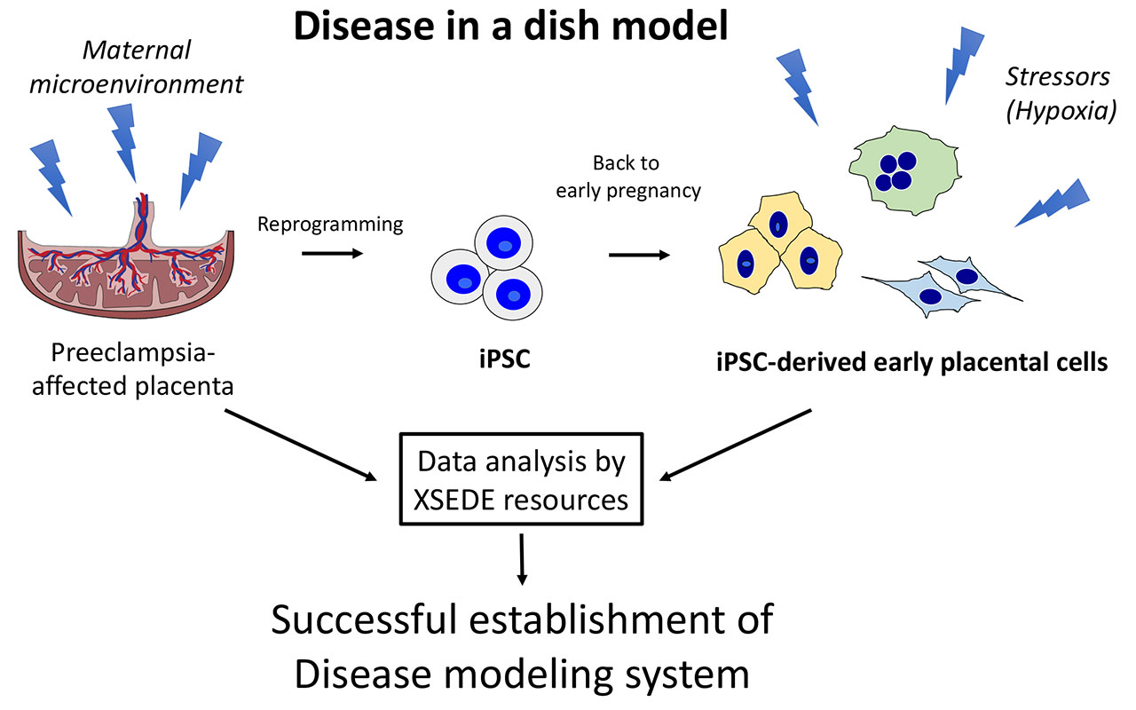
A CIRM supported study using induced pluripotent stem cells (iPSCs), a kind of stem cell that can turn into virtually any cell type, was able to create a “disease in a dish” model in order to better understand preeclampsia.

Credit: UC San Diego

For this study, Mariko Horii, M.D., and her team of researchers at the UC San Diego School of Medicine obtained cells from the placenta of babies born under preeclampsia conditions. These cells were then “reprogrammed” into a stem cell-like state, otherwise known as iPSCs. The iPSCs were then turned into cells resembling placental cells in early pregnancy. This enabled the team to create the preeclampsia “disease in the dish” model. Using this model, they were then able to study the processes that cause, result from, or are otherwise associated with preeclampsia.

The findings revealed that cellular defects observed are related to an abnormal response in the environment in the womb. Specifically, they found that preeclampsia was associated with a low-oxygen environment in the uterus. The researchers used a computer modeling system at UC San Diego known as Comet to detail the differences between normal and preeclampsia placental tissue.

Horii and her team hope that these findings not only shed more light on the environment in the womb observed in preeclampsia, but also provided insight for future development of diagnostic tools and identification of potential medications. Furthermore, they hope that their iPSC disease model can be used to study other placenta-associated pregnancy disorders such as fetal growth restriction, miscarriage, and preterm birth.

The team’s next steps are to develop a 3D model to better study the relationship between environment and development of placental disease.

In a news release from UC San Diego, Horri elaborates more on these future goals.

“Currently, model systems are in two-dimensional cultures with single-cell types, which are hard to study as the placenta consists of maternal and fetal cells with multiple cell types, such as placental cells (fetal origin), maternal immune cells and maternal endometrial cells. Combining these cell types together into a three-dimensional structure will lead to a better understanding of the more complex interactions and cell-to-cell signaling, which can then be applied to the disease setting to further understand pathophysiology.”

The full study was published in Scientific Reports.

Women who have changed, and are changing, the world

The problem with trying to write about something like Women’s History Month is where do you start? Even if you narrow it down to women in science the list is vast.

Marie Curie

I suppose you could always start with Maria Salomea Skłodowska who is better known as Marie Curie. She not only discovered radium and polonium, but she was also the first woman to win a Nobel Prize (in Physics). When she later won another Nobel (in Chemistry) she became the first person ever to win two Nobels and is still the only person ever to win in two different fields. Not a bad place to start.

Agnes Pockels

Or how about Agnes Pockels (1862–1935). Even as a child Agnes was fascinated by science but, in Germany at the time, women were not allowed to attend university. So, she depended on her younger brother to send her his physics textbooks when he was finished with them. Agnes studied at home while taking care of her elderly parents. Doing the dishes  Agnes noticed how oils and soaps could impact the surface tension of water. So, she invented a method of measuring that surface tension. She wrote a paper about her findings that was published in Nature, and went on to become a highly respected and honored pioneer in the field.

Jennifer Doudna (left) and Emmanuelle Charpentier: Photo courtesy Nature

Fast forward to today we could certainly do worse than profile the two women who won the 2020 Nobel Prize in Chemistry for their work with the gene-editing tool CRISPR-Cas9; Jennifer Doudna at the University of California, Berkeley, and Emmanuelle Charpentier at the Max Planck Unit for the Science of Pathogens in Berlin. Their pioneering work showed how you could use CRISPR  to make precise edits in genes, creating the possibility of using it to edit human genes to eliminate or cure diseases. In fact, some CIRM-funded research is already using this approach to try and cure sickle cell disease.

In awarding the Nobel to Charpentier and Doudna, Pernilla Wittung Stafshede, a biophysical chemist and member of the Nobel chemistry committee, said: “The ability to cut DNA where you want has revolutionized the life sciences. The ‘genetic scissors’ were discovered just eight years ago but have already benefited humankind greatly.”

Barbara McClintock: Photo courtesy Brittanica

Appropriately enough none of that work would have been possible without the pioneering work of another woman, Barbara McClintock. She dedicated her career to studying the genetics of corn and developed a technique that enabled her to identify individual chromosomes in different strains of corn.

At the time it was thought that genes were stable and were arranged in a linear fashion on chromosomes, like beads on a string. McClintock’s work showed that genes could be mobile, changing position and altering the work of other genes. It took a long time before the scientific world caught up with her and realized she was right. But in 1983 she was awarded the Nobel Prize in Medicine for her work.

Katherine Johnson at her desk at Langley Research Center: Photo courtesy NASA /AFP

Katherine Johnson is another brilliant mind whose recognition came later in life. But when it did, it made her a movie star. Kind of. Johnson was a mathematician, a “computer” in the parlance of the time. She did calculations by hand, enabling NASA to safely launch and recover astronauts in the early years of the space race.

Johnson and the other Black “computers” were segregated from their white colleagues until the last 1950’s, when signs dictating which restrooms and drinking fountains they could use were removed. She was so highly regarded that when John Glenn was preparing for the flight that would make him the first American to orbit the earth he asked for her to manually check the calculations a computer had made. He trusted her far more than any machine.

Johnson and her co-workers were overlooked until the 2016 movie “Hidden Figures” brought their story to life. She was also awarded the Presidential Medal of Freedom, America’s highest civilian honor, by President Obama.

There are so many extraordinary women scientists we could talk about who have made history. But we should also remind ourselves that we are surrounded by remarkable women right now, women who are making history in their own way, even if we don’t recognized it at the moment. Researchers that CIRM funds, Dr. Catriona Jamieson at UC San Diego, Dr. Jan Nolta at UC Davis, Dr. Jane Lebkowski with Regenerative Patch technologies and so many others. They’re all helping to change the world. We just don’t know it yet.

If you would like to learn about other women who have made extraordinary contributions to science you can read about them here and here and here.