Stem cell stories that caught our eye: healing diabetic ulcers, new spinal cord injury insights & an expanding CRISPR toolbox

Here are some stem cell stories that caught our eye this past week. Some are groundbreaking science, others are of personal interest to us, and still others are just fun.

Stem cells heal diabetic foot ulcers in pilot study
Foot ulcers are one of the many long-term complications that diabetics face. About 15 percent of patients develop these open sores which typically appear at the bottom of the foot. In a quarter of these cases, the ulcers lead to serious infection requiring amputation.

diabetic-foot-ulcers

Diabetic foot ulcers are open sores that don’t heal and in many cases leads to amputation. Image source: Izunpharma

But help may be on the horizon in the form of stem cells. Researchers at Mansoura University in Egypt recently presented results of a small study in which 10 patients with diabetic foot ulcers received standard care and another 10 patients received standard care plus injections of mesenchymal stem cells that had been collected from each patient’s own bone marrow. After just six weeks, the stem cell treated group showed a 50% reduction in the foot ulcers while the group with only standard care had a mere 7% reduction.

These superior results with the stem cells were observed even though the group receiving the stem cells had larger foot ulcers to begin with compared to the untreated patients. There are many examples of mesenchymal stem cells’ healing power which make them an extremely popular cell source for hundreds of on-going clinical trials. Mesenchymal stem cells are known to reduce inflammation and increase blood vessel formation, two properties that may be at work to give diabetic foot ulcers the chance to get better.

Medscape Medical News reported on these results which were presented at the 2016 annual meeting of the European Association for the Study of Diabetes (EASD) 2016 Annual Meeting

Suppressing nerve signals to help spinal cord injury victims
Losing the use of one’s limbs is a profound life-altering change for spinal cord injury victims. But their quality of life also suffers tremendously from the loss of bladder control and chronic pain sensations. So much so, victims often say that just improving these secondary symptoms would make a huge improvement in their lives.

While current stem cell-based clinical trials, like the CIRM-funded Asterias study, aim to reverse paralysis by restoring loss nerve signals, recent CIRM-funded animal data published in Cell Stem Cell from UC San Francisco suggest that nerve cells that naturally suppress nerve signals may be helpful for these other symptoms of spinal cord injury.
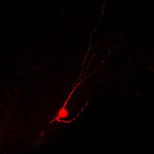
mgecell-integrated.jpg

Mature inhibitory neuron derived from human embryonic stem cells is shown after successfully migrated and integrated into the injured mouse spinal cord.
Photo by Jiadong Chen, UCSF

It turns out that the bladder control loss and chronic pain may be due to overactive nerve signals. So the lab of Arnold Kriegstein transplanted inhibitory nerve cells – derived from human embryonic stem cells – into mice with spinal cord injuries. The scientists observed that these human inhibitory nerve cells, or interneurons, successfully made working connections in the damaged mouse spinal cords. The rewiring introduced by these interneurons also led to reduced pain behaviors in the mice as well as improvements in bladder control.


In a Yahoo Finance interview, Kreigstein told reporters he’s eager to push forward with these intriguing results:

3e3a4-kriegsteinsmall

Arnold Kriegstein, UCSF

“As a clinician, I’m very aware of the urgency that’s felt among patients who are often very desperate for treatment. As a result, we’re very interested in accelerating this work toward clinical trials as soon as possible, but there are many steps along the way. We have to demonstrate that this is safe, as well as replicating it in other animals. This involves scaling up the production of these human interneurons in a way that would be compatible with a clinical product.”


Expanding the CRISPR toolbox
If science had a fashion week, the relatively new gene editing technology called CRISPR/Cas9 would be sure to dominate the runway. You can think of CRISPR/Cas9 as a protein and RNA complex that acts as a molecular scissor which directly targets and cuts specific sequences of DNA in the human genome. Scientists are using CRISPR/Cas9 to develop innovative biomedical techniques such as removing disease-causing mutations in stem cells in hopes of developing potential treatments for patients suffering from diseases that have no cures.

What’s particularly interesting about the CRISPR/Cas9 system is that the Cas9 protein responsible for cutting DNA is part of a family of CRISPR associated proteins (Cas) that have similar but slightly different functions. Scientists are currently expanding the CRISPR toolbox by exploring the functions of other CRISPR associated proteins for gene editing applications.

A CIRM-funded team at UC Berkeley is particularly interested in a CRISPR protein called C2c2, which is different from Cas9 in that it targets and cuts RNA rather than DNA. Led by Berkeley professor Jennifer Doudna, the team discovered that the CRISPR/C2c2 complex has not just one, but two, distinct ways that it cuts RNA. Their findings were published this week in the journal Nature.

The first way involves creation: C2c2 helps make the guide RNAs that are used to find the RNA molecules that it wants to cut. The second way involves destruction: after the CRISPR/C2c2 complex finds it’s RNAs of choice, C2c2 can then cut and destroy the RNAs.

Doudna commented on the potential applications for this newly added CRISPR tool in a Berkeley News release:

Jennifer-Doudna

Jennifer Doudna: Photo courtesy of iPSCell.com

“This study expands our molecular understanding of C2c2 to guide RNA processing and provides the first application of this novel RNase. C2c2 is essentially a self-arming sentinel that attacks all RNAs upon detecting its target. This activity can be harnessed as an auto-amplifying detector that may be useful as a low-cost diagnostic.”
 

A Patient Advocate’s Take on Sickle Cell Disease: The Pain and the Promise

September is National Sickle Cell Awareness Month. First officially recognized by the federal government in 1983, National Sickle Cell Awareness Month calls attention to sickle cell disease (SCD), a genetic disease that researchers estimate affects between 90,000 and 100,000 Americans. CIRM is funding a clinical trial focused on curing the disease with a stem cell-based gene therapy. 

People with this debilitating condition face a number of barriers in getting the help they need to keep their pain under control. In addition to the difficulty of accessing medication, they often have to overcome suspicion and discrimination.  Patient Advocate Nancy Rene, of Axis Advocacy  wrote the following blog about the problems families with SCD face.

Sickle Cell Disease Patient Advocates Adrienne Shapiro and Nancy Rene.

Sickle Cell Disease Patient Advocates Adrienne Shapiro and Nancy Rene.

Sickle Cell Disease: The Pain and the Promise

By Nancy M. Rene, co-founder, Axis Advocacy

The Disease

Sickle Cell Disease is a group of inherited red blood cell disorders. It is the most common genetic disease in the US. Close to 100,000 Americans have sickle cell disease.  Although it affects persons of African descent, it can also be found in Latino families and families from the Middle-East and India. World-wide there are at least 20 million people with the disease.

Normal red blood cells are round like doughnuts, and they move through small blood vessels in the body to deliver oxygen. Red blood cells in the person with sickle cell disease become hard, sticky and shaped like sickles. When these hard and pointed red cells go through the small blood vessels, they clog the flow and break apart. This causes pain, inflammation and organ damage.

The Pain and the Promise

In the last 30 years the United States has made great progress in treating sickle cell disease.  All states now have newborn screening and most children are living to adulthood. However, many children with SCD don’t receive important services to prevent serious complications from the disease.

Unfortunately, according the the American Society of Hematology, the mortality rate for adults appears to have increased during the same 30 years! Patients with SCD experience long delays in the ER, and are often accused of being drug seekers. Once admitted to the hospital they are confronted by medical staff with little understanding or empathy. Research from Dr. Michael DeBaun found that adults with this disease lack access to a primary care doctor who is knowledgeable about sickle cell.

The biggest Pain for those with sickle cell disease does not come from the disease itself but from treatment by the medical community.  When, for most people, going to the hospital represents a place to get help and relief from the burdens of a challenging disease, those with sickle cell see going to the hospital as going into battle. They “gear up” with copies of medical records and NIH guidelines, they make sure they have a diary to record inappropriate remarks from medical staff, they ask a friend to come along as an advocate to help them withstand the implied racism and institutional bias with which they are confronted. Even when new hospitals or clinics are built, they often do not live up to expectations, offering no emergency support or 24-hour access.

The promise of course comes from the diligent work of researchers and clinicians who run model programs.  Bone marrow transplants, while limited in use, have actually cured a number of young people, saving them from pain and organ damage that await their adult years. Pharmaceutical companies are completing clinical trials on several drugs that can reduce the symptoms of sickle cell at the molecular level. These drugs could greatly reduce the effects of the sickle cell crisis which often results in a lengthy hospital stay.

Stem cell research, while moving slowly, can be the holy grail of medical practice, curing many of the 100,000 Americans with sickle cell.  A cure would lead to avoiding the dreaded ER, being free of pain and organ damage, living a healthy life, and having children without worrying that they too would be born with this disease.

What is missing is linking research to clinical practice.  It is clear that the CDC, FDA and NIH have finally understood this missing piece.  The NIH published an extensive report, Guidelines for the The Treatment of Sickle Cell Disease, in 2014. NIH convened the 10th Annual Focus on Sickle Cell that brought researchers, clinicians, and other leaders together to make presentations on their work in sickle cell. The Sickle Cell Research Foundation convened an outstanding medical conference in Florida that again brought leaders together to gain knowledge from one another. ASH, the American Society of Hematology, is planning to launch a Sickle Cell Initiative this month.

We in the sickle cell community, patients, care-givers, and advocates, feel that we have finally got some big guns in this fight. Once doctors in all communities understand this disease, once they are aware of their own implicit bias and that of their institutions, there should be improvement in the treatment of people with this painful, debilitating illness.


Related Links:

Gene required for sperm stem cells linked to male infertility, UCSD study suggests

Even in this day and age, when a couple is having trouble conceiving a child, it’s often the woman who is initially suspected of having infertility problems and is likely the first to seek out the advice of doctor. But according to Miles Wilkinson, professor of reproductive medicine at UC San Diego School of Medicine, infertility issues can just as likely be due to problems with sperm production in the male. In fact, about 4 million men of reproductive age in the U.S. are confronted with infertility challenges and in 30% percent of those cases, the cause of the infertility is not well understood.

Through some scientific detective work, Wilkinson and his research team have zeroed in on a gene called RhoX10 that plays an essential role in the development of adult stem cells which give rise to sperm production. The study results, funded in part by CIRM and published yesterday in Cell Reports, may help provide a path toward new treatment options for male infertility.

sperm

The process of sperm formation (spermatogenesis). Image: Wikipedia

To get at a cellular and molecular understanding of infertility, the team focused on the function of spermatogonial stem cells (SSCs). Through a multistep process within the male reproductive system, SSCs form into mature sperm cells capable of fertilizing an egg.  The SSC itself forms from primordial germ cells. The key genetic switches that help these germ cells give rise to SSCs was not well understood.  Earlier studies had shown that a group of adjacent genes, called the Rhox cluster, on the X-chromosome are expressed in the testes, suggesting a role in sperm production.

Using genetic engineering techniques, Wilkinson’s team bred mice lacking the 33 genes of the Rhox cluster. The resulting male mice showed a reduction in the number SSCs leading to low sperm number. Through a process of elimination, the team found that deleting just one of those genes, Rhox10, produced nearly the same flaw.

Rhox10 bus_HW, 9-26-16.jpg.jpg

Rhox 10: a genetic “bus” that “drives” sperm stem cells toward sperm production. Illustration by Hye-Won Song.

Further analysis, indicated Rhox10 was key to driving the development of germ cells into SSCs. So when the gene is deleted, not enough SSCs develop in the testis, leading to low sperm counts.  The researchers also found the Rhox10 is a master regulator of genes that control the germ cells’ movement from one part of the testis to another that, due to a different chemical environment, helps the germ cells transform into SSCs.

In addition to this mouse data, the team also co-authored a recent Human Molecular Genetics report with scientists at the University of Münster in Germany that connects Rhox genes and human male infertility. In the study, three RHOX genes were sequenced in 250 men with extremely low sperm counts and revealed six genetic mutations. Together, these results present solid evidence that mutations in Rhox are the culprit in at least some forms of male infertility. First author Hye-Won Song discussed this point in a university press release:

“Spermatogonial stem cells allow men — even in their 70s — to generate sperm and father children. Our finding that Rhox10 is critical for spermatogonial stem cells, coupled with the finding that human RHOX genes are mutated in infertile men, suggests that mutations in these genes cause human male infertility.”

Stem Cell Experts Discuss the Ethical Implications of Translating iPSCs to the Clinic

Part of The Stem Cellar blog series on 10 years of iPSCs.

This year, scientists are celebrating the 10-year anniversary of Shinya Yamanaka’s Nobel Prize winning discovery of induced pluripotent stem cells (iPSCs). These are cells that are very similar biologically to embryonic stem cells and can develop into any cell in the body. iPSCs are very useful in scientific research for disease modeling, drug screening, and for potential cell therapy applications.

However, with any therapy that involves testing in human patients, there are ethical questions that scientists, companies, and policy makers must consider. Yesterday, a panel of stem cell and bioethics experts at the Cell Symposium 10 Years of iPSCs conference in Berkeley discussed the ethical issues surrounding the translation of iPSC research from the lab bench to clinical trials in patients.

The panel included Shinya Yamanaka (Gladstone Institutes), George Daley (Harvard University), Christine Mummery (Leiden University Medical Centre), Lorenz Studer (Memorial Sloan Kettering Cancer Center), Deepak Srivastava (Gladstone Institutes), and Bioethicist Hank Greely (Stanford University).

iPSC Ethics Panel

iPSC Ethics Panel at the 10 Years of iPSCs Conference

Below is a summary of what these experts had to say about questions ranging from the ethics of patient and donor consent, genetic modification of iPSCs, designer organs, and whether patients should pay to participate in clinical trials.

How should we address patient or donor consent regarding iPSC banking?

Multiple institutes including CIRM are developing iPSC banks that store thousands of patient-derived iPSC lines, which scientists can use to study disease and develop new therapies. These important cell lines wouldn’t exist without patients who consent to donate their cells or tissue. The first question posed to the panel was how to regulate the consent process.

Christine Mummery began by emphasizing that it’s essential that companies are able to license patient-derived iPSC lines so they don’t have to go back to the patient and inconvenience them by asking for additional samples to make new cell lines.

George Daley and Hank Greely discussed different options for improving the informed consent process. Daley mentioned that the International Society for Stem Cell Research (ISSCR) recently updated their informed consent guidelines and now provide adaptable informed consent templates that can be used for obtaining many type of materials for human stem cell research.  Daley also mentioned the move towards standardizing the informed consent process through a single video shared by multiple institutions.

Greely agreed that video could be a powerful way to connect with patients by using talented “explainers” to educate patients. But both Daley and Greely cautioned that it’s essential to make sure that patients understand what they are getting involved in when they donate their tissue.

Greely rounded up the conversation by reminding the audience that patients are giving the research field invaluable information so we should consider giving back in return. While we can’t and shouldn’t promise a cure, we can give back in other ways like recognizing the contributions of specific patients or disease communities.

Greely mentioned the resolution with Henrietta Lack’s family as a good example. For more than 60 years, scientists have used a cancer cell line called HeLa cells that were derived from the cervical cancer cells of a woman named Henrietta Lacks. Henrietta never gave consent for her cells to be used and her family had no clue that pieces of Henrietta were being studied around the world until years later.

In 2013, the NIH finally rectified this issue by requiring that researchers ask for permission to access Henrietta’s genomic data and to include the Lacks family in their publication acknowledgements.

Hank Greely, Stanford University

Hank Greely, Stanford University

“The Lacks family are quite proud and pleased that their mother, grandmother and great grandmother is being remembered, that they are consulted on various things,” said Hank Greely. “They aren’t making any direct money out of it but they are taking a great deal of pride in the recognition that their family is getting. I think that returning something to patients is a nice thing, and a human thing.”

What are the ethical issues surrounding genome editing of iPSCs?

The conversation quickly focused on the ongoing CRISPR patent battle between the Broad Institute, MIT and UC Berkeley. For those unfamiliar with the technique, CRISPR is a gene editing technology that allows you to cut and paste DNA at precise locations in the genome. CRISPR has many uses in research, but in the context of iPSCs, scientists are using CRISPR to remove disease-causing mutations in patient iPSCs.

George Daley expressed his worry about a potential fallout if the CRISPR battle goes a certain way. He commented, “It’s deeply concerning when such a fundamentally enabling platform technology could be restricted for future gene editing applications.”

The CRISPR patent battle began in 2012 and millions of dollars in legal fees have been spent since then. Hank Greely said that he can’t understand why the Institutes haven’t settled this case already as the costs will only continue to rise, but that it might not matter how the case turns out in the end:

“My guess is that this isn’t ultimately going to be important because people will quickly figure out ways to invent around the CRISPR/Cas9 technology. People have already done it around the Cas9 part and there will probably be ways to do the same thing for the CRISPR part.”

 Christine Mummery finished off with a final point about the potential risk of trying to correct disease causing mutations in patient iPSCs using CRISPR technology. She noted that it’s possible the correction may not lead to an improvement because of other disease-causing genetic mutations in the cells that the patient and their family are unaware of.

 Should patients or donors be paid for their cells and tissue?

Lorenz Studer said he would support patients being paid for donating samples as long as the payment is reasonable, the consent form is clear, and patients aren’t trying to make money off of the process.

Hank Greely said the big issue is with inducement and whether you are paying enough money to convince people to do something they shouldn’t or wouldn’t want to do. He said this issue comes up mainly around reproductive egg donation but not with obtaining simpler tissue samples like skin biopsies. Egg donors are given money because it’s an invasive procedure, but also because a political decision was made to compensate egg donors. Greely predicts the same thing is unlikely to happen with other cell and tissue types.

Christine Mummery’s opinion was that if a patient’s iPSCs are used by a drug company to produce new successful drugs, the patient should receive some form of compensation. But she said it’s hard to know how much to pay patients, and this question was left unanswered by the panel.

Should patients pay to participate in clinical trials?

George Daley said it’s hard to justify charging patients to participate in a Phase 1 clinical trial where the focus is on testing the safety of a therapy without any guarantee that there will be beneficial outcome to the patient. In this case, charging a patient money could raise their expectations and mislead them into thinking they will benefit from the treatment. It would also be unfair because only patients who can afford to pay would have access to trials. Ultimately, he concluded that making patients pay for an early stage trial would corrupt the informed consent process. However, he did say that there are certain, rare contexts that would be highly regulated where patients could pay to participate in trials in an ethical way.

Lorenz Studer said the issue is very challenging. He knows of patients who want to pay to be in trials for treatments they hope will work, but he also doesn’t think that patients should have to pay to be in early stage trials where their participation helps the progress of the therapy. He said the focus should be on enrolling the right patient groups in clinical trials and making sure patients are properly educated about the trial they are participating.

Thoughts on the ethics behind making designer organs from iPSCs?

Deepak Srivastava said that he thinks about this question all the time in reference to the heart:

Deepak Srivastava, Gladstone Institutes

Deepak Srivastava, Gladstone Institutes

“The heart is basically a pump. When we traditionally thought about whether we could make a human heart, we asked if we could make the same thing with the same shape and design. But in fact, that’s not necessarily the best design – it’s what evolution gave us. What we really need is a pump that’s electrically active. I think going forward, we should remove the constraint of the current design and just think about what would be the best functional structure to do it. But it is definitely messing with nature and what evolution has given us.”

Deepak also said that because every organ is different, different strategies should be used. In the case of the heart, it might be beneficial to convert existing heart tissue into beating heart cells using drugs rather than transplant iPSC-derived heart cells or tissue. For other organs like the pancreas, it is beneficial to transplant stem cell-derived cells. For diabetes, scientists have shown that injecting insulin secreting cells in multiple areas of the body is beneficial to Diabetes patients.

Hank Greely concluded that the big ethical issue of creating stem cell-derived organs is safety. “Biology isn’t the same as design,” Greely said. “It’s really, really complicated. When you put something into a biological organism, the chances that something odd will happen are extremely high. We have to be very careful to avoid making matters worse.”

For more on the 10 years of iPSCs conference, check out the #CSStemCell16 hashtag on twitter.

Full Steam Ahead: First Patient is Dosed in Expanded CIRM Spinal Cord Injury Trial

Today we bring you more good news about a CIRM-funded clinical trial for spinal cord injury that’s received a lot of attention lately in the news. Asterias Biotherapeutics has treated its first patient in an expanded patient population of spinal cord injury patients who suffer from cervical, or neck, injuries.

In late August, Asterias reported that they had passed the first hurdle in their Phase 1/2a trial and showed that their stem cell therapy is safe to use in patients with a more serious form of cervical spinal cord injuries.

Earlier this month, we received more exciting updates from Asterias – this time reporting that the their embryonic stem cell-based therapy, called AST-OPC1, appeared to benefit treated patients. Five patients with severe spinal cord injuries to their neck were dosed, or transplanted, with 10 million cells. These patients are classified as AIS-A on the ASIA impairment scale – meaning they have complete injuries in which the spinal cord tissue is severed and patients lose all feeling and use of their limbs below the injury site. Amazingly, after three months, all five of the AIS-A patients have seen improvements in their movement.

Today, Asterias announced that it has treated its first patient with an AIS-B grade cervical spinal cord injury with a dose of 10 million cells at the Sheperd Center in Atlanta. AIS-B patients have incomplete neck injuries, meaning that they still have some spinal cord tissue at the injury site, some feeling in their arms and legs, but no movement. This type of spinal cord injury is still severe, but these patients have a better chance at gaining back some of their function and movement after treatment.

In a press release by Asterias, Chief Medical Officer Dr. Edward Wirth said:

“We have been very encouraged by the first look at the early efficacy data, as well as the safety profile, for AST-OPC1 in AIS-A patients, and now look forward to also evaluating efficacy and safety in AIS-B patients. AIS-B patients also have severe spinal cord injuries, but compared to AIS-A patients they have more spared tissue in their spinal cords.  This may allow these patients to have a greater chance of meaningful functional improvement after being treated with AST-OPC1 cells.”

Dr. Donald Peck Leslie, who directs the Sheperd Center and is the lead investigator at the Atlanta clinical trial site, expressed his excitement about the trials’ progress.

“As someone who regularly treats patients who have sustained paralyzing spinal cord injuries, I am encouraged by the progress we’ve seen in evaluations of AST-OPC1 in people with AIS-A injuries, particularly the improvements in hand, finger and arm function. Now, I am looking forward to continuing the evaluation of this promising new treatment in AIS-B patients, as well.”

Asterias has plans to enroll a total of five to eight AIS-B patients who will receive a dose of 10 million cells. They will continue to monitor all patients in this trial (both AIS-A and B) and will conduct long-term follow up studies to make sure that the AST-OPC1 treatment remains safe.

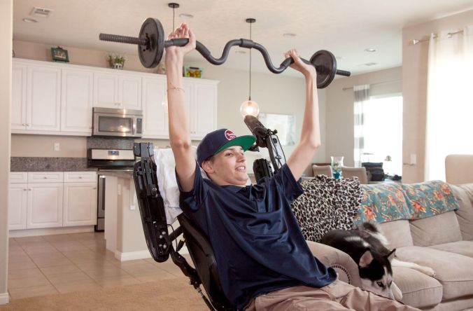
We hope that the brave patients who have participated in the Asterias trial continue to show improvements following treatment. Inspiring stories like that of Kris Boesen, who was the first AIS-A patient to get 10 million cells in the Asterias trial and now has regained the use of his arms and hands (and regaining some sensation in his legs), are the reason why CIRM exists and why we are working so hard to fund promising clinical trials. If we can develop even one stem cell therapy that gives patients back their life, then our efforts here at CIRM will be worthwhile.

Kris Boesen, CIRM spinal cord injury clinical trial patient.

Kris Boesen, CIRM spinal cord injury clinical trial patient.


Related Links:

Stem cell stories that caught our eye: two studies of the heart and cool stem cell art

Here are some stem cell stories that caught our eye this past week. Some are groundbreaking science, others are of personal interest to us, and still others are just fun.

Image from Scope Blog.

Image from Scope Blog.

Understanding Heart Defects. Healthy heart tissue is made up of smooth, solid muscle, which is essential for normal heart function. Patients with a heart defect called left ventricular non-compaction (LVNC), lack normal heart tissue in their left ventricle – the largest, strongest blood-pumping chamber – and instead have spongy-looking tissue.

LVNC occurs during early heart development where pieces of heart muscle fail to condense (compact) and instead form an airy, sponge-like network that can leave patients at risk for heart failure and other complications.

A team at Stanford is interested in learning how LVNC occurs in humans, and they’re using human stem cells for the answer. Led by CIRM grantee Joe Wu, the scientists generated induced pluripotent stem cells (iPSCs) from four patients with LVNC. iPSCs are cells that can be turned into any other cell in the body, so Wu turned these cells into iPSC-derived heart muscle in a dish.

Wu’s team was particularly interested in determining why some LVNC patients have symptoms of disease while others seem perfectly normal. After studying the heart muscle cells derived from the four LVNC patients, they identified a genetic mutation in a gene called TBX20. This gene produces a type of protein called a cardiac transcription factor, which controls the expression of other heart related genes.

Upon further exploration, the scientists found that the genetic mutation in TBX20 prevented LVNC heart muscle cells from dividing at their normal rate. If they blocked the signal of mutant TBX20, the heart cells went back to their normal activity and created healthy looking heart tissue.

This study was published in Nature Cell Biology and covered by the Stanford Medicine Scope blog. In an interview with Scope, Joe Wu highlighted the big picture of their work:

Joseph Wu Stanford

Joseph Wu Stanford

“This study shows the feasibility of modeling such developmental defects using human tissue-specific cells, rather than relying on animal cells or animal models. It opens up an exciting new avenue for research into congenital heart disease that could help literally the youngest — in utero — patients.”

Stem Cell Heart Patch. Scientists from the University of Wisconsin, Madison are creating stem cell-based heart patches that they hope one day could be used to treat heart disease.

In a collaboration with Duke and the University of Alabama at Birmingham, they’re developing 3D stem cell-derived patches that contain the three main cell types found in the heart: cardiomyocytes (heart muscle cells), fibroblasts (support cells), and endothelial cells (cells that line the insides of blood vessels). These patches would be transplanted into heart disease patients to replace damaged heart tissue and improve heart function.

As with all research that has the potential for reaching human patients, the scientists must first determine whether the heart patches are safe in animal models. They plan to transplant the heart patches into a pig model – chosen because pigs have similar sized hearts compared to humans.

In a UW-Madison News release, the director of the UW-Madison Stem Cell and Regenerative Medicine Center Timothy Kamp, hinted at the potential for this technology to reach the clinic.

“The excitement here is we’re moving closer to patient applications. We’re at a stage when we need to see how these cells do in a large animal heart attack model. We’ll be making patches of heart muscle that can be applied to these injured areas.”

Kamp and his team still have a lot of work to do to perfect their heart patch technology, but they are thinking ahead. Two issues that they are trying to address are how to prevent a patient’s immune system from rejecting the heart patch transplant, and how to make sure the heart patches beat in sync with the heart they are transplanted into.

Check out the heart patches in action in this video:

(Video courtesy of Xiaojun Lian)

Cool Stem Cell Art! When I was a scientist, I worked with stem cells all the time. I grew them in cell culture dishes, coaxed them to differentiate into brain cells, and used a technique called immunostaining to take really beautiful, colorful pictures of my final cell products. I took probably thousands of pictures over my PhD and postdoc, but sadly, only a handful of these photos ever made it into journal publications. The rest collected dust either on my hard drive or in my lab notebook.

It’s really too bad that at the time I didn’t know about this awesome stem cell art contest called Cells I See run by the Centre for Commercialization of Regenerative Medicine (CCRM) in Ontario Canada and sponsored by the Stem Cell Network.

The contest “is about the beauty of stem cells and biomaterials, seen directly through the microscope or through the interpretive lens of the artist.” Scientists can submit their most prized stem cell images or art, and the winner receives a cash prize and major science-art street cred.

The submission deadline for this year’s contest was earlier this month, and you can check out the contenders on CCRM’s Facebook page. Even better, you can vote for your favorite image or art by liking the photo. The last date to vote is October 15th and the scientist whose image has the most likes will be the People’s Choice winner. CCRM will also crown a Grand Prize winner at the Till & McCulloch Stem Cell Meeting in October.

I’ll leave you with a few of my favorite photos, but please don’t let this bias your vote =)!

"Icy Astrocytes" by Samantha Yammine

“Icy Astrocytes” by Samantha Yammine (Vote here!)

"Reaching for organoids" by Amy Wong

“Reaching for organoids” by Amy Wong (Vote here!)

"Iris" by Sabiha Hacibekiroglu

“Iris” by Sabiha Hacibekiroglu (Vote here!)

Funding stem cell research targeting a rare and life-threatening disease in children

cystinosis

Photo courtesy Cystinosis Research Network

If you have never heard of cystinosis you should consider yourself fortunate. It’s a rare condition caused by an inherited genetic mutation. It hits early and it hits hard. Children with cystinosis are usually diagnosed before age 2 and are in end-stage kidney failure by the time they are 9. If that’s not bad enough they also experience damage to their eyes, liver, muscles, pancreas and brain.

The genetic mutation behind the condition results in an amino acid, cystine, accumulating at toxic levels in the body. There’s no cure. There is one approved treatment but it only delays progression of the disease, has some serious side effects of its own, and doesn’t prevent the need for a  kidney transplant.

Researchers at UC San Diego, led by Stephanie Cherqui, think they might have a better approach, one that could offer a single, life-long treatment for the problem. Yesterday the CIRM Board agreed and approved more than $5.2 million for Cherqui and her team to do the pre-clinical testing and work needed to get this potential treatment ready for a clinical trial.

Their goal is to take blood stem cells from people with cystinosis, genetically-modify them and return them to the patient, effectively delivering a healthy, functional gene to the body. The hope is that these genetically-modified blood stem cells will integrate with various body organs and not only replace diseased cells but also rescue them from the disease, making them healthy once again.

In a news release Randy Mills, CIRM’s President and CEO, said orphan diseases like cystinosis may not affect large numbers of people but are no less deserving of research in finding an effective therapy:

“Current treatments are expensive and limited. We want to push beyond and help find a life-long treatment, one that could prevent kidney failure and the need for kidney transplant. In this case, both the need and the science were compelling.”

The beauty of work like this is that, if successful, a one-time treatment could last a lifetime, eliminating or reducing kidney disease and the need for kidney transplantation. But it doesn’t stop there. The lessons learned through research like this might also apply to other inherited multi-organ degenerative disorders.

Science and Improv: Spotlight on CIRM Bridges Scholar Jill Tsai

As part of our CIRM scholar series, we’re featuring the research and career accomplishments of CIRM funded students.

What do science and improv have in common? The answer is not a whole lot. However, I recently met a talented student from our CIRM Bridges master’s program who one day is going to change this.

Jill Tsai

Jill Tsai, CIRM Bridges scholar

Meet Jill Tsai. She recently graduated from the CIRM Bridges program at the Scripps Research Institute in San Diego and is now starting a PhD program in cancer biology at the City of Hope in Duarte California.

Jill received her Bachelors from UC Merced general biology and went to Cal Poly Pomona for a Master’s program in cancer research. While at Cal Poly Pomona, she successfully applied for a CIRM Bridges internship that allowed her to finish her Master’s degree at Scripps in the lab of Dr. Lazzerini Denchi.

I met Jill at the 2016 Bridges Conference in July and was immediately impressed by her passion for science and communications. I was also intrigued by her interest in improv and how she balances her time between two very different passions. I’m thrilled that Jill agreed to an interview for the Stem Cellar as I think it’s valuable to read about scientists who are pursuing multiple passions not necessarily related to science.

Enjoy!

Q: What did you study during your Bridges internship?

JT: I was a research intern in the lab of Dr. Lazzerini Denchi. In his lab, we study telomeres, which are the pieces of DNA at the end of chromosomes that help protect them from being degraded. We’re specifically looking at proteins that help maintain telomere function in mouse stem cells. We do big protein pull downs to try to figure out what new and novel proteins are surrounding the mechanisms that maintain telomere function, and then we do functional assays to figure out what these proteins do.

Lazzerini Denchi’s lab focuses on basic research and how certain proteins affect telomere length and also the telomere deprotection response. One function of telomeres is that they suppress the double and single stranded DNA repair mechanism. If you don’t suppress those mechanisms, then the ends of those linear chromosomes look exactly like double stranded DNA breaks and repair proteins try to fix them by fusing those chromosomes together.

There are great pictures from Lazzerini Denchi’s first author publication showing chromosomes hooked end to end to end like long strings of spaghetti as a result of telomere deprotection. We are studying novel proteins that assist telomeres with the deprotection response and determining whether these proteins have some other kind of function as well.

Telomere deprotection results in chromosomes that are linked together (right) instead of separate (left). (Source Denchi et al. Nature)

Telomere deprotection results in chromosomes that are linked together (right) instead of separate (left). (Source Nature: Denchi et al., 2007)

Our larger focus in the lab is being able to understand cancer and specific telomere related genetic disorders that are associated with cancer.

Q: What was your CIRM Bridges experience like?

JT: CIRM was really amazing, and I credit it a lot for being able to start a PhD this fall. I’d been working in my lab at Cal Poly Pomona for five years, and my research unfortunately wasn’t working out. I was probably going to have to quit the program or take an out with an easier project. When I applied to CIRM, I was hoping to get the internship because if I didn’t get it, I was going to go down a completely different career path.

The CIRM internship was very valuable to me. It provided training through stem cell classes and lectures and allowed me to immerse myself in a real lab that had real equipment and personnel. The experience took my research knowledge to the next level and then some. And I knew for sure it had when I was at the poster session during the Bridges conference. I was walking around and asking students about their research, and I understood clearly the path of their research. I knew what questions were good to ask and what the graphs meant without having to take them home and dissect them. It was extremely satisfying to be able to understand other’s scientific research by just listening to them.

I am so excited to start my PhD in the fall. For the first time, I feel confident about my foundational biology and research skills. I also have a better understanding of myself and where I need to improve in comprehension and technique. I am ready to jump into grad school and improve as a scientist.

Q: What are your future career steps?

JT: I want to do something that involves teaching or being able to educate people. I’ve worked as a TA in my master’s program for a few years, and I really enjoy that experience of clarifying complex subjects for people. But to be honest, I also don’t know what I want to do right now so I’m keeping my options open.

Q: What’s your favorite thing about being scientist?

JT: Being a scientist forces you to never be complacent in what you understand. If I had never gotten my master’s, there would be this whole level of critical thinking that I wouldn’t have right now. Learning more is one of the biggest reasons why I want to get my PhD even if I don’t know exactly what I want to do yet.

I want to be able to think at a higher level because I think it’s valuable. And I see my Professor at Scripps: he has all these publications under his belt, but he’s always tinkering with things and he’s always learning new software and he’s always reading new papers. As a scientist, you can’t be stagnant in your learning, and I think because of that you’re always pushing yourself to your best potential.
Q: Do you have advice for future Bridges students?

JT: For anyone who is interested in doing a PhD, this is the world’s best preparatory program. After you start a PhD, you hit the ground running. If I were to give advice, I’d say to not be too hard on yourself. There’s going to be expectations put on you that you might not be ready for and you might not do the best job. But you should try your best and know it’s going to help you grow.

Usually people who go into PhD programs are people that have always done well in school. But it’s important to know that learning in grad school is very different than how we are taught to learn elsewhere. Every other time it’s just like show up, listen, take the test you’re done. A PhD relies on a little bit of luck, getting the right project, and doing everything meticulously.

Q: What are your hobbies?

JT: My favorite hobby is improv comedy. What I really like about improv is that it is so different from science and it helps me to relax after work.

Improv is performing comedic scenes on stage with a bunch of people without a script. Skills that it requires are not being stuck in your own head and really paying attention to what’s going on around you. You also need to take big risks and not worry so much about what the end result is going to be, which is very different from research. It’s a nice break to be able to make big giant mistakes and know that after that day it doesn’t matter.

As a researcher, it’s hard to make friends, and even if you have friends, it’s hard to find the time to hang out with them. I love improv because it’s a built in activity. All of my friends outside of work are in improv. We show up and we play make believe together on stage – it’s just a really nice atmosphere. In improv we teach a philosophy that everything you have is enough. Everything you come in with is enough. It’s really nice, because being an adult is hard and life is hard. So it’s a nice thing to hear.

Jill's Improv team.

Flyspace Improv team.

Q: Do you see yourself combining your passions for science and improve in the future?

JT: I do. I don’t know what I want to do yet as a career, but improv is such a big part of my identity that it will always play a role in my life. Improv is so important in communication and interpersonal connections. I believe everyone in science could benefit from it. Ideally, I will find a career that allows me to use both of these passions to help people.

Faulty fat stem cells & obesity-related diabetes

You see it in the news all the time: more and more people around the world are obese and as a result they’re at a higher risk for diabetes, heart disease and cancer. In fact, 90% of individuals with type 2 diabetes are overweight or obese.

yellow_adipose_tissue_in_paraffin_section_-_lipids_washed_out

Fat cells (Image: Wikimedia Commons)

“Healthy” obese individuals protected from diabetes and other complications
A fascinating observation is that despite this tight association between weight and diabetes, some obese people are somehow shielded from the increased risks for diabetes and other associated diseases. Considering these conditions are among the leading causes of preventable death in the U.S., understanding how exactly these “healthy” obese individuals are protected could benefit millions of people.

A new study by researchers at the University of Bristol and Anti-Doping Laboratory Qatar (ADLQ) suggests that fat stem cells may hold the key to unlocking this mystery. Reporting in Diabetologia, the team found that fat stem cells from “healthy” obese people were better at storing fat compared to these same cells in people with increased risk for diabetes.

Belly fat and the development of diabetes
To delve deeper into the study, let’s take a closer look at the cellular biology of obesity and diabetes. The accumulation of fat in obese individuals initially leads to bigger fat cells but eventually causes the recruitment of fat stem cells. These additional fat cells can deposit as so-called visceral fat (aka belly fat) which accumulates within larger organs like the liver, heart and muscle instead of under the skin. Now, when a carbohydrate meal is eaten, the food is broken down into simple sugars which enter the blood. This rise in blood sugar is temporary because our organs like the liver and muscle use the sugar for energy. The blood sugar enters muscle and liver cells with the help of the hormone, insulin. But visceral fat mucks up these organs’ ability to sense insulin – they’re called insulin resistant – and blood sugar levels stay elevated which is the hallmark of type 2 diabetes (in type 1 diabetes the body doesn’t make any insulin).

In the study, the research team collected blood samples and isolated fat stem cells from 57 severely obese individuals undergoing liposuction.  Some of the volunteers were insulin resistant (their organs had a hard time taking up blood sugar despite the presence of insulin) and had obesity-related conditions like diabetes, hypertension and heart disease. The others were insulin sensitive (their organs could take in blood sugar) and had no signs of obesity-related conditions.

Obesity-related complications and faulty fat stem cells
It turned out that the fat stem cells from obese individuals with insulin resistance (increased risk of complications) did not store fat as well as the fat stem cells from the “healthy” obese subjects. It’s this inefficient fat storage that likely leads to the build-up of visceral fat.

wael-diabetes-article

So why the difference? A comparison of various proteins in the blood of the two groups, showed that IL-6 – a protein secreted by the white blood cells of our immune system – was higher in the insulin resistant subjects. Back in the lab, the team found that the elevated IL-6 played a role in the cells’ reduced ability to store fat. Mohamed Elrayess, one of the authors from ADLQ, summarized the results in a press release:

“In this study we have shown that the impaired ability of fat stem cells to store excess fat was partially due to increased levels of the inflammatory marker interleukin-6 in the blood. Indeed, when fat stem cells isolated from healthy obese individuals were exposed to interleukin-6 in the laboratory, they behaved like those obtained from individuals with risk of diabetes.”

With this new piece of the obesity puzzle, the researchers are now focused on how they can make the fat stem cells from at risk individuals better at storing fat as a means to prevent the onset of diabetes.

CIRM Grantees Reflect on Ten Years of iPS Cells

For the fourth entry for our “Ten Years of Induced Pluripotent Stem (iPS) Cells” series, which we’ve been posting all month, I reached out to three of our CIRM grantees to get their perspectives on the impact of iPSC technology on their research and the regenerative medicine field as a whole:

granteesStep back in time for us to August 2006 when the landmark Takahashi/Yamanaka Cell paper was published which described the successful reprogramming of adult skin cells into an embryonic stem cell-like state, a.k.a. induced pluripotent stem (iPS) cells. What do you remember about your initial reactions to the study?

Sheng Ding, MD, PhD
Senior Investigator, Gladstone Institute of Cardiovascular Disease
Shinya had talked about the (incomplete) iPS cell work well before his 2006 publication in several occasions, so seeing the paper was not a total surprise.

Alysson Muotri, PhD
Associate Professor, UCSD Dept. of Pediatrics/Cellular & Molecular Medicine
At that time, I was a postdoc. I was in a meeting when Shinya first presented his findings. I think he did not give the identity of the 4 factors at that time. I was very excited but remember hearing rumors in the corridors saying the data was too good to be true. Soon after, the publication come out and it was a lot of fun reading it.

Joseph Wu, MD, PhD
Director, Stanford Cardiovascular Institute
I remember walking to the parking lot after work. One of my colleagues called me on my cell phone and he asked if I had seen “the Cell paper” published earlier that day. I said I haven’t and I would look it up when I get back home. I read it that night and found it quite interesting because the concept was simple but yet powerful.

How soon after the publication did you start using the iPSC technique in your own research? At that time, what research questions were you able to start exploring that weren’t possible in the “pre-iPS” era?

Ding:
I think many of us in the (pluripotent stem cell) field quickly jumped on this seminal discovery and started working on the iPSC technology itself as, at the time, there were many aspects of the discovery that would need to be better understood and further improved for its applications.

Muotri:
Immediately after the first mouse Cell paper, but I started with human cells. There were some concerns if the 4 factors will also work in humans. Nonetheless, I start using the mouse cDNA factors in human cells and it worked! I was amazed to witness the transformation and see the iPSC colonies in my dish – I showed the results to everyone in the lab.

Soon after, the papers showing that the procedure worked in human cells were published but I already knew that. Thus, I started to apply this to model disease, my main focus. In 2010, we published the modeling of the first neurodevelopmental disease using the iPSC technology. It is still a landmark publication, and I am very happy to be among the pioneers who believed in the Yamanaka technology.

Wu:
We started working on iPS cells about a couple of months after the initial publication. To our surprise, it was incredibly easy to reproduce, and we were able to get successful clones after a few initial attempts, in part because we had already been working on human embryonic stem (ES) cells for several years.

I think the biggest advantage of iPS cells is that we can know the medical record of the donor. So we can study the correlation between the donor’s underlying genetic makeup and their resulting cellular and whole-body characteristics using iPS cells as a platform for integrating these analyses. Examining these correlations is simply not possible with ES cells since no adult donor exists.

Dr. Ding, what do you think made you and your research team especially skilled at pioneering the use of small molecules to replace the “Yamanaka” reprogramming factors?

Ding:
We had been working on identifying and using small molecules to modulate stem cell fate (including cell proliferation, differentiation, and reprogramming) before iPS cell technology was reported. So when the iPS cell work was reported, it was obvious to us that we could apply our expertise in small molecule discovery to better understand and improve iPS cell reprogramming and replace the genetic factors by pharmacological approaches.

Now, come back to the present and reflect on how the paper has impacted your research over the past 10 years. Describe some of the key findings your lab has made over the past 10 years through iPSC studies

Ding:
We’ve worked on three aspects that are related to iPS cell research: one is to identify small molecule drugs that can functionally replace the genetic reprogramming factors, and enhance reprogramming efficiency and iPS cell quality (to mitigate risks associated with genetic manipulation, to make the iPS cell generation process more robust and efficient, and reduce the cost etc).

Second is to better understand the reprogramming mechanisms, that would allow us to improve reprogramming and better utilize cellular reprogramming technology. For example, we had uncovered and characterized several fundamental mechanisms underlying the reprogramming process.

The third is to “repurpose/re-direct” the iPS cell reprogramming into directly generating tissue/organ-specific precursor cells without generating iPS cell (itself, which is tumorigenic and needs to be differentiated for most of its applications). This so-called “Cell-Activation and Signaling-Directed/CASD” reprogramming approach allowed us to directly generate cells in the brain, heart, pancreas, liver, and blood vessels.

Muotri:
My lab has focused on the use of iPS cells to model autism spectrum disorder, a condition that is very heterogeneous both clinically and genetically. Previous models for autism, such as animals and postmortem tissues, were limited because we could not have access to live neurons to test experimentally several hypotheses. Thus, the attractiveness of the iPS cell model, by capturing the genome of patients in pluripotent stem cells and then guide them to become neural networks.

While the modeling in a dish was a great potential, there were some clear limitations too: the variability in the system was too high for example. My lab has worked hard to develop a chemically-defined culture media (iDEAL) to grow iPS cells and reduce the variability in the system. Moreover, we have developed robust protocols to analyze the morphology and electrophysiological properties of cortical neurons derived from iPS cells. We have used these methods to learn more about how genes impact neuronal networks and to screen drugs for several diseases.

We also used these methods to create cerebral organoids or “mini-brains” in a dish and have applied this technology to test the impact of several genetic and environmental factors. For example, we recently showed that the Zika virus could target neural progenitor cells in these organoids, leading to defects in the human developing cortex. Without this technology, we would be limited to mouse models that do not recapitulate the microcephaly of the babies born in Brazil.

Wu:
Our lab has taken advantage of the iPS cell platform to better understand cardiovascular diseases and to advance the precision medicine initiative. For example, we have used iPS cells to elucidate the molecular mechanisms of diseases related to an enlarged heart, cardiac arrhythmias, viral- and chemotherapy-induced heart disease, the genetics of coronary artery disease, among other diseases. We have also used iPS cells for testing the safety and efficacy of various cardiovascular drugs (i.e., “clinical trial in a dish”).

How are your findings important in terms of accelerating stem cell treatments to patients with unmet medical needs?

Ding:
Better understanding the reprogramming process and developing small molecule drugs for enhancing reprogramming would allow more effective generation of safe stem cells with reduced cost for treating diseases or doing research.

Muotri:
We work with two concepts. First, we screen drugs that could repair the disorder at a cellular level in a dish, hoping these drugs will be useful for a large fraction of autistic individuals. This approach can also be used to stratify the autistic population, finding subgroups that are more responsive to a particular drug. This strategy should help future clinical trials.

In parallel, we also work with the idea of personalized medicine by using patient-derived cells to create “disease in a dish” models in the lab. We then examine the genomic information of these cells to help us find drugs that are more specific to that individual. This approach should allow us to better design the treatment, testing ideal drugs and dosage, before prescribing it to the patient.

Wu:
The iPS cell technology provides us with an unprecedented glimpse into cardiovascular developmental biology. With this knowledge, we should be able to better understand how cardiac and vascular cells regenerate in the heart during different phases of human life and also during times of stress such as in the case of a heart attack. However, to be able to translate this knowledge into clinical care for patients will take a significant amount of time. This is because we still need to tackle the issues of immunogenicity, tumorigenicity, and safety for products that are derived from ES and iPS cells. Equally importantly, we need to understand how transplanted cells integrate into the patient because based on our experience so far, most of the injected cells die upon transplant into the heart. Finally, the economics of this type of personalized regenerative medicine is a daunting challenge.

Finally, it’s foolhardy to predict the future but, just for fun, imagine that I revisit you in August 2026. What key iPSC-related accomplishments do you think your lab will achieve by then?

Ding:
We are hoping to have cell-based therapy and small molecule drugs developed based on iPS cell-related research for treating human diseases. Particularly, we are also hoping our cellular reprogramming research would lead us to identify and develop small molecule drugs that control tissue/organ regeneration in vivo [in an animal].

Muotri:
We hope to have improved several steps on the neural differentiation, dramatically reducing costs and increasing efficiency.

Wu:
We would like to use the iPS cell platform to discover several new drugs (or repurpose existing drugs) for our cardiovascular patients; to replace the current industry standard of drug toxicity testing using the hERG assay (which I believe is outdated); to predict what medications patients should be taking (i.e., precision cardiovascular medicine); and to elucidate risk index of genetic variants (in combination with genome editing approach).