It’s hard to be modest when people keep telling you how good you are

THIS BLOG IS ALSO AVAILABLE AS AN AUDIO CAST

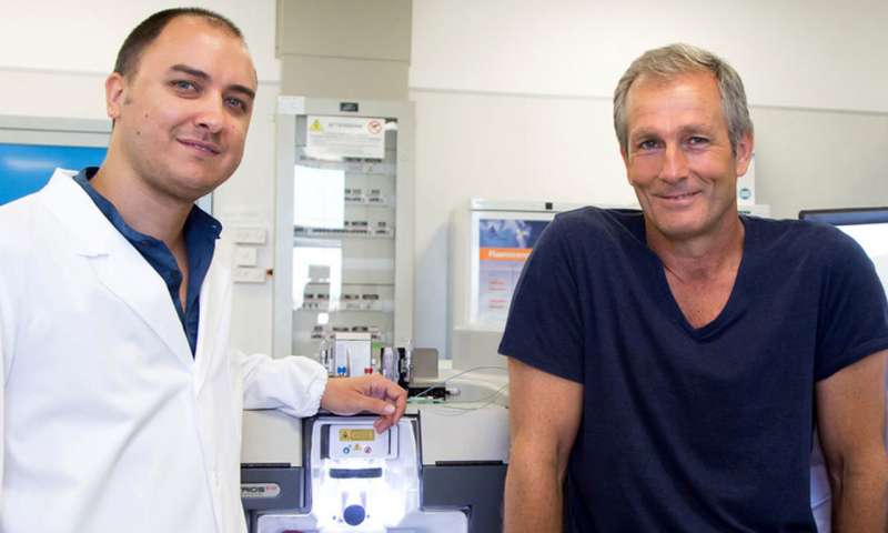
I have a confession. Deep down I’m shallow. So when something I am part of is acknowledged as one of the best, I delight in it (my fellow bloggers Katie and Esteban also delight in it, I am just more shameless about letting everyone know.)

And that is just what happened with this blog, The Stem Cellar. We have been named as one of the “22 best biology and stem cell blogs of 2022”. And not just by anyone. We were honored by Dr. Paul Knoepfler, a stem cell scientist, avid blogger and all-round renaissance man (full disclosure, Paul is a recipient of CIRM funding but that has nothing to do with this award. Obviously.)

We are particularly honored to be on the list because Paul includes some heavy hitters including The Signals Blog, a site that he describes this way:

“This one from our friends in Canada is fantastic. They literally have dozens of authors, which is probably the most of any stem cell-related website, and their articles include many interesting angles. They post really often too. I might rank Signal and The Stem Cellar as tied for best stem cell blog in 2021.”

Now I’m really blushing.

Other highly regarded blogs are EuroStemCell, the Mayo Clinic Regenerative Medicine Blog and Stem Cell Battles (by Don Reed, a good friend of CIRM’s)

Another one of the 22 is David Jensen’s California Stem Cell report which is dedicated to covering the work of, you guessed it, CIRM. So, not only are we great bloggers, we are apparently great to blog about. 

As a further demonstration of my modesty I wanted to point out that Paul regularly produces ‘best of’ lists, including his recent “50 influencers on stem cells on Twitter to follow” which we were also on.

Stories that caught our eye: National Geographic takes a deep dive into iPS cells; Japanese researchers start iPS cell clinical trial for spinal cord injury; and do high fat diets increase your risk of colorectal cancer

Can cell therapy beat the most difficult diseases?

That’s the question posed in a headline in National Geographic. The answer; maybe, but it is going to take time and money.

The article focuses on the use of iPS cells, the man-made equivalent of embryonic stem cells that can be turned into any kind of cell or tissue in the body. The reporter interviews Kemal Malik, the member of the Board of Management for pharmaceutical giant Bayer who is responsible for innovation. When it comes to iPS cells, it’s clear Malik is a true believer in their potential.

“Because every cell in our bodies can be produced from a stem cell, the applicability of cell therapy is vast. iPSC technology has the potential to tackle some of the most challenging diseases on the planet.”

But he also acknowledges that the field faces some daunting challenges, including:

  • How to manufacture the cells on a large scale without sacrificing quality and purity
  • How do you create products that have a stable shelf life and can be stored until needed?
  • How do you handle immune reactions if you are giving these cells to patients?

Nonetheless, Malik remains confident we can overcome those challenges and realize the full potential of these cells.

“I believe human beings are on the cusp of the next big wave of pharmaceutical innovation. The use of living cells to make people better.”

As if to prove Malik right there was also news this week that researchers at Japan’s Keio University have been given permission to start a clinical trial using iPS cells to treat people with spinal cord injuries. This would be the first of its kind anywhere in the world.

Japan launches iPSC clinical trial for spinal cord injury

An article in Biospace says that the researchers plan to treat four patients who have suffered varying degrees of paralysis due to a spinal cord injury.  They will take cells from the patients and, using the iPS method, turn them into the kind of nerve cells found in the spinal cord, and then transplant two million of them back into the patient. The hope is that this will create new connections that restore movement and feeling in the individuals.

This trial is expected to start sometime this summer.

CIRM has already funded a first-of-its-kind clinical trial for spinal cord injury with Asterias Biotherapeutics. That clinical trial used embryonic stem cells turned into oligodendrocyte progenitor cells – which develop into cells that support and protect nerve cells in the central nervous system. We blogged about the encouraging results from that trial here.

High fat diet drives colorectal cancer

Finally today, researchers at Salk have uncovered a possible cause to the rise in colorectal cancer deaths among people under the age of 55; eating too much high fat food.

Our digestive system works hard to break down the foods we eat and one way it does that is by using bile acids. Those acids don’t just break down the food, however, they also break down the lining of our intestines. Fortunately, our gut has a steady supply of stem cells that can repair and replace that lining. Unfortunately, at least according to the team from Salk, mutations in these stem cells can lead to colorectal cancer.

The study, published in the journal Cell, shows that bile acids affect a protein called FXR that is responsible for ensuring that gut stem cells produce a steady supply of new lining for the gut wall. When someone eats a high fat diet it upsets the balance of bile acids, starting a cascade of events that help cancer develop and grow.

In a news release Annette Atkins, a co-author of the study, says there is a strong connection between bile acid and cancer growth:

“We knew that high-fat diets and bile acids were both risk factors for cancer, but we weren’t expecting to find they were both affecting FXR in intestinal stem cells.”

So next time you are thinking about having that double bacon cheese burger for lunch, you might go for the salad instead. Your gut will thank you. And it might just save your life.

Stem Cell Stories that Caught Our Eye: Human Eggs From Stem Cells, A New Way to Heal Broken Bones and A Lab Grown Esophagus

Stem cell image of the week:  Immature human eggs (pink) were created by Japanese researchers using stem cells that were derived from blood cells.

steinstory-custom-9e57980ac5d757b1440c9f20a55b81d352283a93_0

Photo Courtesy of Saitou Lab

A team of Japanese scientists say they have taken an important step toward creating human eggs in a lab dish.

Their work, which was reported Thursday in the journal Science, outlined their research and explained how they were able to turn human blood cells into stem cells, which they then transformed into very immature human eggs.

They say the eggs are too immature to be fertilized or make a baby. And much more research would be needed to create eggs that could be useful, and safe for human reproduction. But they believe the technique could someday help millions of people suffering from infertility.

In their paper, the Japanese scientists say the next step will be to try to make mature human eggs and produce human sperm this way.

“It’s the beginning of a paradigm change,” says Kyle Orwig, a professor in the department of obstetrics, gynecology and reproductive sciences at the University of Pittsburgh School of Medicine.

In addition to helping infertile people, such a development could enable same sex couples to have babies with sperm and eggs made from their own skin cells.

But such a possibility would also have much broader implications, say others following the field.

Newly discovered stem cells may help heal broken bones and arthritic joints. (Todd Dubnicoff)

Oh, to be a newt. This semi-aquatic salamander is able to regenerate an entire limb after injury. The regenerative ability of our human bodies just doesn’t measure up: we can heal a bone fracture though that ability weakens as we age, and some bone fractures called nonunions are unable to heal. And we have no ability to regrow lost cartilage leaving 75 million Americans suffering with painful, debilitating arthritis.

image.img.620.high

A small bone structure arising from the human skeletal stem cell contains cartilage (blue), bone marrow (brown) and bone (yellow). Image credit: Longaker and Chan labs, Stanford University.

CIRM-funded research published this week in Cell may one day give doctors a leg up on treating bone-related disease and injury. The Stanford team behind the study reports that they’ve identified a stem cell that gives the three main components of our skeleton: the outer bone, the spongy interior and cartilage that provides cushion in our joints. The scientists showed that these skeletal stem cells are separate from mesenchymal stem cells which can also specialize, or differentiate, into skeletal tissues as well as fat and muscle. One of the lead authors, Dr. Charles Chan, PhD, explained the important distinction between the two cell types in a press release:

“Mesenchymal stem cells are loosely characterized and likely to include many populations of cells, each of which may respond differently and unpredictably to differentiation signals. In contrast, the skeletal stem cell we’ve identified possesses all of the hallmark qualities of true, multipotential, self-renewing, tissue-specific stem cells. They are restricted in terms of their fate potential to just skeletal tissues, which is likely to make them much more clinically useful.”

The researchers located skeletal stem cells at the end of developing bone and found them in increasing numbers at the site of healing broken bones. The scientists were also able to derive them by reprogramming readily available human fat cells as well as embryonic stem cell-like induced pluripotent stem cells (iPSCs). With these skeletal stem cells now in hand, the team is excited with the prospect of combining cartilage-repair surgeries with an injection of the stem cells to boost healing. Senior author Michael Longaker envisions the impact of such therapies on healthcare in the U.S.:

“I would hope that, within the next decade or so, this cell source will be a game-changer in the field of arthroscopic and regenerative medicine. The United States has a rapidly aging population that undergoes almost 2 million joint replacements each year. If we can use this stem cell for relatively noninvasive therapies, it could be a dream come true.”

Cincinnati Children’s researchers report progress growing a human esophagus in a lab (Adonica Shaw) 

Cincinnati-Childrens-Hospital-esophageal-organoid

A confocal microscopic image shows a two-month-old human esophageal organoid bioengineered by Cincinnati Children’s Hospital researchers from pluripotent stem cells.      Image courtesy of Cincinnati Children’s Hospital

Scientists from Cincinnati Children’s Center for Stem Cell and Organoid Medicine (CuSTOM) have successfully grown human esophageal tissue entirely from pluripotent stem cells (PSCs).

Their research, which was published in the journal Cell Stem Cell, is the latest advancement from (CuSTOM). They believe it will open the door for other scientists  to form any tissue type in the body from stem cells.

The center is developing new ways to study birth defects and diseases that affect millions of people with gastrointestinal disorders, such as gastric reflux, and this research is a milestone for them.

“Disorders of the esophagus and trachea are prevalent enough in people that organoid models of human esophagus could be greatly beneficial. In addition to being a new model to study birth defects like esophageal atresia, the organoids can be used to study diseases like eosinophilic esophagitis and Barrett’s metaplasia, or to bioengineer genetically matched esophageal tissue for individual patients, ” said Jim Wells, PhD, chief scientific officer at CuSTOM and study lead investigator.

The resulting human esophageal organoids were fully formed and grew to a length of about 300-800 micrometers in about two months. Compared biochemically with esophageal tissues from patient biopsies, the bioengineered tissues were similar.

The research team plans to further the technology’s therapeutic potential through projects including using the organoids to examine the progression of specific diseases and congenital defects affecting the esophagus.

Stem cell stories that caught our eye: Reprogramming cells in vivo may help heal ulcers, CIRM-funded clinical trial shows promise and a New report, clears up an old question.

Stem cell image of the week:  New Research out of the Salk Institute could bring us closer to reprogramming stem cells without taking them out of the body (Adonica Shaw)

Our stem cell image of the week could be a step towards reprogramming cells in vivo.

The image represents the first proof of principle for the successful regeneration of a functional organ (the skin) inside a mammal, by a technique known as AAV-based in vivo reprogramming. Epithelial (skin) tissues were generated by converting one cell type (red: mesenchymal cells) to another (green: basal keratinocytes) within a large ulcer in a laboratory mouse model.

Sep6_2016_SalkInst_EpithelialInVivoRegeneration1822142291-1

Photo courtesy of the Salk Institute

In large patches of ulcerous skin, surviving cells prioritize inflammation and wound closure. That’s what they’re programmed to do. Cells, on the other hand, can be reprogrammed in living tissue, wounded tissue, to expedite healing.

A group of scientists at the Salk Institute developed a new approach to cellular reprogramming. They found a way to directly convert the cells in an open wound into new skin cells. Their findings were published on Wednesday in Nature.

Reprogramming wound-resident cells could be useful for healing skin damage, countering the effects of aging, and helping us to better understand skin cancer. It could also supplant plastic surgery and the application of skin grafts as a way to treat large cutaneous ulcers, including those seen in people with severe burns, bedsores, or chronic diseases such as diabetes.

When an ulcer is especially large, it can be difficult for surgeons to graft enough skin. In these cases, researchers can isolate skin stem cells from a patient, grow them in the lab and transplant them back into the patient. However, such a procedure requires an extensive amount of time, which may put the patient’s life at risk and is sometimes not effective.

 “Our observations constitute an initial proof of principle for in vivo regeneration of an entire three-dimensional tissue like the skin, not just individual cell types as previously shown,” says Dr. Izpisua Belmonte. “This knowledge might not only be useful for enhancing skin repair but could also serve to guide in vivo regenerative strategies in other human pathological situations, as well as during aging, in which tissue repair is impaired.”

Positive news from a CIRM-funded clinical trial targeting a deadly blood cancer. (Kevin McCormack)  Multiple myeloma is a type of blood cancer where certain cells in the bone marrow grow out of control, crowding out the healthy cells and forming tumors. There is no cure but there are many treatments that can slow down or even halt the progression. Over time, however, many of those treatments lose their effectiveness and the cancer returns. Now a new CIRM-funded clinical trial targeting this kind of relapsing multiple myeloma is showing promise.

The trial, by Poseida Therapeutics, takes an immunotherapy approach that uses the patient’s own engineered immune system T cells to seek and destroy the myeloma cells. This product, called P-BCMA-101, is a stem cell memory chimeric antigen receptor T-cell (CAR-T).

In the first eleven patients treated there were no serious side effects and only one patient had a suspected case of cytokine release syndrome. That’s where large amounts of cytokines, immune substances, are rapidly released into the body causing fever, nausea, rapid heartbeat etc. However, even in this patient the symptoms quickly passed.

In a news release, Eric Ostertag, the CEO of Poseida said that even though the goal of this Phase 1 study was just to make sure it was safe and to identify the best dose to give patients, they have already seen a very good partial response in some patients.

“We believe our advantages of a purified product, where all cells express the CAR molecule, and a product with high levels of stem cell memory T cells, producing a more gradual and prolonged immune response against tumor cells, provide a significantly better therapeutic index when compared with other CAR-T therapeutics. We are also encouraged that P-BCMA-101 is demonstrating significant efficacy even at doses that have been ineffective for other anti-BCMA CAR-T therapies and that our response rates continue to improve as the dose increases.”

The clinical trial will eventually treat 40 patients with relapsing or remitting multiple myeloma and we will bring more results as they become available.

Techniques used in ecology help rewrite basic fact about blood stem cells (Todd Dubnicoff) It’s been over half a century since the first blood stem cell transplantation was performed. And yet, some fundamental facts about these cells – which give rise to all the cell types of our blood – have remained cloudy, like the number of blood stem cells present in the human body. This week, researchers at Wellcome Sanger Institute and Wellcome – MRC Cambridge Stem Cell Institute report that they’ve cleared up this long-lasting question.

blood_cell_colony

A blood cell colony grown from a single cell isolated from a 59-year-old man. Image Credit: Mairi Shepherd, Kent Lab

And the results were surprising. Using whole genome DNA sequencing and techniques found in ecology for tracking population sizes, the team determined that a healthy adult has between 50,000 and 200,000 blood stem cells at any given time. That’s about 10 times more than what was previously thought.

Dr. David Kent, a co-senior author on the report, described the implications of this discovery in a press release:

“This new approach is hugely flexible. Not only can we measure how many stem cells exist, we can also see how related they are to each other and what types of blood cells they produce. Applying this technique to samples from patients with blood cancers, we should now be able to learn how single cells outcompete normal cells to expand their numbers and drive a cancer.”

The study was published in Nature.


Stem cell stories that caught our eye: CIRM-funded scientist wins prestigious prize and a tooth trifecta

CIRM-grantee wins prestigious research award

Do we know how to pick ‘em or what? For a number of years now we have been funding the work of Stanford’s Dr. Marius Wernig, who is doing groundbreaking work in helping advance stem cell research. Just how groundbreaking was emphasized this week when he was named as the winner of the 2018 Ogawa-Yamanaka Stem Cell Prize.

WernigMarius_Stanford

Marius Wernig, MD, PhD. [Photo: Stanford University]

The prestigious award, from San Francisco’s Gladstone Institutes, honors Wernig for his innovative work in developing a faster, more direct method of turning ordinary cells into, for example, brain cells, and for his work advancing the development of disease models for diseases of the brain and skin disorders.

Dr. Deepak Srivastava, the President of Gladstone, announced the award in a news release:

“Dr. Wernig is a leader in his field with extraordinary accomplishments in stem cell reprogramming. His team was the first to develop neuronal cells reprogrammed directly from skin cells. He is now investigating therapeutic gene targeting and cell transplantation–based strategies for diseases with mutations in a single gene.”

Wernig was understandably delighted at the news:

“It is a great honor to receive this esteemed prize. My lab’s goal is to discover novel biology using reprogrammed cells that aids in the development of effective treatments.”

Wernig will be presented with the award, and a check for $150,000, at a ceremony on Oct. 15 at the Gladstone Institutes in San Francisco.

A stem cell trifecta for teeth research

It was a tooth trifecta among stem cell scientists this week. At Tufts University School of Medicine, researchers made an important advance in the development of bioengineered teeth. The current standard for tooth replacement is a dental implant. This screw-shaped device acts as an artificial tooth root that’s inserted into the jawbone. Implants have been used for 30 years and though successful they can lead to implant failure since they lack many of the properties of natural teeth. By implanting postnatal dental cells along with a gel material into mice, the team demonstrated, in a Journal of Dental Research report, the development of natural tooth buds. As explained in Dentistry Today, these teeth “include features resembling natural tooth buds such as the dental epithelial stem cell niche, enamel knot signaling centers, transient amplifying cells, and mineralized dental tissue formation.”

Another challenge with the development of a bioengineered tooth replacement is reestablishing nerve connections within the tooth, which plays a critical role in its function and protection but doesn’t occur spontaneously after an injury. A research team across the “Pond” at the French National Institute of Health and Medical Research, showed that bone marrow-derived mesenchymal stem cells in the presence of a nerve fiber can help the nerve cells make connections with bioengineered teeth. The study was also published in the Journal of Dental Research.

And finally, a research report about stem cells and the dreaded root canal. When the living soft tissue, or dental pulp, of a tooth becomes infected, the primary course of action is the removal of that tissue via a root canal. The big downside to this procedure is that it leaves the patient with a dead tooth which can be susceptible to future infections. To combat this side effect, researchers at the New Jersey Institute of Technology report the development of a potential remedy: a gel containing a fragment of a protein that stimulates the growth of new blood vessels as well as a fragment of a protein that spurs dental stem cells to divide and grow. Though this technology is still at an early stage, it promises to help keep teeth alive and healthy after root canal. The study was presented this week at the National Meeting of the American Chemical Society.

Here’s an animated video that helps explain the research:

Stem cell stories that caught our eye: 3 blind mice no more and a tale of two tails

Stem cell image of the week: The demise of Three Blind Mice nursery rhyme (Todd Dubnicoff)
Our stem cell image of the week may mark the beginning of the end of the Three Blind Mice nursery rhyme and, more importantly, usher in a new treatment strategy for people suffering from vision loss. That’s because researchers from Icahn School of Medicine at Mount Sinai, New York report in Nature the ability to reprogram support cells in the eyes of blind mice to become photoreceptors, the light-sensing cells that enable sight. The image is an artistic rendering of the study results by team led Dr. Bo Chen, PhD.

Aug16_2018_BoChen_MullerGlia_Eye3930249103

An artist’s rendering incorporates the images of the Müller glia-derived rod photoreceptors. Image credit: Bo Chen, Ph.D.

The initial inspiration for this project came from an observation in zebrafish. These creatures have the remarkable ability to restore vision after severe eye injuries. It turns out that, in response to injury, a type of cell in the eye called Muller glia – which helps maintain the structure and function of the zebrafish retina – transforms into rod photoreceptors, which allow vision in low light.

Now, Muller glia are found in humans and mice too, so the research team sought to harness this shape-shifting, sight-restoring ability of the Muller glia but in the absence of injury. They first injected a gene into the eyes of mice born blind that stimulated the glia cells to divide and grow. Then, to mimic the reprogramming process seen in zebrafish, specific factors were injected to cause the glia to change identity into photoreceptors.

The researchers showed that the glia-derived photoreceptors functioned just like those observed in normal mice and made the right connections with nerve cells responsible for sending visual information to the brain. The team’s next steps are to not only show the cells are functioning properly in the eye and brain but to also do behavioral studies to confirm that the mice can do tasks that require vision.

If these studies pan out, it could lead to a new therapeutic strategy for blinding diseases like retinitis pigmentosa and macular degeneration. Rather than transplanting replacement cells, this treatment approach would spur our own eyes to repair themselves. In the meantime, CIRM-funded researchers have studies currently in clinical trials testing stem cell-based treatments for retinitis pigmentosa and macular degeneration.

A tale of two tails: one regenerates, the other, not quite so much (Kevin McCormack) One of the wonders of nature, well two if you want to be specific, is how both salamanders and lizards are able to regrow their tails if they lose them. But there is a difference. While salamanders can regrow a tail that is almost identical to the original, lizard’s replacements are rather less impressive. Now researchers have found out why.

081518_LR_regeneration_inline_730

In these fluorescence microscopy images, cross sections of original lizard and salamander tails (left) show cartilage (green) and nerve cells (red). In the regenerated tails (right), the lizard’s is made up mostly of cartilage, while the salamander also has developed new nerve cells. Image: Thomas Lozito

The study, published in the Proceedings of the National Academy of Sciences, shows how a lizard’s new tail doesn’t have bone but instead has cartilage, and also lacks nerve cells. The key apparently is the stem cells both use to regenerate the tail. Salamanders use neural stem cells from their spinal cord and turn them into other types of nervous system cell, such as neurons. Lizards neural stem cells are not able to do this.

The researchers, from the University of Pittsburgh, tested their findings by placing neural stem cells from the axolotl salamander into tail stumps from geckos. They noted that, as those tails regrew, some of those transplanted cells turned into neurons.

In an interview in Science News, study co-author Thomas Lozito says the team hope to take those findings and, using the CRISPR/Cas9 gene-editing tool, see if they can regenerate body parts in other animals:

 “My goal is to make the first mouse that can regenerate its tail. We’re kind of using lizards as a stepping-stone.”

Stem Cell Roundup: Knowing the nose, stem cell stress and cell fate math.

The Stem Cellar’s Image of the Week.
Our favorite image this week, comes to us from researchers at Washington University School of Medicine in St. Louis. Looking like a psychedelic Rorschach test, the fluorescence microscopy depicts mouse olfactory epithelium (in green), a sheet of tissue that develops in the nose. The team identified a new stem cell type that controls the growth of this tissue. New insights from the study of these cells could help the team better understand why some animals, like dogs, have a far superior sense of smell than humans.

MouseOlfactoryEpithelium-700x467

Peering into the nasal cavity of a mouse. Olfactory epithelium is indicated by green. Image credit: Lu Yang, Washington University School of Medicine in St. Louis.

A Washington U. press release provides more details about this fascinating study which appears in Developmental Cell.

How stress affects blood-forming stem cells.
Stress affects all of us in different ways. Some people handle it well. Some crack up and become nervous wrecks. So, perhaps it shouldn’t come as a huge surprise that stress also affects some stem cells. What is a pleasant surprise is that knowing this could help people undergoing cancer therapy or bone marrow transplants.

First a bit of background. Hematopoietic, or blood-forming stem cells (HSCs) come from bone marrow and are supported by other cells that secrete growth factors, including one called pleiotrophin or PTN. While researchers knew PTN was present in bone marrow they weren’t sure precisely what role it played.

So, researchers at UCLA set out to discover what PTN did.

In a CIRM-funded study they took mice that lacked PTN in endothelial cells – these line the blood vessels – or in their stromal cells – which make up the connective tissue. They found that a lack of PTN in stromal cells caused a lack of blood stem cells, but a lack of PTN in endothelial cells had no impact.

Chute Combo w Barrier 800x533

Expression of pleiotrophin (green) in bone marrow blood vessels (red) and stromal cells (white) is shown in normal mice (left) and in mice at 24 hours following irradiation (right). Image credit: UCLA

However, as Dr. John Chute explained in a news release, when they stressed the cells, by exposing them to radiation, they found something very different:

“The surprising finding was that pleiotrophin from stromal cells was not necessary for blood stem cell regeneration following irradiation — but pleiotrophin from endothelial cells was necessary.”

In other words, during normal times the stem cells rely on PTN from stromal cells, but after stress they depend on PTN from endothelial cells.

Dr. Chute says, because treatments like chemotherapy and radiation deplete bone marrow stem cells, this finding could have real-world implications for patients.

“These therapies for cancer patients suppress our blood cell systems over time. It may be possible to administer modified, recombinant versions of pleiotrophin to patients to accelerate blood cell regeneration. This strategy also may apply to patients undergoing bone marrow transplants.”

The study appears in the journal Cell Stem Cell.

Predicting the fate of cells with math
Researchers at Harvard Medical School and the Karolinska Institutet in Sweden reported this week that they have devised a mathematical model that can predict the fate of stem cells in the brain.

It may sound like science-fiction but the accomplished the feat by tracking changes in messenger RNA (mRNA), the genetic molecule that translates our DNA code into instructions for building proteins. As a brain stem cell begins specializing into specific cell types, hundreds of genes get turns on and off, which is observed by the rate of changes in mRNA productions.

The team built their predictive model by measuring these changes. In a press release, co-senior author, Harvard professor Peter Kharchenko, described this process using a great analogy:

“Estimating RNA velocity—or the rate of RNA change over time—is akin to observing the cooks in a restaurant kitchen as they line up the ingredients to figure out what dishes they’ll be serving up next.”

The team verified their mathematical model by inputting other data that was not use in constructing the model. Karolinkska Institutet professor, Sten Linnarsson, the other co-senior author on the study, described how such a model could be applied to human biomedical research:

“RNA velocity shows in detail how neurons and other cells acquire their specific functions as the brain develops and matures. We’re especially excited that this new method promises to help reveal how brains normally develop, but also to provide clues as to what goes wrong in human disorders of brain development, such as schizophrenia and autism.”

The study appears in the journal Nature.

Stem Cell Roundup: Artificial Embryos to Study Miscarriage and ALS Insight – Muscle Repair Cells Go Rogue

Stem Cell Image of the Week: Artificial embryos for studying miscarriage (Adonica Shaw)

etxembryos

Mouse embryos artificially generated by combining three types of stem cells.
Image: University of Cambridge.

This week’s stem cell image of the week comes from a team of researchers from The University of Cambridge who published research in Nature Cell Biology earlier this week indicating they’d achieved a breakthrough in stem cell research that resulted in the generation of a key developmental step that’d never before been achieved when trying to generate an artificial embryo.

To create the artificial embryo, the scientists combined mouse embryonic stem cells with two other types of stem cells that are present in the very earliest stages of embryo development. The reseachers grew the three stem cell types into a dish and coaxed them into simulating a process called gastrulation – one of the very first events that happens during a creature’s development in which the early embryo begins reorganizing into more and more complex multilayer organ structures.

In an interview with The Next Web (TNW), Professor Magdalena Zernicka-Goetz, who led the research team, says:

”Our artificial embryos underwent the most important event in life in the culture dish. They are now extremely close to real embryos. To develop further, they would have to implant into the body of the mother or an artificial placenta.”

The goal of this research isn’t to create mice on demand. Its purpose is to gain insights into early life development. And that could lead to a giant leap in our understanding of what happens during the period in a woman’s pregnancy where the risk of miscarriage is highest.

According to professor Zernicka-Goetz,

magda3

Magdalena Zernicka-Goetz, PhD

“We can also now try to apply this to the equivalent human stem cell types and so study the very earliest events in human embryo development without actually having to use natural human embryos.The early stages of embryo development are when a large proportion of pregnancies are lost and yet it is a stage that we know very little about. Now we have a way of simulating embryonic development in the culture dish, so it should be possible to understand exactly what is going on during this remarkable period in an embryo’s life, and why sometimes this process fails.”

Muscle repair cells go rogue – a possible drug target for ALS?
Call it a case of a good cell gone bad. This week researchers at Sanford Burnham Prebys Medical Discovery Institute, report in Nature Cell Biology that fibro-adipogenic progenitors (FAPs) – cells that are critical in coordinating the repair of torn muscles – can turn rogue, causing muscles to wither and scar. This “Dr. Jekyl and Mr. Hype” discovery may lead to novel treatments for a number of incurable disorders like amyotrophic lateral sclerosis (ALS), spinal muscular atrophy (SMA) and spinal cord injury.

drjekyllmrhy

Senior author Pier Lorenzo Puri, M.D. (right) and co-first author Luca Madaro, Ph.D. Credit: Fondazione Santa Lucia IRCCS

When muscle is strained, whether due to an acute injury or even weight-lighting, a consistent order of events occurs within the muscle. FAB cells enter the muscle tissue after immune cells called macrophages come in and gobble up dead tissue but before muscle stem cells are stimulated to regenerate the lost muscle. However, to the researchers’ surprise, something entirely different happens in the case of neuromuscular disorders like ALS where nerve signal connections to the muscles degenerate.

Once nerves are no longer attached to muscle and stop sending movement signals from the brain, the macrophages don’t infiltrate the muscle and instead the FAPs pile up in the muscle and never leave. And as a result, muscle stem cells are never activated. In ALS patients, this cellular train crash leads to progressive loss of muscle control to move the limbs and ultimately even to breathe.

The promising news from these findings, which were funded in part by CIRM, is that the team identified of an out-of-whack cell signaling pathway that is responsible for the breakdown in the rogue function of the FAP cells. The researchers hope further studies of this pathway’s role in muscle degeneration may lead to novel therapies and disease-screening technologies for ALS and other motor neuron diseases.

Stem cell roundup: summer scientists, fat-blocking cells & recent human evolution

Stem cell photo of the week: high schooler becoming a stem cell pro this summer

InstagramAnnaJSPARK

High school student Anna Guzman learning important lab skills at UC Davis

This summer’s CIRM SPARK Programs, stem cell research internships for high school students, are in full swing. Along with research assignments in top-notch stem cell labs, we’ve asked the students to chronicle their internship experiences through Instagram. And today’s stem cell photo of the week is one of those student-submitted posts. The smiling intern in this photo set is Anna Guzman, a rising junior from Sheldon High School who is in the UC Davis SPARK Program. In her post, she describes the lab procedure she is doing:

“The last step in our process to harvest stem cells from a sample of umbilical cord blood! We used a magnet to isolate the CD34 marked stem cells [blood stem cells] from the rest of the solution.”

Only a few days in and Anna already looks like a pro! It’s important lab skills like this one that could land Anna a future job in the stem cell field. Check out #cirmsparklab on Instagram to view the ever-growing number of posts.

Swiss team identifies a cell type that block formation of fat cells

Jun21_2018_EPFL_TwoDifferentAspectsOfFat1871459512

(Left) Mature human fat cells grown in a Petri dish (green, lipid droplets). (Right) A section of mouse fat tissue showing, in the middle, a blood vessel (red circle) surrounded by fat cell blocking cells called Aregs (arrows). [Bart Deplancke/EPFL]

Liposuction surgery helps slim and reshape areas of a person’s body through the removal of excess fat tissue. While the patient is certainly happy to get rid of those extra pounds, that waste product is sought after by researchers because it’s a rich source of regenerative cells including fat stem cells.

The exact populations of cells in this liposuction tissue has been unclear, so a collaboration of Swiss researchers – at Ecole Polytechnique Fédérale de Lausanne (EPFL) and Eidgenössische Technische Hochschule Zürich (ETHZ) – used a cutting-edge technique allowing them to examine the gene activity within single cells.

The analysis was successful in identifying several newly defined subpopulations of cells in the fat tissue. To their surprise, one of those cell types did not specialize into fat cells but instead did the opposite: they inhibited other fat stem cells from giving rise to fat cells. The initial experiments were carried out in mice, but the team went on to show similar fat-blocking cells in human tissue. Further experiments will explore the tantalizing prospect of applying these cells to control obesity and the many diseases, like diabetes, that result from it.

The study was published June 20st in Nature.

Connection identified between recent human evolution & risk for premature birth
Evidence of recent evolution in a human gene that’s critical for maintaining pregnancy may help explain why some populations have a higher risk for giving birth prematurely than others. That’s according to a recent report by researchers at the University of Stanford School of Medicine.

The study, funded in part by CIRM’s Genomics Initiative, compared DNA from people with East Asian, European and African ancestry. They specifically examined the gene encoding the progesterone hormone receptor which helps keep a pregnant woman from going into labor too soon. The gene is also associated with preterm births, the leading cause of infant death in the U.S.

The team was very surprise to find that people with East Asian ancestry had an evolutionarily new version of the gene while the European and African populations had mixtures of new and ancient versions. These differences may explain why the risk for premature birth among East Asian populations is lower than among pregnant women of European and African descent, though environment clearly plays a role as well.

Pediatrics professor Gary Shaw, PhD, one of the team leaders, put the results in perspective:

“Preterm birth has probably been with us since the origin of the human species,” said Shaw in a press release, “and being able to track its evolutionary history in a way that sheds new light on current discoveries about prematurity is really exciting.”

The study was published June 21st in The American Journal of Human Genetics.

Friday Stem Cell Roundup: Making Nerves from Blood; New Clues to Treating Parkinson’s

Stanford lab develops method to make nerve cells from blood.

wernig_ineurons_blood

Induced neuronal (iN) cells derived from adult human blood cells. Credit: Marius Wernig, Stanford University.

Back in 2010, Stanford Professor Marius Wernig and his team devised a method to directly convert skin cells into neurons, a nerve cell. This so-called transdifferentiation technique leapfrogs over the need to first reprogram the skin cells into induced pluripotent stem cells. This breakthrough provided a more efficient path to studying how genetics plays a role in various mental disorders, like autism or schizophrenia, using patient-derived cells. But these types of genetic analyses require data from many patients and obtaining patient skin samples hampered progress because it’s not only an invasive, somewhat painful procedure but it also takes time and money to prepare the tissue sample for the transdifferentiation method.

This week, the Wernig lab reported on a solution to this bottleneck in the journal, PNAS. The study, funded in part by CIRM, describes a variation on their transdifferentiation method which converts T cells from the immune system, instead of skin cells, into neurons. The huge advantage with T cells is that they can be isolated from readily available blood samples, both fresh or frozen. In a press release, Wernig explains this unexpected but very welcomed result:

“It’s kind of shocking how simple it is to convert T cells into functional neurons in just a few days. T cells are very specialized immune cells with a simple round shape, so the rapid transformation is somewhat mind-boggling. We now have a way to directly study the neuronal function of, in principle, hundreds of people with schizophrenia and autism. For decades we’ve had very few clues about the origins of these disorders or how to treat them. Now we can start to answer so many questions.”

Two studies targeting Parkinson’s offer new clues to treating the disease (Kevin McCormack)
Despite decades of study, Parkinson’s disease remains something of a mystery. We know many of the symptoms – trembling hands and legs, stiff muscles – are triggered by the loss of dopamine producing cells in the brain, but we are not sure what causes those cells to die. Despite that lack of certainty researchers in Germany may have found a way to treat the disease.

Mitochondria

Simple diagram of a mitochondria.

They took skin cells from people with Parkinson’s and turned them into the kinds of nerve cell destroyed by the disease. They found the cells had defective mitochondria, which help produce energy for the cells. Then they added a form of vitamin B3, called nicotinamide, which helped create new, healthy mitochondria.

In an article in Science & Technology Research News Dr. Michela Deleidi, the lead researcher on the team, said this could offer new pathways to treat Parkinson’s:

“This substance stimulates the faulty energy metabolism in the affected nerve cells and protects them from dying off. Our results suggest that the loss of mitochondria does indeed play a significant role in the genesis of Parkinson’s disease. Administering nicotinamide riboside may be a new starting-point for treatment.”

The study is published in the journal Cell Reports.

While movement disorders are a well-recognized feature of Parkinson’s another problem people with the condition suffer is sleep disturbances. Many people with Parkinson’s have trouble falling asleep or remaining asleep resulting in insomnia and daytime sleepiness. Now researchers in Belgium may have uncovered the cause.

Working with fruit flies that had been genetically modified to have Parkinson’s symptoms, the researchers discovered problems with neuropeptidergic neurons, the type of brain cell that helps regulate sleep patterns. Those cells seemed to lack a lipid, a fat-like substance, called phosphatidylserine.

In a news release Jorge Valadas, one of the lead researchers, said replacing the missing lipid produced promising results:

“When we model Parkinson’s disease in fruit flies, we find that they have fragmented sleep patterns and difficulties in knowing when to go to sleep or when to wake up. But when we feed them phosphatidylserine–the lipid that is depleted in the neuropeptidergic neurons–we see an improvement in a matter of days.”

Next, the team wants to see if the same lipids are low in people with Parkinson’s and if they are, look into phosphatidylserine – which is already approved in supplement form – as a means to help ease sleep problems.