Stem cell image of the week: Immature human eggs (pink) were created by Japanese researchers using stem cells that were derived from blood cells.
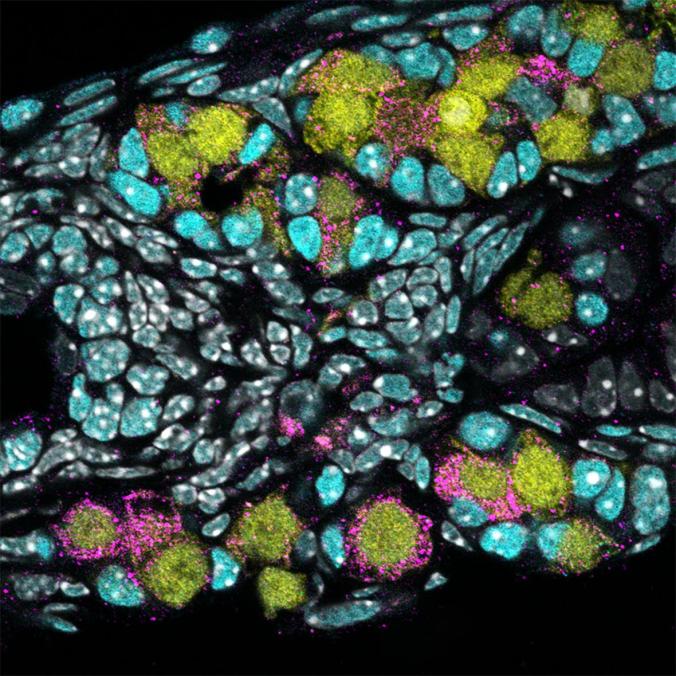
Photo Courtesy of Saitou Lab
A team of Japanese scientists say they have taken an important step toward creating human eggs in a lab dish.
Their work, which was reported Thursday in the journal Science, outlined their research and explained how they were able to turn human blood cells into stem cells, which they then transformed into very immature human eggs.
They say the eggs are too immature to be fertilized or make a baby. And much more research would be needed to create eggs that could be useful, and safe for human reproduction. But they believe the technique could someday help millions of people suffering from infertility.
In their paper, the Japanese scientists say the next step will be to try to make mature human eggs and produce human sperm this way.
“It’s the beginning of a paradigm change,” says Kyle Orwig, a professor in the department of obstetrics, gynecology and reproductive sciences at the University of Pittsburgh School of Medicine.
In addition to helping infertile people, such a development could enable same sex couples to have babies with sperm and eggs made from their own skin cells.
But such a possibility would also have much broader implications, say others following the field.
Newly discovered stem cells may help heal broken bones and arthritic joints. (Todd Dubnicoff)
Oh, to be a newt. This semi-aquatic salamander is able to regenerate an entire limb after injury. The regenerative ability of our human bodies just doesn’t measure up: we can heal a bone fracture though that ability weakens as we age, and some bone fractures called nonunions are unable to heal. And we have no ability to regrow lost cartilage leaving 75 million Americans suffering with painful, debilitating arthritis.
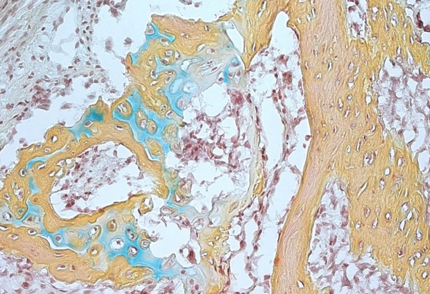
A small bone structure arising from the human skeletal stem cell contains cartilage (blue), bone marrow (brown) and bone (yellow). Image credit: Longaker and Chan labs, Stanford University.
CIRM-funded research published this week in Cell may one day give doctors a leg up on treating bone-related disease and injury. The Stanford team behind the study reports that they’ve identified a stem cell that gives the three main components of our skeleton: the outer bone, the spongy interior and cartilage that provides cushion in our joints. The scientists showed that these skeletal stem cells are separate from mesenchymal stem cells which can also specialize, or differentiate, into skeletal tissues as well as fat and muscle. One of the lead authors, Dr. Charles Chan, PhD, explained the important distinction between the two cell types in a press release:
“Mesenchymal stem cells are loosely characterized and likely to include many populations of cells, each of which may respond differently and unpredictably to differentiation signals. In contrast, the skeletal stem cell we’ve identified possesses all of the hallmark qualities of true, multipotential, self-renewing, tissue-specific stem cells. They are restricted in terms of their fate potential to just skeletal tissues, which is likely to make them much more clinically useful.”
The researchers located skeletal stem cells at the end of developing bone and found them in increasing numbers at the site of healing broken bones. The scientists were also able to derive them by reprogramming readily available human fat cells as well as embryonic stem cell-like induced pluripotent stem cells (iPSCs). With these skeletal stem cells now in hand, the team is excited with the prospect of combining cartilage-repair surgeries with an injection of the stem cells to boost healing. Senior author Michael Longaker envisions the impact of such therapies on healthcare in the U.S.:
“I would hope that, within the next decade or so, this cell source will be a game-changer in the field of arthroscopic and regenerative medicine. The United States has a rapidly aging population that undergoes almost 2 million joint replacements each year. If we can use this stem cell for relatively noninvasive therapies, it could be a dream come true.”
Cincinnati Children’s researchers report progress growing a human esophagus in a lab (Adonica Shaw)
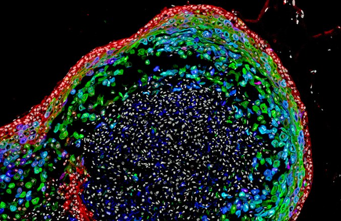
A confocal microscopic image shows a two-month-old human esophageal organoid bioengineered by Cincinnati Children’s Hospital researchers from pluripotent stem cells. Image courtesy of Cincinnati Children’s Hospital
Scientists from Cincinnati Children’s Center for Stem Cell and Organoid Medicine (CuSTOM) have successfully grown human esophageal tissue entirely from pluripotent stem cells (PSCs).
Their research, which was published in the journal Cell Stem Cell, is the latest advancement from (CuSTOM). They believe it will open the door for other scientists to form any tissue type in the body from stem cells.
The center is developing new ways to study birth defects and diseases that affect millions of people with gastrointestinal disorders, such as gastric reflux, and this research is a milestone for them.
“Disorders of the esophagus and trachea are prevalent enough in people that organoid models of human esophagus could be greatly beneficial. In addition to being a new model to study birth defects like esophageal atresia, the organoids can be used to study diseases like eosinophilic esophagitis and Barrett’s metaplasia, or to bioengineer genetically matched esophageal tissue for individual patients, ” said Jim Wells, PhD, chief scientific officer at CuSTOM and study lead investigator.
The resulting human esophageal organoids were fully formed and grew to a length of about 300-800 micrometers in about two months. Compared biochemically with esophageal tissues from patient biopsies, the bioengineered tissues were similar.
The research team plans to further the technology’s therapeutic potential through projects including using the organoids to examine the progression of specific diseases and congenital defects affecting the esophagus.
