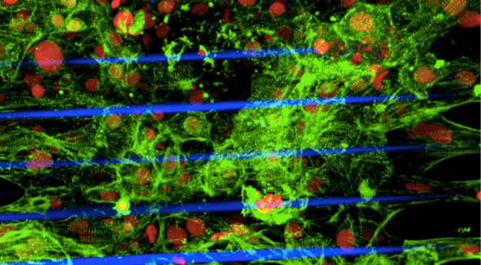
3-D printed cardiac microtissue generated from human stem cells. Image credit to Zhen Ma PhD, courtesy of the Gladstone Institutes
Hank Gathers was a 23-year-old basketball player at Loyola Marymount University with numerous accolades to his name and a promising future in the sport. This all came to an end when he suddenly collapsed in the middle of one of his games and subsequently passed away. It was the beginning of numerous such sudden death instances in the sporting world, that have been traced to hypertrophic cardiomyopathy (HCM)
HCM affects approximately 1 in 200 people and belongs to a family of diseases called cardiomyopathies, all of which impair the ability of heart muscle to effectively pump blood throughout your body. While there are several environmental factors that can contribute to the onset of HCM, one of the most common factors is a genetic mutation in a particular myosin binding protein called MYBPC3. This protein is responsible for both proper development of heart tissue, as well as controlling heart muscle contraction. Mutation in this gene is also associated with the most common type of cardiomyopathy, called dilated cardiomyopathy.
In HCM, mutation of MYBPC3 results in enlarged heart muscle with irregular contraction patterns. There is no cure, but medications to alleviate symptoms are available as well as surgical options. Surgery, however, can lead to serious complications and is not an option for everyone. Better understanding of the disease is necessary to develop treatment options that are effective for all patients.
A CIRM funded scientific collaboration between labs at UC Berkeley and the Gladstone Institutes has found a better way to study heart diseases such as HCM by generating a 3-D model heart tissue. While the genetic mutations that lead to cardiomyopathies are fairly well studied, how exactly the mutation leads to disease symptoms is not well understood, partially because many of those scientific studies relied on insights derived from two-dimensional culture systems. While informative in many ways, these types of cultures do not mimic the 3-D interactions that occur in nature. To overcome this challenge, these scientists used laser-guided 3-D printing system along with human cardiac stem cells to assemble the heart microtissue.
To confirm that the microtissue functioned like a real heart, they changed the structure of the printing scaffold to mimic the stress that hearts undergo during development and in different environmental conditions. They found that the microtissue derived from healthy human cardiac stem cells was able to adapt and contract normally in these changing environmental conditions.
When comparing the structure of WT and MYBPC3 generated microtissues, they surprisingly saw no different in microtissue architecture derived from WT or mutant cells. They did, however, observe functional differences between the normal and mutant tissues: mutant tissues displayed an increased contraction rate in response to stress and dysregulated contraction, both of which are hallmarks of HCM. Thus, this microtissue can mimic both normal and disease states.
Zhen Ma, lead author of the study explains the importance of this technology in a press release:
“With these microtissues we were able to observe how the human heart can develop this syndrome. Even though this is a microscopically tiny part of the heart, we could measure its contraction, the mechanical forces generated, and the calcium flow associated with the electrical signaling that triggers contraction of heart muscle. This advance gives us an opportunity to study cardiac disease in a much more precise manner.”
The most exciting aspect of this bioengineering success is its applicability beyond even heart disease. Bruce Conklin, one of the lead authors of the study explains:
“Some of the worst drug safety issues are due to problems with side effects on the heart, so we need better ways to test drugs for potential cardiac effects. It’s possible that in the future microtissues might become the preferred choice for their capability to capture a fuller range of cardiac physiology.

Pingback: A 3-D model of heart tissue gives scientists a leg up in studying heart disease — The Stem Cellar – Shubert Regenerative Care