The present and future of regenerative medicine

One of the great pleasures of my job is getting to meet the high school students who take part in our SPARK or Summer Internship to Accelerate Regenerative Medicine Knowledge program. It’s a summer internship for high school students where they get to spend a couple of months working in a world class stem cell and gene therapy research facility. The students, many of whom go into the program knowing very little about stem cells, blossom and produce work that is quite extraordinary.

One such student is Tan Ieng Huang, who came to the US from China for high school. During her internship at U.C. San Francisco she got to work in the lab of Dr. Arnold Kriegstein. He is the Founding Director of the Eli and Edythe Broad Center of Regeneration Medicine and Stem Cell Research at the University of California, San Francisco. Not only did she work in his lab, she took the time to do an interview with him about his work and his thoughts on the field.

It’s a fascinating interview and shows the creativity of our SPARK students. You will be seeing many other examples of that creativity in the coming weeks. But for now, enjoy the interview with someone who is a huge presence in the field today, by someone who may well be a huge presence in the not too distant future.

‘a tête-à-tête with Prof. Arnold Kriegstein’

The Kriegstein lab team: Photo courtesy UCSF

Prof. Arnold Kriegstein is the Founding Director of the Eli and Edythe Broad Center of Regeneration Medicine and Stem Cell Research at the University of California, San Francisco. Prof. Kriegstein is also the Co-Founder and Scientific Advisor of Neurona Therapeutics which seeks to provide effective and safe cell therapies for chronic brain disorder. A Clinician by training, Prof. Kriegstein has been fascinated by the intricate workings of the human brain. His laboratory focuses on understanding the transcriptional and signaling networks active during brain development, the diversity of neuronal cell types, and their fate potential. For a long time, he has been interested in harnessing this potential for translational and therapeutic intervention.

During my SEP internship I had the opportunity to work in the Kriegstein lab. I was in complete awe. I am fascinated by the brain. During the course of two months, I interacted with Prof. Kriegstein regularly, in lab meetings and found his ideas deeply insightful. Here’s presenting some excerpts from some of our discussions, so that it reaches many more people seeking inspiration!

Tan Ieng Huang (TH): Can you share a little bit about your career journey as a scientist?

Prof. Arnold Kriegstein (AK): I wanted to be a doctor when I was very young, but in high school I started having some hands-on research experience. I just loved working in the lab. From then on, I was thinking of combining those interests and an MD/PhD turned out to be an ideal course for me. That was how I started, and then I became interested in the nervous system. Also, when I was in high school, I spent some time one summer at Rockefeller University working on a project that involved operant conditioning in rodents and I was fascinated by behavior and the role of the brain in learning and memory. That happened early on, and turned into an interest in cortical development and with time, that became my career.

TH: What was your inspiration growing up, what made you take up medicine as a career?

AK: That is a little hard to say, I have an identical twin brother. He and I used to always share activities, do things together. And early on we actually became eagle scouts, sort of a boy scout activity in a way. In order to become an eagle scout without having to go through prior steps, we applied to a special program that the scouts had, which allowed us to shadow physicians in a local hospital. I remember doing that at a very young age. It was a bit ironic, because one of the evenings, they showed us films of eye surgery, and my brother actually fainted when they made an incision in the eye. The reason it makes me laugh now is because my brother became an eye surgeon many years later. But I remember our early experience, we both became very fascinated by medicine and medical research.

Tan Ieng and Dr. Arnold Kriegstein at UCSF

TH: What inspired you to start the Eli and Edythe Broad Center of Regeneration Medicine and Stem Cell Research Institute?

AK: My interest in brain development over the years became focused on earlier stages of development and eventually Neurogenesis, you know, how neurons are actually generated during early stages of in utero brain development. In the course of doing that we discovered that the radial glial cells, which have been thought for decades to simply guide neurons as they migrate, turned out to actually be the neural stem cells, they were making the neurons and also guiding them toward the cortex. So, they were really these master cells that had huge importance and are now referred to as neural stem cells. But at that time, it was really before the stem cell field took off. But because we studied neurogenesis, because I made some contributions to understanding how the brain develops from those precursors or progenitor cells, when the field of stem cells developed, it was very simple for me to identify as someone who studied neural stem cells. I became a neural stem cell scientist. I started a neural stem cell program at Columbia University when I was a Professor there and raised 15 million dollars to seed the program and hired new scientists. It was shortly after that I was approached to join UCSF as the founder of a new stem cell program. And it was much broader than the nervous system; it was a program that covered all the different tissues and organ systems.

TH: Can you tell us a little bit about how stem cell research is contributing to the treatment of diseases? How far along are we in terms of treatments?

AK: It’s taken decades, but things are really starting to reach the clinic now. The original work was basic discovery done in research laboratories, now things are moving towards the clinic. It’s a really very exciting time. Initially the promise of stem cell science was called Regenerative medicine, the idea of replacing injured or worn-out tissues or structures with new cells and new tissues, new organs, the form of regeneration was made possible by understanding that there are stem cells that can be tweaked to actually help make new cells and tissues. Very exciting process, but in fact the main progress so far hasn’t been replacing worn out tissues and injured cells, but rather understanding diseases using human based model of disease. That’s largely because of the advent of induced pluripotent stem cells, a way of using stem cells to make neurons or heart cells or liver cells in the laboratory, and study them both in normal conditions during development and in disease states. Those platforms which are relatively easy to make now and are pretty common all over the world allow us to study human cells rather than animal cells, and the hope is that by doing that we will be able to produce conventional drugs and treatments that work much better than ones we had in the past, because they will be tested in actual human cells rather than animal cells.

TH: That is a great progress and we have started using human models because even though there are similarities with animal models, there are still many species-specific differences, right?

AK: Absolutely, in fact, one of the big problems now in Big Pharma, you know the drug companies, is that they invest millions and sometimes hundreds of millions of dollars in research programs that are based on successes in treating mice, but patients don’t respond the same way. So the hope is that by starting with a treatment that works on human cells it might be more likely that the treatment will work on human patients.

TH: What are your thoughts on the current challenges and future of stem cell research?

AK: I think this is an absolute revolution in modern medicine, the advent of two things that are happening right now, first the use of induced pluripotent stem cells, the ability to make pluripotent cells from adult tissue or cells from an individual allows us to use models of diseases that I mentioned earlier from actual patients. That’s one major advance. And the other is gene editing, and the combination of gene editing and cell-based discovery science allows us to think of engineering cells in ways that can make them much more effective as a form of cell therapy and those cell therapies have enormous promise. Right now, they are being used to treat cancer, but in the future, they might be able to treat heart attack, dementia, neurodegenerative diseases, ALS, Parkinson’s disease, a huge list of disorders that are untreatable right now or incurable. They might be approached by the combination of cell-based models, cell therapies, and gene editing.

TH: I know there are still some challenges right now, like gene editing has some ethical issues because people don’t know if there can be side effects after the gene editing, what are your thoughts?

AK: You know, like many other technologies there are uncertainties, and there are some issues. Some of the problems are off-target effects, that is you try to make a change in one particular gene, and while doing that you might change other genes in unexpected ways and cause complications. But we are understanding that more and more now and can make much more precise gene editing changes in just individual genes without affecting unanticipated areas of the genome. And then there are also the problems of how to gene-edit cells in a safe way. There are certain viral factors that can be used to introduce the gene editing apparatus into a cell, and sometimes if you are doing that in a patient, you can also have unwanted side effects from the vectors that you are using, often they are modified viral vectors. So, things get complicated very quickly when you start trying to treat patients, but I think these are all tractable problems and I think in time they will all be solved. It will be a terrific, very promising future when it comes to treating patients who are currently untreatable.

TH: Do you have any advice for students who want to get into this field?

AK: Yes, I think it’s actually never been a better time and I am amazed by the technologies that are available now. Gene editing that I mentioned before but also single cell approaches, the use of single cell multiomics revealing gene expression in individual cells, the molecular understanding of how individual cells are formed, how they are shaped, how they change from one stage to another, how they can be forced into different fates. It allows you to envision true Regenerative medicine, improving health by healing or replacing injured or diseased tissues. I think this is becoming possible now, so it’s a very exciting time. Anyone who has an interest in stem cell biology or new ways of treating diseases, should think about getting into a laboratory or a clinical setting. I think this time is more exciting than it’s ever been.

TH: So excited to hear that, because in school we have limited access to the current knowledge, the state-of-art. I want to know what motivates you every day to do Research and contribute to this field?

AK: Well, you know that I have been an MD/PhD, as I mentioned before, in a way, there are two different reward systems at play. In terms of the PhD and the science, it’s the discovery part that is so exciting. Going in every day and thinking that you might learn something that no one has ever known before and have a new insight into a mechanism of how something happens, why it happens. Those kinds of new insights are terrifically satisfying, very exciting. On the MD side, the ability to help patients and improve peoples’ lives is a terrific motivator. I always wanted to do that, was very driven to become a Neurologist and treat both adult and pediatric patients with neurological problems. In the last decade or so, I’ve not been treating patients so much, and have focused on the lab, but we have been moving some of our discoveries from the laboratory into the clinic. We have just started a clinical trial, of a new cell-based therapy for epilepsy in Neurona Therapeutics, which is really exciting. I am hoping it will help the patients but it’s also a chance to actually see something that started out as a project in the laboratory become translated into a therapy for patients, so that’s an achievement that has really combined my two interests, basic science, and clinical medicine. It’s a little late in life but not too late, so I’m very excited about that.

Tan Ieng Huang, Kriegstein Lab, SEP Intern, CIRM Spark Program 2022

City of Hope scientists use stem cells to develop ‘mini-brains’ to study Alzheimer’s and to test drugs in development

Alzheimer’s is a progressive disease that destroys memory and other important mental functions. According to the non-profit HFC, co-founded by CIRM Board member Lauren Miller Rogen and her husband Seth Rogen, more than 5 million Americans are living with Alzheimer’s. It is the 6th leading cause of death in the U.S and it is estimated that by 2050 as many as 16 million Americans will have the disease. Alzheimer’s is the only cause of death among the top 10 in the U.S. without a way to prevent, cure, or even slow its progression, which is it is crucial to better understand the disease and to develop and test potential treatments.

It is precisely for this reason that researchers led by Yanhong Shi, Ph.D. at City of Hope have developed a ‘mini-brain’ model using stem cells in order to study Alzheimer’s and to test drugs in development.

The team was able to model sporadic Alzheimer’s, the most common form of the disease, by using human induced pluripotent stem cells (iPSCs), a kind of stem cell that can be created from skin or blood cells of people through reprogramming and has the ability to turn into virtually any other kind of cell. The researchers used these iPSCs to create ‘mini-brains’, also known as brain organoids, which are 3D models that can be used to analyze certain features of the human brain. Although they are far from perfect replicas, they can be used to study physical structure and other characteristics. 

The scientists exposed the ‘mini-brains’ to serum that mimics age-associated blood-brain barrier (BBB) breakdown. The BBB is a protective barrier that surrounds the brain and its breakdown has been associated with Alzheimer’s and other age-related neurodegenerative diseases . After exposure, the team tested the ‘mini-brains’ for various Alzheimer’s biomarkers. These markers included elevated levels of proteins known as amyloid and tau that are associated with the disease and synaptic breaks linked to cognitive decline.

Research using brain organoids has shown that exposure to serum from blood could induce multiple Alzheimer’s symptoms. This suggests that combination therapies targeting multiple areas would be more effective than single-target therapies currently in development.

The team found that attempting a single therapy, such as inhibiting only amyloid or tau proteins, did not reduce the levels of tau or amyloid, respectively. These findings suggest that amyloid and tau likely cause disease progression independently. Furthermore, exposure to serum from blood, which mimics BBB breakdown, could cause breaks in synaptic connections that help brains remember things and function properly.

Image Description: Yanhong Shi, Ph.D.

In a press release from the Associated Press, Dr. Shi elaborated on the importance of their model for studying Alzheimer’s.

“Drug development for Alzheimer’s disease has run into challenges due to incomplete understanding of the disease’s pathological mechanisms. Preclinical research in this arena predominantly uses animal models, but there is a huge difference between humans and animals such as rodents, especially when it comes to brain architecture. We, at City of Hope, have created a miniature brain model that uses human stem cell technology to study Alzheimer’s disease and, hopefully, to help find treatments for this devastating illness.”

The full results of this study were published in Advance Science.

Dr. Shi has previously worked on several CIRM-funded research projects, such as looking at a potential link between COVID-19 and a gene for Alzheimer’s as well as the development of a therapy for Canavan disease.

Scientists use stem cell ‘mini-brains’ to better understand signs of frontotemporal dementia

Dementia is a general term that describes a set of diseases that impair the ability to remember, think, or make decisions that interfere with doing everyday activities. According to the World Health Organization (WHO), around 50 million people worldwide have dementia with nearly 10 million new cases every year. Although it primarily affects older people it is not a normal part of aging. As our population ages its critical to better understand why this occurs.

Frontotemporal dementia is a rare form of dementia where people start to show signs between the ages of 40 and 60. It affects the front and side (temporal) areas of the brain, hence the name. It leads to behavior changes and difficulty with speaking and thinking. This form of the disease is caused by a genetic mutation called tau, which is known to be associated with Alzheimer’s disease and other dementias.

A CIRM supported study using induced pluripotent stem cells (iPSCs) led by Kathryn Bowles, Ph.D. and conducted by a team of researchers at Mount Sinai were able to recreate much of the damage seen in a widely studied form of the frontotemporal dementia by growing special types of ‘mini-brains’, also known as cerebral organoids.

iPSCs are a kind of stem cell that can be created from skin or blood cells through reprogramming and have the ability to turn into virtually any other kind of cell. The team used iPSCs to create thousands of tiny, 3D ‘mini-brains’, which mimic the early growth and development of the brain.

The researchers examined the growth and development of these ‘mini-brains’ using stem cells derived from three patients, all of whom carried a mutation in tau. They then compared their results with those observed in “normal” mini-brains which were derived from patient stem cells in which the disease-causing mutation was genetically corrected.

After six months, signs of neurodegeneration were seen in the patient ‘mini-brains’. The patient-derived ‘mini-brains’ had fewer excitatory neurons compared to the “normal” ones which demonstrates that the tau mutation was sufficient to cause higher levels of cell death of this specific class of neurons. Additionally, the patient-derived ‘mini-brains’ also had higher levels of harmful versions of tau protein and elevated levels of inflammation.

In a news release from Mount Sinai, Dr. Bowles elaborated on the results of this study.

“Our results suggest that the V337M mutant tau sets off a vicious cycle in the brain that puts excitatory neurons under great stress. It hastens the production of new proteins needed for maturation but prevents disposal of the proteins that are being replaced.”

The full results of this study were published in Cell.

UCSD researchers use stem cell model to better understand pregnancy complication

A team of UC San Diego researchers recently published novel preeclampsia models to aid in understanding this pregnancy complication that occurs in one of 25 U.S. pregnancies. Researchers include (left to right): Ojeni Touma, Mariko Horii, Robert Morey and Tony Bui. Credit: UC San Diego

Pregnant women often tread uncertain waters in regards to their health and well-being as well as that of their babies. Many conditions can arise and one of these is preeclampsia, a type of pregnancy complication that occurs in approximately one in 25 pregnancies in the United States according to the Center for Disease Control (CDC). It occurs when expecting mothers develop high blood pressure, typically after 20 weeks of pregnancy, and that in turn reduces the blood supply to the baby. This can lead to serious, even fatal, complications for both the mother and baby.

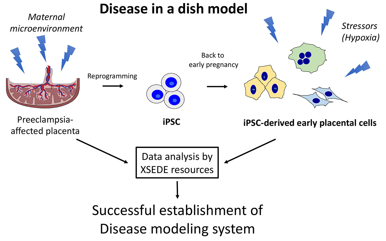
A CIRM supported study using induced pluripotent stem cells (iPSCs), a kind of stem cell that can turn into virtually any cell type, was able to create a “disease in a dish” model in order to better understand preeclampsia.

Credit: UC San Diego

For this study, Mariko Horii, M.D., and her team of researchers at the UC San Diego School of Medicine obtained cells from the placenta of babies born under preeclampsia conditions. These cells were then “reprogrammed” into a stem cell-like state, otherwise known as iPSCs. The iPSCs were then turned into cells resembling placental cells in early pregnancy. This enabled the team to create the preeclampsia “disease in the dish” model. Using this model, they were then able to study the processes that cause, result from, or are otherwise associated with preeclampsia.

The findings revealed that cellular defects observed are related to an abnormal response in the environment in the womb. Specifically, they found that preeclampsia was associated with a low-oxygen environment in the uterus. The researchers used a computer modeling system at UC San Diego known as Comet to detail the differences between normal and preeclampsia placental tissue.

Horii and her team hope that these findings not only shed more light on the environment in the womb observed in preeclampsia, but also provided insight for future development of diagnostic tools and identification of potential medications. Furthermore, they hope that their iPSC disease model can be used to study other placenta-associated pregnancy disorders such as fetal growth restriction, miscarriage, and preterm birth.

The team’s next steps are to develop a 3D model to better study the relationship between environment and development of placental disease.

In a news release from UC San Diego, Horri elaborates more on these future goals.

“Currently, model systems are in two-dimensional cultures with single-cell types, which are hard to study as the placenta consists of maternal and fetal cells with multiple cell types, such as placental cells (fetal origin), maternal immune cells and maternal endometrial cells. Combining these cell types together into a three-dimensional structure will lead to a better understanding of the more complex interactions and cell-to-cell signaling, which can then be applied to the disease setting to further understand pathophysiology.”

The full study was published in Scientific Reports.

Stem cell treatment improves motor function in monkeys modeling Parkinson’s Disease

Neurodegenerative diseases impact millions of people worldwide with the risk of being affected by one of these diseases increasing as you get older. For many of these diseases, there are very few treatments available to patients. As life expectancy increases and the population continues to age, it is crucial to try and find treatments that can potentially slow the progression of these diseases or cure them entirely. This is one of the reasons why CIRM has committed directing around $1.5 billion in funding over the next few years to research related to neurological disorders.

One of the most common neurodegenerative diseases is Parkinson’s Disease (PD), a movement disorder that affects one million people in the U.S alone and leads to shaking, stiffness, insomnia, fatigue, and problems with walking, balance, and coordination.  It is caused by the breakdown and death of dopaminergic neurons, special nerve cells in the brain responsible for the production of dopamine, a chemical messenger that is crucial for normal brain activity.

A recent study published in Nature Medicine has shown improved motor function and growth of neurons over a two year period in monkeys modeling PD. The study was conducted by Su-Chun Zhang, M.D., Ph.D. and his team at the University of Wisconsin using induced pluripotent stem cells (iPSCs), a kind of stem cell that can become virtually any type of cell that can be made from skin cells. The hope is that these results can pave the way for starting human clinical trials.

In order to replicate PD in humans, the team injected 10 adult monkeys with a neurotoxin that produces PD like symptoms. As a result of this, all 10 monkeys developed slow movements, imbalances, tremors, and impaired coordination in the hand on the opposite side of the injection. Additionally, scans revealed that on the injected side, monkeys lost most brain activity involving dopamine in two key brain areas. The team then waited three years after injecting the neurotoxin before administering the therapy, during which time the monkeys’ symptoms persisted.

To generate iPSC lines, the team obtained skin cells from five of the monkeys. The iPSCs were then turned into dopamine neural progenitor cells, which have the ability to create dopamine. These newly created cells were then administered into the brains of the five monkeys, with each monkey receiving a treatment derived from their own skin cells. A sixth iPSC line from a donor monkey was used for the remaining five monkeys to see how the treatment would work if it was not derived from their own skin cells.

The results showed that the monkeys that received the treatment derived from their own skin cells recovered. These animals moved more, moved faster, and were nimbler than before the treatment. They gained the ability to grasp treats, use all four limbs for walking, and climb their cages with ease and increased agility. However, the monkeys that received iPSCs derived from a donor did not recover. Their symptoms remained unchanged or worsened compared to before the treatment.

In a news article, Zhang emphasizes how he and his team are proceeding with a treatment derived from one’s own cells (autologous) vs. one from a donor (allogeneic).

“I initially wanted to do allogeneic transplants in patients because the autologous approach is too expensive. However, after seeing [our] data, I changed my mind. I want to go with the autologous first… because I feel the chance of success is really, really high.”

CIRM is currently funding a human clinical trial ($5.5 million) that is using a gene therapy approach for PD.

Friday Round Up

Here’s a look at a couple of stories that caught our eye this week:

Jasper Therapeutics has had a busy couple of weeks. Recently they announced data from their Phase 1 clinical trial treating people with Myelodysplastic syndromes (MDS). This is a group of disorders in which immature blood-forming cells in the bone marrow become abnormal and leads to low numbers of normal blood cells, especially red blood cells. We blogged about that here.

The data showed that six patients were given JSP191 – in combination with low-dose radiation five of the six had no detectable levels of disease and the sixth patient had reduced levels.

This was a big deal for us because CIRM funded the early stage research and even a clinical trial  that led to the development of JSP191.

Now Jasper has announced it is partnering with the National Institute of Allergy and Infectious Disease in a Phase 1/2 clinical trial using JSP191, as part of a treatment for chronic granulomatous disease (CGD). Congratulations to Jasper. And congratulations to us for helping them get there.

Oh, and just to toot our horn a little bit more – it is Friday after all – we have funded other approaches to CGD including one that resulted in curing Brenden Whittaker.

OK, enough about us.

To say that this last year has been a stressful one would be something of an understatement. But it’s not just people who get stressed. Stem cells do too. And, like people, when stem cells get stressed they don’t always behave in the way you would like them to. When some people get stressed they find a cocktail can help take the edge of it. Apparently that works for stem cells as well!

Now we are not talking about slipping a Manhattan or Mai Tai into a petri dish filled with stem cells. We are talking about a very different kind of cocktail.

Researchers at the National Institutes of Health have developed what they describe as a “four-part small molecule cocktail” that can help protect a specific kind of stem cell from stress. The cell is an induced pluripotent stem cell (iPSC), which has the ability to turn into any other kind of cell in the body. iPSC’s have great potential for treating a variety of different diseases and conditions, but they’re also sensitive and without the right conditions and environment they can get stressed and that in turn can damage their DNA and lead to them dying.

In a news release Dr. Ilyas Singeç, the lead researcher, says this NIH “cocktail” could help prevent that: “The small-molecule cocktail is safeguarding cells and making stem cell use more predictable and efficient. In preventing cellular stress and DNA damage that typically occur, we’re avoiding cell death and improving the quality of surviving cells. The cocktail will become a broadly used staple of the stem cell field and boost stem cell applications in both research and the clinic.”  

The team hope this could enhance the potential therapeutic uses of iPSCs in finding treatments for diseases such as diabetes, Parkinson’s and spinal cord injury.

The study is published in the journal Nature Methods.

CIRM funded study uses drug development in a dish for treatment of heart arrhythmias

Image Credit: Center for Disease Control and Prevention (CDC)

Cardiac (heart) arrhythmias occur when electrical impulses that coordinate your heartbeats don’t work properly, causing your heart to beat too fast, too slow, or in an irregular manner. In the U.S. alone, almost one million individuals are hospitalized every year for heart arrhythmias. Close to 300,000 individuals die of sudden arrhythmic death syndrome every year, which occurs when there is a sudden loss of blood flow resulting from the failure of the heart to pump effectively. Unfortunately, drugs to treat arrhythmias have liabilities and several drugs have been pulled from the market due to serious side effects. Mexiletine is one potential drug for heart arrhythmias that has liabilities and potential side effects.

That is why a CIRM funded study ($6.3 million) conducted by John Cashman, Ph.D. at the Human BioMolecular Research Institute in San Diego looked at re-engineering mexiletine in a way that the drug could still produce a desired result and not be as toxic.

The study used induced pluripotent stem cells (iPSCs), a type of stem cell “reprogrammed” from the skin or blood of patients that can be used to make virtually any kind of cell. iPSCs obtained for the study were from a healthy patient and from one with a type of heart arrhythmia. The healthy and arrhythmia iPSCs were then converted into cardiomyocytes, a type of cell that makes up the heart muscle.

By using their newly created healthy cardiomyocytes and those with the arrhythmia defect, Cashman and his team were able to carry out drug development in a dish. This enabled them to attempt to lessen drug toxicity while still potentially treating heart arrhythmias. The team was able to modify mexiletine such that is was less toxic and found that it could potentially decrease a patient’s risk of developing ventricular tachycardia (a fast, abnormal heart rate) and ventricular fibrillation (an abnormal heart rhythm), both of which are types of heart arrhythmias.

“The new compounds may lead to treatment applications in a whole host of cardiovascular conditions that may prove efficacious in clinical trials,” said Cashman in a press release. “As antiarrhythmic drug candidate drug development progresses, we expect the new analogs to be less toxic than current therapeutics for arrhythmia in congenital heart disease, and patients will benefit from improved safety, less side effects and possibly with significant cost-savings.”

The team hopes that their study can pave the way for future research in which cells in a dish can be used to lessen the toxicity of a potential drug candidate while still producing a desired result for different diseases and conditions.

The full study was published in ACS Publications.

CIRM funded researchers discover link between Alzheimer’s gene and COVID-19

Dr. Yanhong Shi (left) and Dr. Vaithilingaraja Arumugaswami (right)

All this month we are using our blog and social media to highlight a new chapter in CIRM’s life, thanks to the voters approving Proposition 14. We are looking back at what we have done since we were created in 2004, and also looking forward to the future. Today we focus on groundbreaking CIRM funded research related to COVID-19 that was recently published.

It’s been almost a year since the world started hearing about SARS-CoV-2, the virus that causes COVID-19.  In our minds, the pandemic has felt like an eternity, but scientists are still discovering new things about how the virus works and if genetics might play a role in the severity of the virus.  One population study found that people who have ApoE4, a gene type that has been found to increase the risk of developing Alzheimer’s, had higher rates of severe COVID-19 and hospitalizations.

It is this interesting observation that led to important findings of a study funded by two CIRM awards ($7.4M grant and $250K grant) and conducted by Dr. Yanhong Shi at City of Hope and co-led by Dr. Vaithilingaraja Arumugaswami, a member of the UCLA Broad Stem Cell Research Center.  The team found that the same gene that increases the risk for Alzheimer’s disease can increase the susceptibility and severity of COVID-19.

At the beginning of the study, the team was interested in the connection between SARS-CoV-2 and its effect on the brain.  Due to the fact that patients typically lose their sense of taste and smell, the team theorized that there was an underlying neurological effect of the virus.  

The team first created neurons and astrocytes.  Neurons are cells that function as the basic working unit of the brain and astrocytes provide support to them.  The neurons and astrocytes were generated from induced pluripotent stem cells (iPSCs), which are a kind of stem cell that can become virtually any type of cell and can be created by “reprogramming” the skin cells of patients.  The newly created neurons and astrocytes were then infected with SARS-CoV-2 and it was found that they were susceptible to infection.

Next, the team used iPSCs to create brain organoids, which are 3D models that mimic certain features of the human brain.  They were able to create two different organoid models: one that contained astrocytes and one without them.  They infected both brain organoid types with the virus and discovered that those with astrocytes boosted SARS-CoV-2 infection in the brain model. 

The team then decided to further study the effects of ApoE4 on susceptibility to SARS-CoV-2.  They did this by generating neurons from iPSCs “reprogrammed” from the cells of an Alzheimer’s patient.  Because the iPSCs were derived from an Alzheimer’s patient, they contained ApoE4.  Using gene editing, the team modified some of the ApoE4 iPSCs created so that they contained ApoE3, which is a gene type considered neutral.  The ApoE3 and ApoE4 iPSCs were then used to generate neurons and astrocytes.

The results were astounding.  The ApoE4 neurons and astrocytes both showed a higher susceptibility to SARS-CoV-2 infection in comparison to the ApoE3 neurons and astrocytes.  Moreover, while the virus caused damage to both ApoE3 and ApoE4 neurons, it appeared to have a slightly more severe effect on ApoE4 neurons and a much more severe effect on ApoE4 astrocytes compared to ApoE3 neurons and astrocytes. 

“Our study provides a causal link between the Alzheimer’s disease risk factor ApoE4 and COVID-19 and explains why some (e.g. ApoE4 carriers) but not all COVID-19 patients exhibit neurological manifestations” says Dr. Shi. “Understanding how risk factors for neurodegenerative diseases impact COVID-19 susceptibility and severity will help us to better cope with COVID-19 and its potential long-term effects in different patient populations.”

In the last part of the study, the researchers tested to see if the antiviral drug remdesivir inhibits virus infection in neurons and astrocytes.  They discovered that the drug was able to successfully reduce the viral level in astrocytes and prevent cell death.  For neurons, it was able to rescue them from steadily losing their function and even dying. 

The team says that the next steps to build on their findings is to continue studying the effects of the virus and better understand the role of ApoE4 in the brains of people who have COVID-19.  Many people that developed COVID-19 have recovered, but long-term neurological effects such as severe headaches are still being seen months after. 

“COVID-19 is a complex disease, and we are beginning to understand the risk factors involved in the manifestation of the severe form of the disease” says Dr. Arumugaswami.  “Our cell-based study provides possible explanation to why individuals with Alzheimer’s’ disease are at increased risk of developing COVID-19.”

The full results to this study were published in Cell Stem Cell.

“Mini-brains” model an autism spectrum disorder and help test treatments

Alysson Muotri, PhD, professor and director of the Stem Cell Program at UC San Diego School of Medicine
and member of the Sanford Consortium for Regenerative Medicine.
Image credit: UC San Diego Health

Rett syndrome is a rare form of autism spectrum disorder that impairs brain development and causes problems with movement, speech, and even breathing. It is caused by mutations in a gene called MECP2 and primarily affects females. Although there are therapies to alleviate symptoms, there is currently no cure for this genetic disorder.

With CIRM funding ($1.37M and $1.65M awards), Alysson Muotri, PhD and a team of researchers at the University of California San Diego School of Medicine and Sanford Consortium for Regenerative Medicine have used brain organoids that mimic Rett syndrome to identify two drug candidates that returned the “mini-brains” to near-normal. The drugs restored calcium levels, neurotransmitter production, and electrical impulse activity.

Brain organoids, also referred to as “mini-brains”, are 3D models made of cells that can be used to analyze certain features of the human brain. Although they are far from perfect replicas, they can be used to study changes in physical structure or gene expression over time.

Dr. Muotri and his team created induced pluripotent stem cells (iPSCs), a type of stem cell that can become virtually any type of cell. For the purposes of this study, they were created from the skin cells of Rett syndrome patients. The newly created iPSCs were then turned into brain cells and used to create “mini-brains”, thereby preserving each Rett syndrome patient’s genetic background. In addition to this, the team also created “mini-brains” that artificially lack the MECP2 gene, mimicking the issues with the same gene observed in Rett syndrome.

Lack of the MECP2 gene changed many things about the “mini-brains” such as shape, neuron subtypes present, gene expression patterns, neurotransmitter production, and decreases in calcium activity and electrical impulses. These changes led to major defects in the emergence of brainwaves.

To correct the changes caused by the lack of the MECP2 gene, the team treated the brain organoids with 14 different drug candidates known to affect various brain cell functions. Of all the drugs tested, two stood out: nefiracetam and PHA 543613. The two drugs resolved nearly all molecular and cellular symptoms observed in the Rett syndrome “mini-brains”, with the number active neurons doubling post treatment.

The two drugs were previously tested in clinical trials for the treatment of other conditions, meaning they have been shown to be safe for human consumption.

In a news release from UC San Diego Health, Dr. Muotri stresses that although the results for the two drugs are promising, the end treatment for Rett syndrome may require a multi-drug cocktail of sorts.

“There’s a tendency in the neuroscience field to look for highly specific drugs that hit exact targets, and to use a single drug for a complex disease. But we don’t do that for many other complex disorders, where multi-pronged treatments are used. Likewise, here no one target fixed all the problems. We need to start thinking in terms of drug cocktails, as have been successful in treating HIV and cancers.”

The full results of this study were published in EMBO Molecular Medicine.

CIRM-funded study discovers potential therapy for one of the leading causes of heart disease

Dr. Deepak Srivastava and his team found a drug candidate that could help prevent tens of thousands of heart surgeries every year. Image Credit: Gladstones Institute

According to the Center for Disease Control and Prevention (CDC), heart disease is the leading cause of death for men, women, and people of most racial and ethnic groups in the United States. About 655,000 Americans die from heart disease each year, which is about one in every four deaths.

Calcific aortic valve disease, the third leading cause of heart disease overall, occurs when calcium starts to accumulate in the heart valves and vessels over time, causing them to gradually harden like bone. This leads to obstruction of blood flow out of the heart’s pumping chamber, causing heart failure. Unfortunately there is no treatment for this condition, leaving patients only with the option of surgery to replace the heart valve once the hardening is severe enough.

But thanks to a CIRM-funded ($2.4 million) study conducted by Dr. Deepak Srivastava and his team at the Gladstone Institutes, a potential drug candidate for heart valve disease was discovered. It has been found to function in both human cells and animals and is ready to move toward a clinical trial.

For this study, Dr. Srivastava and his team looked for drug-like molecules that had the potential to correct the mechanism in heart valve disease that leads to gradual hardening. To do so, the team first had to determine the network of genes that are turned on or off in the diseased cells.

Once the genes were identified, they used an artificial intelligence method to train a machine learning program to detect whether a cell was healthy or diseased based on the network of genes identified. They proceeded to treat the diseased human cells with nearly 1,600 molecules in order to identify any drugs that would cause the machine learning program to reclassify diseased cells as healthy. The team successfully identified a few molecules that could correct diseased cells back to a healthy state.

Dr. Srivastara then collaborated with Dr. Anna Malashicheva, from the Russian Academy of Sciences, who had collected valve cells from over 20 patients at the time of surgical replacement. Using the valve cells that Dr. Malashicheva had collected, Dr. Srivastara and his team conducted a “clinical trial in a dish” in which they tested the molecules they had previously identified in the cells from the 20 patients with aortic valve hardening. The results were remarkable, as the molecule that seemed most effective in the initial study was able to restore these patients’ cells as well.

The final step taken was to determine whether the drug-like molecule would actually work in a whole, living organ. To do this, Dr. Srivastava and his team did a “pre-clinical trial” in a mouse model of the disease. The team found that the therapeutic candidate could successfully prevent and treat aortic valve disease. In young mice who had not yet developed the disease, the therapy prevented the hardening of the valve. In mice that already had the disease, the therapy was able to halt the disease and, in some cases, reverse it. This finding is especially important since most patients aren’t diagnosed until hardening of the heart valve has already begun.

Dr. Deepak Srivastava (left) and Dr. Christina V. Theodoris (right)
Image Credit: Gladstones Institute

Dr. Christina V. Theodoris, a lead author of the study who is now completing her residency in pediatric genetics, was a graduate student in Dr. Srivastava’s lab and played a critical role in this research. Her first project was to convert the cells from patient families into induced pluripotent stem cells (iPSCs), which have the potential of becoming any cell in the body. The newly created iPSCs were then turned into cells that line the valve, allowing the team to understand why the disease occurs. Her second project was to make a mouse model of calcific aortic valve disease, which enabled them to start using the models to identify a therapy.

In a press release from Gladstone Institutes, Dr. Theodoris, discusses the impact of the team’s research.

“Our strategy to identify gene network–correcting therapies that treat the core disease mechanism may represent a compelling path for drug discovery in a range of other human diseases. Many therapeutics found in the lab don’t translate well to humans or focus only on a specific symptom. We hope our approach can offer a new direction that could increase the likelihood of candidate therapies being effective in patients.”

In the same press release, Dr. Srivastava emphasizes the scientific advances that have driven the team’s research to this critical point.

“Our study is a really good example of how modern technologies are facilitating the kinds of discoveries that are possible today, but weren’t not so long ago. Using human iPSCs and gene editing allowed us to create a large number of cells that are relevant to the disease process, while powerful machine learning algorithms helped us identify, in a non-biased fashion, the important genes for distinguishing between healthy and diseased cells.”

The full results of this study were published in Science.