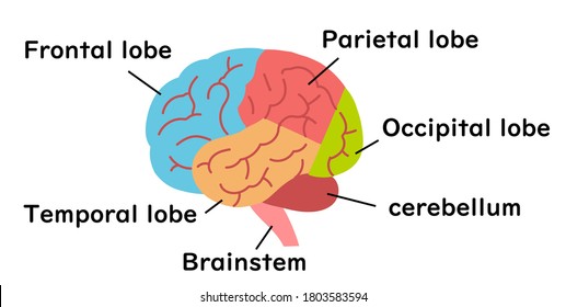
City of Hope researchers discover potential therapy to treat brain tumors

Glioblastoma (GBM) is a common type of aggressive brain tumor that is found in adults.  Survival of this type of brain cancer is poor with just 40% survival in the first-year post diagnosis and 17% in the second year, according to the American Association of Neurological Surgeons.  This disease has taken the life of former U.S. Senator John McCain and Beau Biden, the late son of U.S. President Joe Biden.

In a CIRM supported lab that conducted the study, Dr. Yanhong Shi and her team at City of Hope, a research and treatment center for cancer, have discovered a potential therapy that they have tested that has been shown to suppress GBM tumor growth and extend the lifespan of tumor-bearing mice. 

Dr. Shi and her team first started by looking at PUS7, a gene that is highly expressed in GBM tissue in comparison to normal brain tissue.  Dr. Qi Cui, a scientist in Dr. Shi’s team and the first author of the study, analyzed various databases and found that high levels of PUS7 have also been associated with worse survival in GBM patients.  The team then studied different glioblastoma stem cells (GSCs), which play a vital role in brain tumor growth, and found that shutting off the PUS7 gene prevented GSC growth and self-renewal. 

The City of Hope team then transplanted two kinds of GSCs, some with the PUS7 gene and some with the PUS7 gene turned off, into immunodeficient mice.  What they found was that the mice implanted with the PUS7-lacking GSCs had less tumor growth and survived longer compared to the mice with the control GSCs that had PUS7 gene.

The team then proceeded to look for an inhibitor of PUS7 from a database of thousands of different compounds and drugs approved by the Food and Drug Administration (FDA).  After identifying a promising compound, the researchers tested the potential therapy in mice implanted with GSCs with the PUS7 gene.  What they found was remarkable.  The therapy inhibited the growth of brain tumors in the mice and their survival was significantly prolonged.

“This is one of the most important studies in my lab in recent years and the first paper to show a causal link between PUS7-mediated modification and cancer in general and GBM in particular” says Dr. Shi.  “It will be a milestone study for RNA modification in cancer.”

The full study was published in Nature Cancer.

Dr. Shi has previously worked on several CIRM-funded research projects, such as looking at a potential link between COVID-19 and a gene for Alzheimer’s as well as the development of a therapy for Canavan disease.

City of Hope scientists use stem cells to develop ‘mini-brains’ to study Alzheimer’s and to test drugs in development

Alzheimer’s is a progressive disease that destroys memory and other important mental functions. According to the non-profit HFC, co-founded by CIRM Board member Lauren Miller Rogen and her husband Seth Rogen, more than 5 million Americans are living with Alzheimer’s. It is the 6th leading cause of death in the U.S and it is estimated that by 2050 as many as 16 million Americans will have the disease. Alzheimer’s is the only cause of death among the top 10 in the U.S. without a way to prevent, cure, or even slow its progression, which is it is crucial to better understand the disease and to develop and test potential treatments.

It is precisely for this reason that researchers led by Yanhong Shi, Ph.D. at City of Hope have developed a ‘mini-brain’ model using stem cells in order to study Alzheimer’s and to test drugs in development.

The team was able to model sporadic Alzheimer’s, the most common form of the disease, by using human induced pluripotent stem cells (iPSCs), a kind of stem cell that can be created from skin or blood cells of people through reprogramming and has the ability to turn into virtually any other kind of cell. The researchers used these iPSCs to create ‘mini-brains’, also known as brain organoids, which are 3D models that can be used to analyze certain features of the human brain. Although they are far from perfect replicas, they can be used to study physical structure and other characteristics. 

The scientists exposed the ‘mini-brains’ to serum that mimics age-associated blood-brain barrier (BBB) breakdown. The BBB is a protective barrier that surrounds the brain and its breakdown has been associated with Alzheimer’s and other age-related neurodegenerative diseases . After exposure, the team tested the ‘mini-brains’ for various Alzheimer’s biomarkers. These markers included elevated levels of proteins known as amyloid and tau that are associated with the disease and synaptic breaks linked to cognitive decline.

Research using brain organoids has shown that exposure to serum from blood could induce multiple Alzheimer’s symptoms. This suggests that combination therapies targeting multiple areas would be more effective than single-target therapies currently in development.

The team found that attempting a single therapy, such as inhibiting only amyloid or tau proteins, did not reduce the levels of tau or amyloid, respectively. These findings suggest that amyloid and tau likely cause disease progression independently. Furthermore, exposure to serum from blood, which mimics BBB breakdown, could cause breaks in synaptic connections that help brains remember things and function properly.

Image Description: Yanhong Shi, Ph.D.

In a press release from the Associated Press, Dr. Shi elaborated on the importance of their model for studying Alzheimer’s.

“Drug development for Alzheimer’s disease has run into challenges due to incomplete understanding of the disease’s pathological mechanisms. Preclinical research in this arena predominantly uses animal models, but there is a huge difference between humans and animals such as rodents, especially when it comes to brain architecture. We, at City of Hope, have created a miniature brain model that uses human stem cell technology to study Alzheimer’s disease and, hopefully, to help find treatments for this devastating illness.”

The full results of this study were published in Advance Science.

Dr. Shi has previously worked on several CIRM-funded research projects, such as looking at a potential link between COVID-19 and a gene for Alzheimer’s as well as the development of a therapy for Canavan disease.

Scientists use stem cell ‘mini-brains’ to better understand signs of frontotemporal dementia

Dementia is a general term that describes a set of diseases that impair the ability to remember, think, or make decisions that interfere with doing everyday activities. According to the World Health Organization (WHO), around 50 million people worldwide have dementia with nearly 10 million new cases every year. Although it primarily affects older people it is not a normal part of aging. As our population ages its critical to better understand why this occurs.

Frontotemporal dementia is a rare form of dementia where people start to show signs between the ages of 40 and 60. It affects the front and side (temporal) areas of the brain, hence the name. It leads to behavior changes and difficulty with speaking and thinking. This form of the disease is caused by a genetic mutation called tau, which is known to be associated with Alzheimer’s disease and other dementias.

A CIRM supported study using induced pluripotent stem cells (iPSCs) led by Kathryn Bowles, Ph.D. and conducted by a team of researchers at Mount Sinai were able to recreate much of the damage seen in a widely studied form of the frontotemporal dementia by growing special types of ‘mini-brains’, also known as cerebral organoids.

iPSCs are a kind of stem cell that can be created from skin or blood cells through reprogramming and have the ability to turn into virtually any other kind of cell. The team used iPSCs to create thousands of tiny, 3D ‘mini-brains’, which mimic the early growth and development of the brain.

The researchers examined the growth and development of these ‘mini-brains’ using stem cells derived from three patients, all of whom carried a mutation in tau. They then compared their results with those observed in “normal” mini-brains which were derived from patient stem cells in which the disease-causing mutation was genetically corrected.

After six months, signs of neurodegeneration were seen in the patient ‘mini-brains’. The patient-derived ‘mini-brains’ had fewer excitatory neurons compared to the “normal” ones which demonstrates that the tau mutation was sufficient to cause higher levels of cell death of this specific class of neurons. Additionally, the patient-derived ‘mini-brains’ also had higher levels of harmful versions of tau protein and elevated levels of inflammation.

In a news release from Mount Sinai, Dr. Bowles elaborated on the results of this study.

“Our results suggest that the V337M mutant tau sets off a vicious cycle in the brain that puts excitatory neurons under great stress. It hastens the production of new proteins needed for maturation but prevents disposal of the proteins that are being replaced.”

The full results of this study were published in Cell.

Improving a special kind of cell to help combat immune related problems

Regulatory T cells (Tregs) are a type of immune cell that play an extremely important role in maintaining stability in the body and preventing the body’s immune system from attacking its own cells and organs. This unique property makes Tregs extremely valuable to researchers as a potential treatment for immune related issues. One of these is autoimmune disease, which is a disease in which the body’s own immune system attacks healthy cells. Some examples of this are type 1 diabetes, multiple sclerosis, and lupus. Another immune related issue is graft vs. host disease (GvHD), which can occur after receiving a transplantation where the donated bone marrow or stem cells start attacking the recipient.

For this reason, researchers at the La Jolla Institute for Immunology (LJI) and Emory University School of Medicine, partially supported by a CIRM training grant , have been working to generate stable induced Tregs (iTregs) for treating autoimmune diseases and rejection of a transplanted organ. The teams were led by LJI professor Anjana Rao, Ph.D, and Emory instructor Benjamin G. Barwick, Ph.D. The two team study showed evidence that vitamin C and and specific proteins called TET can be combined to give Tregs their life-saving power. Studies have previously found that vitamin C can enhance the activity of TET proteins and prompt the generation of stable iTregs under lab conditions.

For this study, the researchers also analyzed gene expression patterns as well as changes that altered the physical structure of DNA in the induced Tregs. The team found a major modification involving the DNA itself and showed that TET enzymes were also involved. All of these interactions can eventually change how cells “read” the DNA code. They also observed the alteration of DNA accessibility which depends on whether DNA is loosely or tightly coiled. As the DNA coils unwind, regulatory regions become exposed which subsequently influence gene expression.

In a news release, LJI instructor Xiaojing Yue, Ph.D elaborated on the results of this study.

“Vitamin C can be used to stabilize iTregs generated in vitro. We hope that these kinds of induced Tregs can be used in the future for treatment of autoimmune diseases and organ transplantation.”

The full study was published in EMBO reports.

Endothelial cell treatment reverses lung damage in mice with emphysema

Emphysema is a condition that causes damage to the alveoli, the air sacs in your lungs. The walls of the damaged air sacs become stretched out and cause your lungs to get bigger. This makes it harder to move your air in and out. It is the most common form of the condition known as chronic obstructive pulmonary disease (COPD) and is typically triggered by long-term cigarette smoking. Estimates show that approximately 200 million people around the world are affected. Unfortunately, there is no cure for this disease of the lungs.

A study conducted by researchers at Weill Cornell Medicine and NewYork-Presbyterian found that specialized endothelial cells may hold the key to treating emphysema. Endothelial cells line the inner surface of blood vessels and have been shown to play an important role in protecting and restoring the health of key organs. Specifically, lung endothelial cells line the inner surface of the lung’s network of blood vessels.

As part of their research, the team studied lung tissue from human emphysema patients while also looking at lung issue from mice with an induced form of the disease. What they found that was that changes in the activity of certain genes in lung endothelial cells and the loss of those cells was associated with decreased lung function and other indicators of emphysema progression.

The researchers then infused mice with induced emphysema with healthy lung endothelial cells from genetically identical mice and the results were astounding. The team showed that they could prevent and/or reverse most of the lung damage that was seen in untreated mice. By contrast, injecting other cell types, including endothelial cells from other tissues, did not have the same effect.

The team believes that this treatment effect might have to do with differences in the molecules secreted by diseased versus healthy lung endothelial cells. To back up this claim, they found that lung endothelial cells in both humans and mice with emphysema showed sharp increases in production of LRG1, a molecule that promotes new blood vessel growth that has been linked to retinal and kidney diseases as well as some cancers. Additionally, when the researchers deleted the gene for LRG1 from lung endothelial cells in mice, the lungs were largely protected from the lung damage of induced emphysema, much as they had been by the endothelial cell therapy.

In a news release from Cornell, Dr. Alexandra Racanelli, a co-first author on this study and an instructor of medicine in the Division of Pulmonary and Critical Care Medicine at Weill Cornell Medicine and a pulmonologist at NewYork-Presbyterian/Weill Cornell Medical Center, had this to say about the results.

“Taken together, our data strongly suggest the critical role of endothelial cell function in mediating the pathogenesis of COPD/emphysema. Targeting endothelial cell biology by administering healthy lung endothelial cells and/or inhibiting the LRG1 pathway may therefore represent strategies of immense potential for the treatment of patients with advanced COPD or emphysema.”

The full study was published in the Journal of Experimental Medicine.

Board Funds Fifteen Bridges to Stem Cell Research and Therapy Programs Across California and New Sickle Cell Disease Trial

Yesterday the governing Board of the California Institute for Regenerative Medicine (CIRM) awarded $8.39 million to the University of California, San Francisco (UCSF) to fund a clinical trial for sickle cell disease (SCD).  An additional $51.08 million was awarded to fifteen community colleges and universities across California to fund undergraduate and master’s level programs that will help train the next generation of stem cell researchers. 

SCD is an inherited blood disorder caused by a single gene mutation that changes a single base in the B globin gene leading to the production of defective hemoglobin that polymerizes and damages red blood cells thus the “sickle” shaped red blood cells.  The damaged cells cause blood vessels to occlude/close up and that can lead to multiple organ damage as well as reduced quality of life and life expectancy. 

Mark Walters, M.D., and his team at UCSF Benioff Children’s Hospital Oakland will be conducting a clinical trial that uses CRISPR-Cas9 gene editing technology to correct the genetic mutation in the blood stem cells of patients with severe SCD.  The corrected blood stem cells will then be reintroduced back into patients with the goal of correcting the defective hemoglobin and thus producing functional, normal shaped red blood cells.

This clinical trial will be eligible for co-funding under the landmark agreement between CIRM and the National Heart, Lung, and Blood Institute (NHLBI) of the NIH.  The CIRM-NHLBI agreement is intended to co-fund cell and gene therapy programs under the NHLBI’s “Cure Sickle Cell” initiative.  The goal is to markedly accelerate the development of cell and gene therapies for SCD. CIRM has previously funded the preclinical development of this therapy through a Translational award as well as its IND-enabling studies through a Late Stage Preclinical award in partnership with NHLBI.

The CIRM Bridges to Stem Cell Research and Therapy program provides undergraduate and master’s students with the opportunity to take stem cell related courses and receive hands on experience and training in a stem cell research related laboratory at a university or biotechnology company.  Fifteen institutions received a total of $51.08 million to carry out these programs to train the next generation of scientists.

The awards are summarized in the table below.

ApplicationTitleInstitutionAward Amount
  EDUC2-12607Bridges to Stem Cell Research and Therapy at Pasadena City College  Pasadena City College$3,605,500
  EDUC2-12611CIRM Bridges to Stem Cell Research and Therapy Training Grant  CSU San Marcos$3,606,500
  EDUC2-12617Bridges to Stem Cell Research Internship Program  San Diego State University$3,606,500
EDUC2-12620CIRM Bridges 3.0  Humboldt State$3,605,495
  EDUC2-12638CIRM Regenerative Medicine and Stem Cell Research Biotechnology Training Program  CSU Long Beach$3,276,500
    EDUC2-12677Stem Cell Internships in Laboratory-based Learning (SCILL) continue to expand the scientific workforce for stem cells research and therapies.  San Jose State University$3,606,500
  EDUC2-12691Strengthening the Pipeline of Master’s-level Scientific and Laboratory Personnel in Stem Cell Research  CSU Sacramento$2,946,500
EDUC2-12693CIRM Bridges Science Master’s Program  San Francisco State University$3,606,500
      EDUC2-12695CIRM Graduate Student Training in Stem Cell Sciences in the Stem Cell Technology and Lab Management Emphasis of the MS Biotechnology Program  CSU Channel Islands$3,606,500
  EDUC2-12718CSUN CIRM Bridges 3.0 Stem Cell Research & Therapy Training Program  CSU Northridge$3,606,500
      EDUC2-12720Stem Cell Scholars: a workforce development pipeline, educating, training and engaging students from basic research to clinical translation.  CSU San Bernardino$3,606,500
  EDUC2-12726Training Master’s Students to Advance the Regenerative Medicine Field  Cal Poly San Luis Obispo$3,276,500
  EDUC2-12730Building Career Pathways into Stem Cell Research and Therapy Development  City College of San Francisco$2,706,200
      EDUC2-12734Bridges to Stem Cell Research and Therapy: A Talent Development Program for Training Diverse Undergraduates for Careers in Regenerative Medicine  CSU Fullerton$3,606,500
  EDUC2-12738CIRM Bridges to Stem Cell Research and Therapy  Berkeley City College  $2,806,896

“We are pleased to fund a promising trial for sickle cell disease that uses the Nobel Prize winning gene editing technology CRISPR-Cas9,” says Maria T. Millan, M.D., President and CEO of CIRM.  “This clinical trial is a testament to how the CIRM model supports promising early-stage research, accelerates it through translational development, and advances it into the clinics. As the field advances, we must also meet the demand for promising young scientists.  The CIRM Bridges programs across the state of California will provide students with the tools and resources to begin their careers in regenerative medicine.”

UCSD researchers use stem cell model to better understand pregnancy complication

A team of UC San Diego researchers recently published novel preeclampsia models to aid in understanding this pregnancy complication that occurs in one of 25 U.S. pregnancies. Researchers include (left to right): Ojeni Touma, Mariko Horii, Robert Morey and Tony Bui. Credit: UC San Diego

Pregnant women often tread uncertain waters in regards to their health and well-being as well as that of their babies. Many conditions can arise and one of these is preeclampsia, a type of pregnancy complication that occurs in approximately one in 25 pregnancies in the United States according to the Center for Disease Control (CDC). It occurs when expecting mothers develop high blood pressure, typically after 20 weeks of pregnancy, and that in turn reduces the blood supply to the baby. This can lead to serious, even fatal, complications for both the mother and baby.

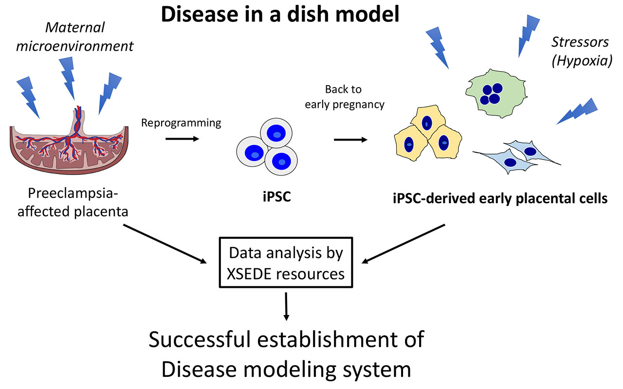
A CIRM supported study using induced pluripotent stem cells (iPSCs), a kind of stem cell that can turn into virtually any cell type, was able to create a “disease in a dish” model in order to better understand preeclampsia.

Credit: UC San Diego

For this study, Mariko Horii, M.D., and her team of researchers at the UC San Diego School of Medicine obtained cells from the placenta of babies born under preeclampsia conditions. These cells were then “reprogrammed” into a stem cell-like state, otherwise known as iPSCs. The iPSCs were then turned into cells resembling placental cells in early pregnancy. This enabled the team to create the preeclampsia “disease in the dish” model. Using this model, they were then able to study the processes that cause, result from, or are otherwise associated with preeclampsia.

The findings revealed that cellular defects observed are related to an abnormal response in the environment in the womb. Specifically, they found that preeclampsia was associated with a low-oxygen environment in the uterus. The researchers used a computer modeling system at UC San Diego known as Comet to detail the differences between normal and preeclampsia placental tissue.

Horii and her team hope that these findings not only shed more light on the environment in the womb observed in preeclampsia, but also provided insight for future development of diagnostic tools and identification of potential medications. Furthermore, they hope that their iPSC disease model can be used to study other placenta-associated pregnancy disorders such as fetal growth restriction, miscarriage, and preterm birth.

The team’s next steps are to develop a 3D model to better study the relationship between environment and development of placental disease.

In a news release from UC San Diego, Horri elaborates more on these future goals.

“Currently, model systems are in two-dimensional cultures with single-cell types, which are hard to study as the placenta consists of maternal and fetal cells with multiple cell types, such as placental cells (fetal origin), maternal immune cells and maternal endometrial cells. Combining these cell types together into a three-dimensional structure will lead to a better understanding of the more complex interactions and cell-to-cell signaling, which can then be applied to the disease setting to further understand pathophysiology.”

The full study was published in Scientific Reports.

Stem cell treatment improves motor function in monkeys modeling Parkinson’s Disease

Neurodegenerative diseases impact millions of people worldwide with the risk of being affected by one of these diseases increasing as you get older. For many of these diseases, there are very few treatments available to patients. As life expectancy increases and the population continues to age, it is crucial to try and find treatments that can potentially slow the progression of these diseases or cure them entirely. This is one of the reasons why CIRM has committed directing around $1.5 billion in funding over the next few years to research related to neurological disorders.

One of the most common neurodegenerative diseases is Parkinson’s Disease (PD), a movement disorder that affects one million people in the U.S alone and leads to shaking, stiffness, insomnia, fatigue, and problems with walking, balance, and coordination.  It is caused by the breakdown and death of dopaminergic neurons, special nerve cells in the brain responsible for the production of dopamine, a chemical messenger that is crucial for normal brain activity.

A recent study published in Nature Medicine has shown improved motor function and growth of neurons over a two year period in monkeys modeling PD. The study was conducted by Su-Chun Zhang, M.D., Ph.D. and his team at the University of Wisconsin using induced pluripotent stem cells (iPSCs), a kind of stem cell that can become virtually any type of cell that can be made from skin cells. The hope is that these results can pave the way for starting human clinical trials.

In order to replicate PD in humans, the team injected 10 adult monkeys with a neurotoxin that produces PD like symptoms. As a result of this, all 10 monkeys developed slow movements, imbalances, tremors, and impaired coordination in the hand on the opposite side of the injection. Additionally, scans revealed that on the injected side, monkeys lost most brain activity involving dopamine in two key brain areas. The team then waited three years after injecting the neurotoxin before administering the therapy, during which time the monkeys’ symptoms persisted.

To generate iPSC lines, the team obtained skin cells from five of the monkeys. The iPSCs were then turned into dopamine neural progenitor cells, which have the ability to create dopamine. These newly created cells were then administered into the brains of the five monkeys, with each monkey receiving a treatment derived from their own skin cells. A sixth iPSC line from a donor monkey was used for the remaining five monkeys to see how the treatment would work if it was not derived from their own skin cells.

The results showed that the monkeys that received the treatment derived from their own skin cells recovered. These animals moved more, moved faster, and were nimbler than before the treatment. They gained the ability to grasp treats, use all four limbs for walking, and climb their cages with ease and increased agility. However, the monkeys that received iPSCs derived from a donor did not recover. Their symptoms remained unchanged or worsened compared to before the treatment.

In a news article, Zhang emphasizes how he and his team are proceeding with a treatment derived from one’s own cells (autologous) vs. one from a donor (allogeneic).

“I initially wanted to do allogeneic transplants in patients because the autologous approach is too expensive. However, after seeing [our] data, I changed my mind. I want to go with the autologous first… because I feel the chance of success is really, really high.”

CIRM is currently funding a human clinical trial ($5.5 million) that is using a gene therapy approach for PD.

CIRM funded trial for AMD shows promising results

This upcoming July is healthy vision month, a time to remember the importance of making vision and eye health a priority. It’s also a time to think about the approximately 12 million people, 40 and over in the United States, that have a vision impairment. Vision can be something that many of us take for granted, but losing even a portion of it can have a profound impact on our everyday life. It can impact your ability to do everyday things, from basic hygiene routines and driving to hobbies such as reading, writing, or watching a film.

It is because of this that CIRM has made vision related problems a priority, providing over $69 million in funding for six clinical trials related to vision loss. There is reason to be hopeful as these trials have demonstrated promising results. One of these trials, conducted by Regenerative Patch Technologies LLC (RPT), announced today results from its CIRM funded clinical trial ($16.3 million) for advanced, dry age-related macular degeneration (AMD).

AMD is a progressive disease resulting in death of the retinal pigment epithelium (RPE), an area of the eye that plays a key role in maintaining vision. Damage to the RPE causes distortion to central vision and eventually leads to legal blindness. Thanks to CIRM funding, RPT and scientists at the University of Southern California (USC) and UC Santa Barbara (UCSB) are growing specialized RPE cells from human embryonic stem cells (hESCs), placing them on a single layer scaffold, and implanting the combination device in the back of the eye to try to reverse the blindness caused by AMD.

One of the trial participants is Anna Kuehl, a USC alumna and avid nature lover. She was diagnosed with AMD in her mid 30s and gradually began losing the central vision in her left eye. Although her peripheral vision remained intact, she could no longer make out people’s faces clearly, drive a car, or read the time on her watch. This also meant she would have much more difficulty going on the nature hikes she enjoys so much. After receiving treatment, she noticed improvements in her vision.

Anna was not alone in these improvements post treatment. The implant, known as CPCB-RPE1, was delivered to the worst eye of 15 patients with AMD. All treated eyes were legally-blind having a best corrected visual acuity (BCVA) of 20/200 or worse (20/20 indicates perfect vision).

Patients in the clinical trial were assessed for visual function and the results were as follows:

  • At an average of 34 months post-implantation (range 12-48 months), 27% (4/15) showed a greater than 5 letter improvement in BCVA and 33% (5/15) remained stable with a BCVA within 5 letters of baseline value. The improvements ranged from 7-15 letters or 1-3 lines on an eye chart.
  • In contrast, BCVA in the fellow, untreated eye declined by more than 5 letters (range 8-21 letters or 1-4 lines on an eye chart) in 80% (12/15) of subjects. There was no improvement in BCVA in the untreated eye of any subject. 
  • The implant was delivered safely and remained stably in place throughout the trial.
  • Refinements to the implantation procedure during the trial further improved its efficiency and safety profile.

In a news release from RPT, Mark Humayun, M.D., Ph.D., founder and co-owner of RPT, Director of the USC Ginsburg Institute for Biomedical Therapeutics and Co-Director of the USC Roski Eye Institute, Keck Medicine of USC, had this to say about the trial results.

“The improvements in best corrected visual acuity observed in some eyes receiving the implant are very promising, especially considering the very late stage of their disease. Improvements in visual acuity are exceedingly rare in geographic atrophy as demonstrated by the large decline in vision in many of the untreated eyes which also had disease. There are currently no approved therapies for this level of advanced dry age-related macular degeneration”. 

The full presentation can be found on RPT’s website linked here.

Watch the video below to learn more about Anna’s story.

CIRM Board Approves New Clinical Trial for ALS

This past Friday the governing Board of the California Institute for Regenerative Medicine (CIRM) awarded $11.99 million to Cedars-Sinai to fund a clinical trial for amyotrophic lateral sclerosis (ALS), also known as Lou Gehrig’s disease. 

ALS is a neurodegenerative disease that results in the death of nerve cells in the brain and spinal cord, causing the muscles in the body to gradually weaken, leading to loss of limb function, difficulty breathing, paralysis, and eventually death.  There are medications that can slow down the progression of ALS, but unfortunately there is no cure for the disease.

Clive Svendsen, Ph.D., executive director of Cedars-Sinai’s Board of Governors Regenerative Medicine Institute, and his team will be conducting a trial that uses a combined cell and gene therapy approach as a treatment for ALS.  The trial builds upon the Stem Cell Agency’s first ALS trial, also conducted by Cedars-Sinai and Svendsen.

Genetically engineered stem cells will be transplanted into the motor cortex, an area of the brain responsible for voluntary movements.  These transplanted cells then become astrocytes, a type of support cell that help keep nerve cells functioning.  The astrocytes have been genetically altered to deliver high doses of a growth factor which has been shown to protect nerve cells.  The goal of this approach is to protect the upper motor neurons controlling muscle function and meaningfully improve the quality of life for ALS patients.

“ALS is a devastating disease that attacks the spinal cord and brain and results in the progressive loss of the ability to move, to swallow and eventually to breathe. ” says Maria T. Millan, M.D., President and CEO of CIRM.  “This clinical trial builds on Dr. Svendsen’s work previously funded by CIRM. We are fortunate to be able to support this important work, which was made possible by California citizens who voted to reauthorize CIRM under Proposition 14 this past November.”