Helping stem cells sleep can boost their power to heal

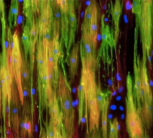
Mouse muscle

Mighty mouse muscle cells

We are often told that sleep is one of the most important elements of a healthy lifestyle, that it helps in the healing and repair of our heart and blood vessels – among other things.

It turns out that sleep, or something very similar, is equally important for stem cells, helping them retain their power or potency, which is a measure of their effectiveness and efficiency in generating the mature adult cells that are needed to repair damage. Now researchers from Stanford, with a little help from CIRM, have found a way to help stem cells get the necessary rest before kicking in to action. This could pave the way for a whole new approach to treating a variety of genetic disorders such as muscular dystrophy.

Inside out

One problem that has slowed down the development of stem cell therapies has been the inability to manipulate stem cells outside of the body, without reducing their potency. In the body these cells can remain quiescent or dormant for years until called in to action to repair an injury. That’s because they are found in a specialized environment or niche, one that has very particular physical, chemical and biological properties. However, once the stem cells are removed from that niche and placed in a dish in the lab they become active and start proliferating and changing into other kinds of cells.

You might think that’s good, because we want those stem cells to change and mature, but in this case we don’t, at least not yet. We want them to wait till we return them to the body to do their magic. Changing too soon means they have less power to do that.

Researchers at Stanford may have found a way to stop that happening, by creating an environment in the lab that more closely resembles that in the body, so the stem cells remain dormant longer.

As senior author, Thomas Rando, said in a Stanford news release, they have found a way to keep the stem cells dormant longer:

Dr. Thomas Rando, Stanford

Dr. Thomas Rando, Stanford

“Normally these stem cells like to cuddle right up against their native muscle fibers. When we disrupt that interaction, the cells are activated and begin to divide and become less stemlike. But now we’ve designed an artificial substrate that, to the cells, looks, smells and feels like a real muscle fiber. When we also bathe these fibers in the appropriate factors, we find that the stem cells maintain high-potency and regenerative capacity.”

Creating an artificial home

When mouse muscle stem cells (MuSCs) are removed from the mouse they lose their potency after just two days. So the Stanford team set out to identify what elements in the mouse niche helped the cells remain dormant. They identified the molecular signature of the quiescent MuSCs and used that to help screen different compounds to see which ones could help keep those cells dormant, even after they were removed from the mouse and collected in a lab dish.

They whittled down the number of potential compounds involved in this process from 50 to 10, and then tested these in different combinations until they found a formulation that kept the stem cells quiescent for at least 2 days outside of the mouse.

But that was just the start. Next they experimented with different kinds of engineered muscle fibers, to simulate the physical environment inside the mouse niche. After testing various materials, they found that the one with the greatest elasticity was the most effective and used that to create a kind of scaffold for the stem cells.

The big test

The artificial niche they created clearly worked in helping keep the MuSCs in a dormant state outside of the mouse. But would they work when transplanted back into the mouse? To answer this question they tested these stem cells to see if they retained their ability to self-renew and to change into other kinds of cells in the mouse. The good news is they did, and were far more effective at both than MuSCs that had not been stored in the artificial niche.

So, great news for mice but what about people, would this same approach work with human muscle stem cells (hMuSCs)? They next tested this approach using hMuSCs and found that the hMuSCs cultured on the artificial niche were more effective at both self-renewal and retaining their potency than hMuSCs kept in more conventional conditions, at least in the lab.

In the study, published in the journal Nature Biotechnology, the researchers say this finding could help overcome some of the challenges that have slowed down the development of effective therapies:

“Research on MuSCs, hematopoietic stem cells and neural stem cells has shown that very small numbers of quiescent stem cells, even single cells, can replace vast amounts of tissue; culture systems that that maintain stem cell quiescence may allow these findings to be translated to clinical practice. In addition, the possibility of culturing hMuSCs for longer time periods without loss of potency in order to correct mutations associated with genetic disorders, such as muscular dystrophy, followed by transplantation of the corrected cells to replace the pathogenic tissue may enable improved stem cell therapeutics for muscle disorders.”

Stem cell stories that caught our eye: reducing radiation damage, making good cartilage, watching muscle repair and bar coding cells

Here are some stem cell stories that caught our eye this past week. Some are groundbreaking science, others are of personal interest to us, and still others are just fun.

A bomb blastaStem cells key to reducing radiation damage. With the anniversary of Hiroshima and President Obama’s historic visit to the site all over the news this week, it was nice to read about research that could result in many more people surviving a major radiation event—either from a power plant accident or the unthinkable repeat of history.

Much of the life-threatening damage that occurs early after radiation exposure happens in the gut, so a way to reduce that damage could buy time for other medical care. A team at the University of Texas Medical Branch at Galveston has discovered a drug that activates stem cells in the gut, which help maintain a healthy population of crypt cells that can repair gut damage.

A single injection of the small protein drug in mice significantly increased their survival, even if it was given 24 hours after exposure to radiation. The researchers published their work in the journal Laboratory Investigation and in a story written for MedicalNewsToday the lead author, Carla Kantara suggested the role the drug might have:

 “The current results suggest that the peptide may be an effective emergency nuclear countermeasure that could be delivered within 24 hours after exposure to increase survival and delay mortality, giving victims time to reach facilities for advanced medical treatment.”

The small protein, or peptide, named TP508, has already been tested in humans for diabetic foot ulcers so could be tested in humans fairly quickly.


Making good cartilage for your knees. Rarely a week goes by that I don’t tell a desperate osteoarthritis patient with painful knees that I am treating my own rotten knees with physical therapy until we learn how to use stem cells to make the right kind of cartilage needed for lasting knee repair. So, I was thrilled to read this week that the National Institutes of Health awarded Case Western Reserve University in Cleveland $6.7 million to develop a center to create standardized systems for monitoring stem cells as they convert into cartilage and for evaluating the resulting cartilage.

ear_wakeforest There are a couple problems with existing attempts to use stem cells for knee and other cartilage repair. First not all cartilage is equal and too often stem cells form the soft kind like in your earlobe, not the hard kind needed to protect knees. Also, it has been hard to generate enough cells to replace the entire area that tends to be eroded away in osteoarthritis, one of the leading causes of disability.
The new center, which will be available to researchers anywhere in the world, will develop tools for them to measure four things:

  • which genes are turned on or off as stem cells take the many steps toward becoming various forms of cartilage;
  • predict the best makeup of the extracellular matrix, the support structures outside cells that help them organize as they become a specific tissue;
  • evaluate the biochemical environment around the cells that helps direct their growth;
  • measure the mechanical properties of the resulting cartilage—is it more like the ear or the knee.

NewsWise posted the university’s press release


Damaged muscle grabs stem cells.  All our tissues have varying skills in self repair. Muscles generally get pretty high marks in that department, but we don’t really know how they do it. A team at Australia’s Monash University used the transparent Zebra fish and fancy microscopes to actually watch the process.

When they injured mature muscle cells they saw those cells send out projections that actually grabbed nearby muscle stem cells, which regenerated the damaged muscle. They published their findings in Science, the university issued a press release and a news site for Western Australia, WAtoday wrote a story quoting the lead researcher Peter Currie:

 “A significant finding is that the wound site itself plays a pivotal role in coordinating the repair of damaged tissue. If that response could be sped up, we are going to get better, or more timely, regeneration and healing.”

The online publication posted four beautiful florescent images of the cells in action.


muscle stem cells Monash

Muscle stem cells in action

“Bar coding” cells points to better transplants.  A team at the University of Southern California, partially funded by CIRM, developed a way genetically “bar code” stem cells so they can be tracked after transplant. In this case they watched the behavior of blood-forming stem cells and found the dose of cells transplanted had a significant impact on what the cells became as they matured.

The general dogma has blood stem cells producing all the various types of cells in our blood system including all the immune cells needed by cancer patients after certain therapies. But the USC tracking showed that only 20 to 30 percent of the stem cells displayed this do-it-all behavior. The type of immune cells created by the remaining 70 to 80 percent varied depending on whether there was a low dose of cells or a high dose, which can be critical to the effectiveness of the transplant.

 “The dose of transplanted bone marrow has strong and lasting effects on how HSCs specialize and coordinate their behavior,” said Rong Lu, senior author, in a USC press release posted by ScienceDaily. “This suggests that altering transplantation dose could be a tool for improving outcomes for patients — promoting bone marrow engraftment, reducing the risk of infection and ultimately saving lives.”

Outsmarting cancer’s deadly tricks

Cancer cells are devious monsters that kill people by sabotaging normal cell functions toward a path of uncontrolled cell growth. Without an effective treatment, aggressive cancers can crowd out healthy tissue and ultimately cause organ failure and death. This devastation by design makes it seem as though a cancer cell has a mind of its own but in reality it’s all due to mindless mutations in DNA. Gaining a deep understanding of those mutations provides scientists with insights into the molecular mechanisms of cancer which can help pinpoint targets for potential cancer treatments.

A team at The Scripps Research Institute (TSRI) followed the trail of such a mutation in a gene called POT1. Today in Cell Reports the researchers, funded in part by CIRM, describe their identification of a novel mechanism for cancer progression in cells carrying the POT1 mutation and they also speculate on the development on a unique therapeutic strategy.

Chromosomes go to pot without POT1
The POT1 protein is one component of shelterin, a multi-protein structure that binds to and protects telomeres, a region of DNA found at the ends of chromosomes. The team found that when POT1’s function is disrupted by mutation, the telomeres become vulnerable to damage which leads to chromosome instability. As a result, many regions of DNA on the chromosomes get rearranged leading to further gene mutations that in turn can accelerate the process of cancerous growth.

Telomere_caps

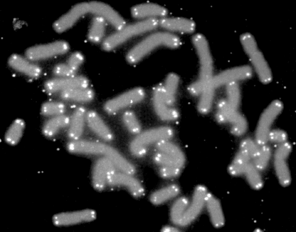
Human chromosomes (grey) capped by telomeres (white) Wikipedia

However, in the case of POT1 mutations, the DNA damage in the unstable chromosomes stimulates an enzyme called ATR that’s known to shut down cell division and initiate apoptosis, or programmed cell death. Now, unless I’m missing something, cells that have either stopped dividing or even died would seem to be the opposite of cancer progression. So why then are POT1 mutations found in a number of cancers such as leukemia, melanoma (skin cancer) and glioma (brain cancer)? As TSRI Associate Professor Eros Lazzerini Denchi, a co-leader on the publication, mentions in a press release, this conundrum presented an opportunity to better understand POT1 related cancers:

lazzerini_denchi

Eros Lazzerini Denchi

“Somehow those cells found a way to survive—and thrive. We thought that if we could understand how that happens, maybe we could find a way to kill those cells.”


Mutant POT1 and p53: diabolical partners in cancer progression
The team looked for answers by studying the POT1 mutation in the presence of a mutated form of the p53 tumor suppressor gene, found in over 50% of all human cancers. Mice bred with the POT1 mutation alone formed no cancers while those animals with the p53 mutation alone developed T cell lymphomas, a type of immune system cancer, by 20 weeks and survived 24 weeks. Mice with both mutations fared much worse with median survival times of just 17 weeks. So somehow the p53 mutation was bringing out the potential of the POT1 mutation to cause aggressive cancer growth.

Further experiments revealed that the p53 mutation quashed the ATR enyzme’s programmed cell death signal which the team had shown was stimulated by the POT1 mutation. As a result, the cells avoided programmed cell death. Because the cells had no mechanism to die, more cancer-causing mutations had the opportunity to develop from the chromosome instability caused by the POT1 mutation.

The bright side to this diabolical cooperation between mutant POT1 and p53 is that it presents a possible opening for new treatment strategies. It turns out that no cell, not even a cancerous one, can survive in the complete absence of ATF. Since cells with the POT1 mutations already have a reduced level of ATF, the authors suggest that delivery of low doses of ATF inhibitors, which have already been developed for the clinic, could kill cancer cells without affecting healthy cells. No doubt the team is eager to follow up on this hypothesis.

It’s comforting to know that there are crafty scientists out there who are closing in on ways to outsmart the sneaky tactics of cancer cells. And it wouldn’t be possible without this fundamental research, as Lazzerini Denchi points out:

“This study shows that by looking at basic biological questions, we can potentially find new ways to treat cancer.”


More Good News From CIRM-Funded Spinal Cord Injury Trial

It’s been less than a year since we last reported on the CIRM-funded Asterias Biotherapeutics trial for spinal cord injury (SCI), and we already have more – still preliminary – but good news to share. The company recently released encouraging long-term follow-up results from their original Phase 1 clinical trial that suggest their stem cell treatment is safe and possibly effective for treating SCI occurring in the back region.

astopc1Back in August 2015, the California-based company reported positive results for the second phase of the clinical trial, the ongoing Phase 1/2a trial, that is testing their AST-OPC1 brain progenitor cell treatment in patients with cervical or neck spinal cord injury. They treated three patients with a low dose of two million AST-OPC1 cells and observed no serious side effects after two months. You can read more about these initial results in our blog.

Asterias plans to expand their Phase 1/2a trial by enrolling more patients and administering higher numbers of cells in hopes that a higher dose might impact or improve motor function in SCI patients. But with any cell transplantation therapy, there is always concerns about whether it’s safe and whether it could cause any long-term consequences in patients.

Good news to those who wait

A news release by Asterias yesterday, puts some of these fears to rest. They report new long-term data on their original Phase 1 trial, which was carried out by Geron, that treated patients with thoracic or back SCI. In this trial, five patients were treated with two million AST-OPC1 cells between 7 and 14 days post injury. The patients were given immunosuppressive drugs for two months so they wouldn’t reject the cell transplant and then were monitored over the next 4-5 years.

During this time, none of the patients showed any signs of transplant rejection, and MRI scans revealed that four out of the five patients showed less cavitation in their spinal cords, a destructive process that occurs after severe spinal cord injury.

Thus it seems that AST-OPC1 does not pose any serious safety issues for SCI patients, at least at the five-year mark. Chief Medical Officer Dr. Edward Wirth explained:

Edward Wirth, CMO at Asterias

Edward Wirth, CMO at Asterias

“This new long term follow-up data continues to support the general safety of AST-OPC1 and indicate minimal risk of the transplanted cells having unintended effects. In detailed immune response monitoring of patients, the results are consistent with long-term cell engraftment, immune system tolerability, and an absence of adverse effects.  In short, AST-OPC1 does not appear to present any immunological or other long-term safety issues when administered to patients suffering from spinal cord injuries.”

These positive long-term results are perfectly timed for Asteria’s expansion of their Phase 1/2a trial where they aim to test doses of AST-OPC1 that they believe would improve motor function in SCI patients. Asterias CEO Steve Cartt commented:

Steve Cartt, CEO of Asterias Biotherapeutics

Steve Cartt, CEO of Asterias

“These new follow-up results are very encouraging and provide important further support for expansion of the ongoing Phase 1/2a clinical study in patients with complete cervical spinal cord injuries announced just last week. We are continuing to enroll patients in the second dose cohort of the current Phase 1/2a trial.  Patients in this cohort are receiving a significantly higher dose of 10 million cells, which we believe corresponds to the doses that showed efficacy in animal studies.”

But that’s not all folks!

Dr. Edward Wirth, Asterias Biotherapeutics

Dr. Edward Wirth from Asterias Biotherapeutics at the CIRM Alpha Clinics Meeting in May

CIRM got the inside scoop on the next steps of this Phase 1/2a trial last week at a CIRM Alpha Stem Cell Clinics Meeting held at UC Irvine. Dr. Edward Wirth was the guest speaker, and during lunch, he explained how their recent successes in both clinical trials has prompted the FDA to grant them clearance to expand their current Phase 1/2a trial from 13 to up to 35 patients.

Asterias can now enroll patients with both AIS A (complete injury) and AIS B injuries and has expanded the age range of trial participants to 18-69 years. Dr. Wirth added that the goal of this trial is to rescue some of the motor function in cervical SCI patients so that they can go from needing full time care to being able to carry out some functions on their own. He also indicated that these patients will be monitored for 15 years to evaluate the safety and success of their treatment.

We at CIRM are encouraged by these early positive results and hopeful that this clinical trial will result in a stem cell treatment that will improve the lives of SCI patients.


Related Links:

Free public event will detail the many ways stem cells are used in clinical trials today

The hundreds of active stem cell clinical trials being run in the US, and indeed around the world, provide ample evidence that our favorite cells are truly multi-talented. There are so many different ways researchers are using them to develop therapies we would be hard-pressed to name them all. However, most fall into five general categories that will be discussed at a free public symposium CIRM is co-hosting in conjunction with the International Society for Stem Cell Research during its annual meeting in San Francisco.

Moscone at dusk

San Francisco’s Moscone Center is close to BART and Muni public transit

The free public event will run from 6:00 to 7:30 on Tuesday evening June 21 at the Moscone West convention center, room 2009, on the corner of Howard and Fourth streets in San Francisco. After a brief overview, four researchers will describe active clinical trials and how stem cells provide hope for therapies in different diseases.  The last half hour will be open for general questions from the audience.

All the details are at a special page on EventBright where you can register to attend. The evening will start with Bruce Conklin of the Gladstone Institutes providing an overview of the many ways to use stem cells, including his own work using them to create laboratory models of heart disease. Then:

  • Malin Parmar of Sweden’s Lund University will discuss a Parkinson’s disease trial where stem cells are used to replace vital brain cells destroyed by the disease;
  • Donald Kohn of the University of California, Los Angeles, will provide details of two trials that combine stem cells and gene therapy, one for sickle cell anemia and one for severe combined immune deficiency, also called Bubble Baby disease;
  • Henry Klassen of University of California, Irvine, will talk about using progenitor stem cells to deliver factors that can protect the photoreceptors in the eyes of patients who have a blinding condition;
  • Catriona Jamieson of the University of California, San Diego will describe the bad boy of the stem cell world, the cancer stem cell, and clinical trials she is conducting to attack those cells.

While some of the hundreds of current stem cell clinical trials will not produce the desired impact on their target diseases, they will all make strides toward learning how to optimize the great potential of stem cell therapies.

Right now CIRM is funding 16 different clinical trials in diseases as varied as HIV/AIDS and type 1 diabetes. Over the next 5 years we hope to add another 50 clinical trials to that list. The field of regenerative medicine is advancing. This event is a chance for you to understand the progress, and the challenges, that we face in bringing potentially life-changing, even life-saving therapies to the people who need it the most, the patients.

Why is a cell therapy that restores sight to the blind against the law?

FDA

A lot of people are frustrated with the US Food and Drug Administration (FDA) and its woefully slow process for approving stem cell therapies. That’s one of the reasons why we started the CIRM Stem Cell Champions campaign, to gather as many like-minded supporters of stem cell research as possible and help to change the way the FDA works, to create a more efficient approval process.

You can read more about that campaign and watch a short video on what being a Stem Cell Champion involves (hint: not very much).

Now Randy Mills, our President and CEO, has teamed up with former US Senator Bill Frist to explain precisely why the FDA needs to change the way it regulates stem cells, and to offer a simple way to create the system that will best serve the needs of patients.

This Op Ed appeared on Fox News’ online Opinion section on Friday, May 20th.


Cell therapy reversed blindness for 47,000 patients in 2015. So why is it against the law?

By C. Randal Mills Ph.D., Sen. Bill Frist M.D.

As medical miracles go, restoring sight to the blind is right up there. A mother seeing her baby for the first time, or a child being able to count the stars is a beautiful gift, and its value cannot be overstated. Last year 47,000 Americans received that gift and had their blindness reversed through the transplantation of cells from a corneal donor’s final selfless act.

It is safe, it is effective, and because it is curative, it is a relatively cost effective procedure. It is medicine at its most beautiful. And according to FDA regulations, the distribution of this cell therapy is in violation of federal law.

That’s right. The regulation says that no matter how competent the surgeon, the FDA must first approve cells from donated corneas as if they were a drug—a process that takes over a decade and can costs billions of dollars — all for a practice that has been successfully restoring sight for more than 50 years. And this is only one example.

The good news: the FDA doesn’t always adhere to its regulations and has not in this case.

The bad news: inconsistent enforcement creates uncertainty, deterring innovation for other unmet medical needs such as arthritis, back pain, and diabetic ulcers.

How did a country known for pioneering medical breakthroughs get here?

Appropriate regulation of living cells that treat disease is inherently complex. Some therapies, like corneal cell transplants, are well-understood. Others are far more sophisticated and can involve forcing cells to change from one type to another, cutting out defective genes, and growing cells in culture to expand their numbers into the billions. Although this may sound like science fiction, it’s the type of very real science that will revolutionize the practice of medicine. And it is a challenging spectrum to regulate.

Unfortunately, what we have today amounts to a regulatory light switch for cell therapy; one that is either OFF or ON. For some cell therapies there is essentially no pre-market regulation. But at some point of added complexity, often arbitrarily decided by the FDA, the switch flips to ON and the cell becomes a drug in the minds of the Agency. And the consequences could not be more profound.

A product can be introduced through the OFF pathway in days with no FDA review and at very little cost. The ON pathway on the other hand, takes 10-20 years and can cost over a billion dollars. For cell therapy, there is no in between.

It is not possible to regulate the continuum of cell therapies fairly and effectively by using this binary approach. The system is broken and is impeding the hunt for safe and effective treatments for suffering patients.

Why? Because sensible people don’t invest significant capital gambling that the FDA will give them a pass out of its rules. They evaluate the time and cost of development assuming they will be forced down the ON pathway. They also assume that this arbitrary approach to regulation will (and often does) work against them by allowing a competitor to enter the market through the OFF pathway, placing them at a prohibitive disadvantage. The results speak for themselves. After 15 years under this paradigm we have had only a few cell therapies approved, all commercial disasters.

This is because the ON-OFF approach fails to adequately account for the difference in cell therapy complexity. To better understand, imagine this methodology applied to the regulation of automobiles. The government might permit low tech cars, say the Model T, to be sold without pre-market regulation. But if a manufacturer wanted to improve the vehicle by adding air conditioning, a radio or other such feature, the car would be subject to massive pre-market regulation. And not just on the new feature. Instead, the addition of the new feature would trigger a bumper-to-bumper evaluation of the entire car, increasing its development cost from basically nothing to that of a Lamborghini. The result would be streets full of hot, radio-less go-karts, except for a few ultra-high-end sports cars whose manufacturers are now defunct because they were never able to recoup the disproportionate costs of satisfying the regulatory system. This is what we see with cell therapies today: progress that is sluggish at best.

How can we move forward?

Ironically, the FDA identified a solution to the problem. In order to account for the broad spectrum inherent to cell therapy, in the late 90’s the FDA proposed a progressive, risk-based approach. The higher the risk, the greater the regulation. This guards against under regulation that might put patients at risk and prevents overregulation that can disincentivize the development of new or improved products.

In the FDA’s own words, the regulation they proposed would abide by a few basic principles:

  • “Under this tiered, risk-based approach, we propose to exert only the type of government regulation necessary to protect the public health.”
  • “The regulation of different types of human cells… will be commensurate with the public health risks…”
  • “These planned improvements will increase the safety of human cells… while encouraging the development of new products.”

It was a remarkably common sense approach that would have balanced safety with the need for innovation over an exceptionally broad range of technological complexity and risk.

It would have.

Unfortunately, the regulatory framework that was promised was never delivered, and it is time to resuscitate it. The burden placed on the development of cell therapies must accurately reflect the risks; must be balanced against the very real consequences of doing nothing (patients continuing to suffer); and must be consistently and fairly applied. In short, the FDA had it right and we need to give them the tools to deliver the regulatory paradigm they originally envisioned.

If we fix this highly fixable problem, we can create a system that will drive new innovations and better outcomes. Europe and Japan have already acted and are seeing the benefits. People with great ideas are coming off the bench, and game changing therapies are entering practice. While challenging the status quo does not sit well with some, particularly those who stand to prosper from the built-in barriers to entry the current structure provides, in the United States we have a responsibility to do better for patients and fix this broken system.

Randal Mills, Ph.D., is the President and CEO of the California Institute for Regenerative Medicine

William “Bill” H. Frist, M.D. is a nationally-acclaimed heart and lung transplant surgeon, former U.S. Senate Majority Leader, and chairman of the Executive Board of the health service private equity firm Cressey & Company.

In the Stem Cellar: making better blood stem cells, a heart guard, iPS model points to ALS drug and tracking cells

Here are some stem cell stories that caught our eye this past week. Some are groundbreaking science, others are of personal interest to us, and still others are just fun.

Major step in creating blood stem cells. If you track stem cells in any online news search, your feed perpetually will have numerous posts about attempts to find a bone marrow stem cell match for a desperate cancer patient. The power of those cells to reconstitute a person’s immune system after aggressive therapy saves the lives of thousands of cancer patients every year, but too many patients still die waiting to find an immunologically compatible donor.

Since pluripotent stem cells, both embryonic and iPS cells, can create any cell type in the body, they should be able to produce the needed blood forming stem cells. But that feat has been one of the toughest to accomplish in the young history of stem cell science. Blood stem cells created from pluripotent cells don’t self-renew like they should and don’t take up residence in the bone marrow properly. A CIRM-funded team at the University of California, Los Angeles, has made a major stride toward making this possible.

The team, led by Hanna Mikkola, started by looking at what genes were turned on in blood stem cells they created in the lab compared to natural ones. They pinpointed one set of genes, the HOXA genes, that is linked to the ability to self-renew. Next, they found that mimicking the effects of retinoic acid, a derivative of vitamin A, can turn on the HOXA genes.

 “Inducing retinoic acid activity at a very specific time in cell development makes our lab-created cells more similar to the real hematopoietic stem cells found in the body,” said Diana Dou, a graduate student in Mikkola’s lab in a UCLA press release.

While this is one major hurdle leaped, the team acknowledges they have more work to do before they can create lab-grown blood stem cell that fully match the functions of natural blood stem cells.


Turned-off gene protects hearts.  When Nobel Prize winner Shinya Yamanaka reprogrammed skin cells into embryonic-like iPS cells, he activated four genes that are very involved in embryo development, but have been assumed to be inactive in adults. Researchers at the University of Virginia published data overturning that dogma, and more importantly suggested that one of those genes, Oct4, is not just active in adults, it protects people from heart disease.

They found that Oct4 plays a role in the formation of atherosclerotic plaque. When that plaque buildup in arteries ruptures it causes heart attacks and strokes.  But Oct4 instructs smooth muscle cells to create protective fibrous caps that make the plaques less likely to rupture. The team leader, Gary Owen, speculated that Oct4 might also be involved in other aspects of the body’s effort to repair damage and heal wounds.

 “Finding a way to augment the expression of this gene in adult cells may have profound implications for promoting health and possibly reversing some of the detrimental effects with ageing,” said Owen in a story in Scicasts adapted from a university press release.

The researchers are now looking for ways to selectively activate Oct4 for therapeutic purposes.


neuronfromiPS

Nerves grown from iPS cells

Stem cell model leads to potential ALS drug.  In amyotrophic lateral sclerosis (ALS or Lou Gehrig’s disease) motor nerves that allow all forms of movement die off. But, some nerves seem to be resistant to this damage. Researchers at Sweden’s Karolinska Institute and at the University of Milan in Italy have found that a specific nerve growth factor can protect motor nerves from ALS.

That factor, insulin-like growth factor 2 (IGF-2), was able to rescue human motor nerves grown in the lab from iPS-type stem cells made by reprogramming skin of ALS patients. The researchers then provided IGF-2 to mice with ALS-like disease through gene therapy and the animals lived longer than without the growth factor.

 “We can see that motor neurons are preserved and that IGF-2 treatment causes the axons to regenerate and recreate vital connections with muscles that were previously lost,” said Karolinska’s Eva Hedlund in a press release posted by MedicalXpress.

 Prior attempts to treat ALS patients with a related compound, IGF-1, by injecting it under the skin failed.  The current team suggests that direct delivery to motor nerves via gene therapy could provide a better outcome.


Labeling and tracking stem cells.  Numerous studies have shown stem cells grown in the lab function more like normal stem cells the closer the lab environment comes to mimicking the natural environment where the cells would grow in the body. Using that strategy a team at Carnegie Mellon University in Pittsburg succeeded in loading stem cells with an FDA-approved iron nanoparticle that will allow them to track the cells after transplant.


MSCs with iron nanoparticles

MSCs labeled with iron nanoparticles

They focused on a type of stem cell found in bone marrow, mesenchymal stem cells (MSCs), which are being used in more than half of the 600+ active stem cell clinical trials. To date, MSC trials have produced a very mixed bag of results, with much of the poorer outcomes attributed to the cells not going to, and staying, where they are needed.  So this tracking technique could help develop strategies to improve those outcomes.

Up to this point, researchers could not get the tracking agent into cells without using an agent to help get the particles across the cell membrane and those agents tend to disrupt the normal cell function. But, in their normal environment cells will engulf small particles on their own.  So the Carnegie team added other cells types found in bone marrow to their lab cultures, the MSCs felt more at home, and took up the nanoparticles. A neat little trick written up in a university press release posted at Science Daily.

What’s the big idea? Or in this case, what’s the 19 big ideas?

supermarket magazineHave you ever stood in line in a supermarket checkout line and browsed through the magazines stacked conveniently at eye level? (of course you have, we all have). They are always filled with attention-grabbing headlines like “5 Ways to a Slimmer You by Christmas” or “Ten Tips for Rock Hard Abs” (that one doesn’t work by the way).

So with those headlines in mind I was tempted to headline our latest Board meeting as: “19 Big Stem Cell Ideas That Could Change Your Life!”. And in truth, some of them might.

The Board voted to invest more than $4 million in funding for 19 big ideas as part of CIRM’s Discovery Inception program. The goal of Inception is to provide seed funding for great, early-stage ideas that may impact the field of human stem cell research but need a little support to test if they work. If they do work out, the money will also enable the researchers to gather the data they’ll need to apply for larger funding opportunities, from CIRM and other institutions, in the future

The applicants were told they didn’t have to have any data to support their belief that the idea would work, but they did have to have a strong scientific rational for why it might

As our President and CEO Randy Mills said in a news release, this is a program that encourages innovative ideas.

Randy Mills, Stem Cell Agency President & CEO

Randy Mills, CIRM President & CEO

“This is a program supporting early stage ideas that have the potential to be ground breaking. We asked scientists to pitch us their best new ideas, things they want to test but that are hard to get funding for. We know not all of these will pan out, but those that do succeed have the potential to advance our understanding of stem cells and hopefully lead to treatments in the future.”

So what are some of these “big” ideas? (Here’s where you can find the full list of those approved for funding and descriptions of what they involve). But here are some highlights.

Alysson Muotri at UC San Diego has identified some anti-retroviral drugs – already approved by the Food and Drug Administration (FDA) – that could help stop inflammation in the brain. This kind of inflammation is an important component in several diseases such as Alzheimer’s, autism, Parkinson’s, Lupus and Multiple Sclerosis. Alysson wants to find out why and how these drugs helps reduce inflammation and how it works. If he is successful it is possible that patients suffering from brain inflammation could immediately benefit from some already available anti-retroviral drugs.

Stanley Carmichael at UC Los Angeles wants to use induced pluripotent stem (iPS) cells – these are adult cells that have been genetically re-programmed so they are capable of becoming any cell in the body – to see if they can help repair the damage caused by a stroke. With stroke the leading cause of adult disability in the US, there is clearly a big need for this kind of big idea.

Holger Willenbring at UC San Francisco wants to use stem cells to create a kind of mini liver, one that can help patients whose own liver is being destroyed by disease. The mini livers could, theoretically, help stabilize a person’s own liver function until a transplant donor becomes available or even help them avoid the need for liver transplantation in the first place. Considering that every year, one in five patients on the US transplant waiting list will die or become too sick for transplantation, this kind of research could have enormous life-saving implications.

We know not all of these ideas will work out. But all of them will help deepen our understanding of how stem cells work and what they can, and can’t, do. Even the best ideas start out small. Our funding gives them a chance to become something truly big.


Related Links:

Adding new stem cell tools to the Parkinson’s disease toolbox

Understanding a complicated neurodegenerative disorder like Parkinson’s disease (PD) is no easy task. While there are known genetic risk factors that cause PD, only about 10 percent of cases are linked to a genetic cause. The majority of patients suffer from the sporadic form of PD, where the causes are unknown but thought to be a combination of environmental, lifestyle and genetic factors.

Unfortunately, there is no cure for PD, and current treatments only help PD patients manage the symptoms of their disease and inevitably lose their effectiveness over time. Another troubling issue is that doctors and scientists don’t have good ways to predict who is at risk for PD, which closes an important window of opportunity for delaying the onset of this devastating disease.

Scientists have long sought relevant disease models that mimic the complicated pathological processes that occur in PD. Current animal models have failed to truly represent what is going on in PD patients. But the field of Parkinson’s research is not giving up, and scientists continue to develop new and improved tools, many of them based on human stem cells, to study how and why this disease happens.

New Stem Cell Tools for Parkinson’s

Speaking of new tools, scientists from the Buck Institute for Research on Aging published a study that generated 10 induced pluripotent stem cell (iPS cell) lines derived from PD patients carrying well known genetic mutations linked to PD. These patient cell lines will be a useful resource for studying the underlying causes of PD and for potentially identifying therapeutics that prevent or treat this disorder. The study was partly funded by CIRM and was published today in the journal PLOS ONE.

Dr. Xianmin Zeng, the senior author on the study and Associate Professor at Buck Institute, developed these disease cell lines as tools for the larger research community to use. She explained in a news release:

Xianmin Zeng, Buck Institute

Xianmin Zeng, Buck Institute

“We think this is the largest collection of patient-derived lines generated at an academic institute. We believe the [iPS cell] lines and the datasets we have generated from them will be a valuable resource for use in modeling PD and for the development of new therapeutics.”


The datasets she mentions are part of a large genomic analysis that was conducted on the 10 patient stem cell lines carrying common PD mutations in the SNCA, PARK2, LRRK2, or GBA genes as well as control stem cell lines derived from healthy patients of the same age. Their goal was to identify changes in gene expression in the Parkinson’s stem cell lines as they matured into the disease-affected nerve cells of the brain that could yield clues into how PD develops at the molecular level.

Using previous methods developed in her lab, Dr. Zeng coaxed the iPS cell lines into neural stem cells (brain stem cells) and then further into dopaminergic neurons – the nerve cells that are specifically affected and die off in Parkinson’s patients. Eight of the ten patient lines were able to generate neural stem cells, and all of the neural stem cell lines could be coaxed into dopaminergic neurons – however, some lines were better at making dopaminergic neurons than others.

Dopaminergic neurons derived from induced pluripotent stem cells. (Xianmin Zeng, Buck Institute)

Dopaminergic neurons derived from induced pluripotent stem cells. (Xianmin Zeng, Buck Institute)

When they analyzed these lines, surprisingly they found that the overall gene expression patterns were similar between diseased and healthy cell lines no matter what cell stage they were at (iPS cells, neural stem cells, and neurons). They next stressed the cells by treating them with a drug called MPTP that is known to cause Parkinson’s like symptoms in humans. MPTP treatment of dopaminergic neurons derived from PD patient iPS cell lines did cause changes in gene expression specifically related to mitochondrial function and death, but these changes were also seen in the healthy dopaminergic neurons.

Parkinson’s, It’s complicated…

These interesting findings led the authors to conclude that while their new stem cell tools certainly display some features of PD, individually they are not sufficient to truly model all aspects of PD because they represent a monogenic (caused by a single mutation) form of the disease.

They explain in their conclusion that the power of their PD patient iPS cell lines will be achieved when combined with additional patient lines, better controls, and more focused data analysis:

“Our studies suggest that using single iPSC lines for drug screens in a monogenic disorder with a well-characterized phenotype may not be sufficient to determine causality and mechanism of action due to the inherent variability of biological systems. Developing a database to increase the number of [iPS cell] lines, stressing the system, using isogenic controls [meaning the lines have identical genes], and using more focused strategies for analyzing large scale data sets would reduce the impact of line-to-line variations and may provide important clues to the etiology of PD.”

Brian Kennedy, Buck Institute President and CEO, also pointed out the larger implications of this study by commenting on how these stem cell tools could be used to identify potential drugs that specifically target certain Parkinson’s mutations:

Brian Kennedy, Buck Institute

Brian Kennedy, Buck Institute

“This work combined with dozens of other control, isogenic and reporter iPSC lines developed by Dr. Zeng will enable researchers to model PD in a dish. Her work, which we are extremely proud of, will help researchers dissect how genes interact with each other to cause PD, and assist scientists to better understand what experimental drugs are doing at the molecular level to decide what drugs to use based on mutations.”

Overall, what inspires me about this study is the author’s mission to provide a substantial number of PD patient stem cell lines and genomic analysis data to the research community. Hopefully their efforts will inspire other scientists to add more stem cell tools to the Parkinson’s tool box. As the saying goes, “it takes an army to move a mountain”, in the case of curing PD, the mountain seems more like Everest, and we need all the tools we can get.


Related links:

You Call It Corn Stem Cells, We Call It An A-Maize-Ing Hope to Feed the World

figure_6A

David Jackson and his team at Cold Spring Harbor Laboratory identify a unique genetic pathway that regulated stem cell growth. Certain mutations in the pathway lead to increased plant yields (two plants on the right). Image: Cold Spring Harbor Laboratory

Here at the Stem Cellar, we’re laser-beam focused on the exciting progress being made to bring stem cell-based treatments to patients with unmet medical needs. But what good will those life-saving treatments be if the patients end up starving from hunger? It’s a serious question to ask considering the world’s diminishing farmlands and yet another record-breaking month for global warming in the books.

Based on a study published yesterday by Cold Spring Harbor Laboratory researchers, our friend the stem cell emerges again as a source of hope. Reporting in Nature Genetics, the team uncovered an important genetic switch in the stem cells of corn that when manipulated can lead to a 50% increase in the size of the corn.

Plants have stem cells too
Plants do indeed have stem cells that reside in an area called the meristem and function similarly to their animal counterparts. The root apical meristem is responsible for providing cells for root growth while the shoot apical meristem gives rise to plant organs like leaves and flowers. Previous research had shown that a signal system within the meristem communicates whether or not to turn on stem cell growth. The current study identified protein signals involved in a similar regulatory circuit but with an intriguing difference which David Jackson, the lead author on the study, explained in a Cold Spring Harbor video (see below):

“In this new study we found that actually the leaves, the developing leaves, send a signal back to the stem cells to control their growth which is really a new finding.”

FCP-1/FEA3: A Leaf to Stem Cell Braking System
The proteins involved in this signal include a receptor protein on the stem cells called FEA3 and a protein from the leaves called FCP-1. When it travels from the leaves to the stem cells, FCP-1 binds to FEA3 causing an inhibition of stem cell growth. So you’d think that disrupting this pathway would release the “brake” on stem cell growth and lead to tractor-sized corn. But when the team tested that idea by growing plants with a fea3 mutation, the resulting crop was short and stubby. The explanation is that too many stem cells is not a good thing and the available water, sunlight, and soil is not enough to support increased growth.

Easing off the brakes is better for crop yields
So as a result of uncontrolled stem cell growth, the corn becomes deformed and leads to a poorer yield. But next, the team analyzed plants with weaker versions, or alleles, of the fea3 mutations. Basically, these mutations still lead to a release of the “brake” on stem cell growth but not as quite as much as the initial fea3 mutation. Under this genetic scenario, the plants grew extra rows of kernels with up to 50% increase in yield.

Because the FCP-1/FEA3 pathway is found throughout the plant kingdom, this result has the tantalizing potential to help increase yields of all sorts of food crops.  As Jackson mentions in an interview with Gizmodo, this future will depend on these laboratory experiments working in a real world setting:

“If the yield increases we have seen in our lab strains hold out when used in agricultural maize strains this would lead to a significant boost in yields, potentially improving agricultural sustainability by requiring less land be devoted to agriculture. The same approaches could also benefit farmers in developing countries growing a wide range of crops.”

Certainly this future crop would be considered a genetically modified organism (GMO) which may concern some. But just today the National Academies of Sciences, Engineering, and Medicine posted evidence on their website that points to GMO foods as being safe and good for the environment.