The 10 Most Popular Stem Cellar Stories of 2017

As the New Year fast approaches, it’s time for us to reflect on our accomplishments these past 12 months. 2017 was an exciting and successful year for California’s Stem Cell Agency. We welcomed Dr. Maria Millan as the new President and CEO of CIRM. We also funded 16 new clinical trials and added two new medical centers (UCSF and UC Davis) to our CIRM Alpha Stem Cell Clinics Network. These are just a few examples of the significant progress that our Agency has made towards accelerating stem cell treatments to patients with unmet medical needs.

As you can imagine, these advances as well as the steady stream of new discoveries in the stem cell field, have kept our communications team very busy. In fact, I took a quick look at how many blogs we published in 2017 and the number is an impressive 242. That translates to blogging about stem cell research 66% of the year! How’s that for dedication?

Todd, Kevin and I love (and I truly mean that) writing for the Stem Cellar. All of the studies, trials, scientists and patients we feature are fascinating, but there are certain stories that steal the spotlight. It’s always fun to see which blogs are the most popular with our readers. So, let’s take a look at the 10 Stem Cellar stories caught your eye in 2017.

  1. Can stem cell therapies help ALS patients?
  2. jCyte gets FDA go-ahead for fast track review process of retinitis pigmentosa stem cell therapy
  3. A stem cell clinical trial for blindness: watch Rosie’s story
  4. Could stem cells help beat multiple sclerosis?
  5. Bye bye bubble baby disease: promising results from the stem cell gene therapy trial for SCID
  6. A clinical trial network focused on stem cell treatments is expanding
  7. Have scientists discovered a natural way to boost muscle regeneration?
  8. Three people left blind by Florida clinic’s unproven stem cell therapy
  9. Good news from Asterias’ CIRM-funded spinal cord injury trial
  10. Scientists make stem cell-derived nerve cells damaged in spinal cord injury

Honorable Mentions (underdog blogs that deserve a second look)

  1. 4 things to know about stem cell clinical trials [Video]
  2. ViaCyte treats first patients in PEC-Direct stem cell trial for type 1 diabetes
  3. Family, faith and funding from CIRM inspire one patient to plan for his future
  4. Texas tries to go it alone in offering unproven stem cell therapies to patients
  5. Has the promise of stem cells been overstated?

See you in January!

From all of us at CIRM, we wish you the happiest of holidays and good luck in the New Year. We’ll see you back here in January with exciting new content from our 2018 Annual Report. Stay tuned and stay curious my friends!

Making beating heart cells from stem cells just got easier

Here’s a heartwarming story for the holidays. Scientists from the Salk Institute in La Jolla, California have figured out a simple, easy way to make beating heart cells from human stem cells that will aid research and therapy development for heart disease. Their study, which received funding support from CIRM, was published last week in the journal Genes & Development.

The Salk team discovered that making beating heart tissue from human stem cells is as simple as turning off a single gene called YAP. You might be wondering how the team settled on this gene and no, it doesn’t involve pulling a random gene name out of a hat.

In previous studies, the researchers found that two cell signaling pathways, Wnt and Activin, are crucial for the development of embryonic stem cells into specialized cells like cardiomyocytes (beating heart cells). This research led to the discovery of a third pathway, controlled by YAP, which sets up a road block for cell specialization and keeps stem cells in their undifferentiated state.

Only hESCs without YAP (right panel) make heart cells (green) in one step. Blue dye marks cell nuclei. (Salk Institute)

The team deleted YAP from these stem cells using CRISPR gene editing technology, and then treated the stem cells to the Activin signaling molecule. Without YAP, exposure to Activin prompted the stem cells to develop immediately into beating cardiomyocytes that you can see beating away in the Salk video below.

Dr. Kathy Jones, Salk professor and senior author on the study, explained why this discovery is important to the field in a news release:

“This discovery is really exciting because it means we can potentially create a reliable protocol for taking normal cells and moving them very efficiently from stem cells to heart cells. Researchers and commercial companies want to easily generate cardiomyocytes to study their capacity for repair in heart attacks and disease—this brings us one step closer to being able to do that.”

First author, Conchi Estarás, emphasized how their new method for making cardiomyocytes is attractive not only for its simplicity, but also for its cost-effectiveness in enabling large-scale manufacturing of these cells for treatment.

“Instead of requiring two steps to achieve specialization, removing YAP cut it to just one step. That would mean a huge savings for industry in terms of reagent materials and expense.”

Looking ahead, Jones and her team do not plan on deleting the YAP gene from stem cells because of the potential side effects cause by the loss of YAP’s other cellular functions. Instead, they will be using commercially available molecules that can temporarily inhibit the function of YAP in hopes that this less permanent action will still readily produce beating heart cells from stem cells.

Kathy Jones and Conchi Estarás. (Image courtesy of Salk Institute)

Harnessing the body’s immune system to tackle cancer

Often on the Stem Cellar we write about work that is in a clinical trial. But getting research to that stage takes years and years of dedicated work. Over the next few months, we are profiling some of the scientists we fund who are doing Discovery (early stage) and Translational (pre-clinical) research, to highlight the importance of this work in developing the treatments that could ultimately save lives. 

This second profile in the series is by Ross Okamura, Ph.D., a science officer in CIRM’s Discovery & Translation Program.

Your immune system is your body’s main protection against disease; harnessing this powerful defense system to target a given disorder is known as immunotherapy.  There are different types of immunotherapies that have been developed over the years. These include vaccines to help generate antibodies against viruses, drugs to direct immune cell function and most recently, the engineering of immune cells to fight cancer.

Understanding How Immunotherapies Work

One of the more recent immunotherapy approaches to fight cancer that has seen rapid development is equipping a subset of immune cells (T cells) with a chimeric antigen receptor (CAR). In brief, CAR T ceIls are first removed from the patient and then engineered to recognize a specific feature of the targeted cancer cells.  This direct targeting of T cells to the cancer allows for an effective anti-cancer therapy made from your own immune system.

Simplified explanation of how CAR T cell therapies fight cancer. (Memorial Sloan Kettering)

For the first time this fall, two therapeutics employing CAR T cells targeting different types of blood cancers were approved for use by the US Food and Drug Administration (FDA) based on remarkable results found during the clinical trials. Specifically, Kymriah (developed by Novartis) was approved for treatment of acute lymphoblastic leukemia and Yescarta (developed by Kite Pharma) was approved for treatment of non-Hodgkin lymphoma.

There are drawbacks to the CAR T approach, however. Revving up the immune system to attack tumors can cause dangerous side effects. When CAR T cells enter the body, they trigger the release of proteins called cytokines, which join in the attack on the tumors. But this can also create what’s referred to as a cytokine storm or cytokine release syndrome (CRS), which can lead to a range of responses, from a mild fever to multi-organ failure and death. Balancing treatments to resolve CRS after it’s detected while still maintaining the treatment’s cancer-killing abilities is a significant challenge that remains to be overcome.  A second issue is that cancer cells can evade the immune system by no longer producing the target that the CAR-T therapy was designed to recognize. When this happens, the patient subsequently experiences a cancer relapse that is no longer treatable by the same cell therapy.

Natural Killer (NK) T cells represent another type of anti-cancer immunotherapy that is also being tested in clinical trials. NK cells are part of the innate immune system responsible for defending your body against both infection and tumor formation.  NK cells target stressed cells by releasing cell-penetrating proteins that poke holes in the cells leading to induced cell death.  As an immunotherapy, NK cells have the potential to avoid both the issues of CRS and cancer cell immune evasion as they release a more limited array of cytokines and do not rely on a specific single target to recognize tumors.  NK cells instead selectively target tumor cells due to the presence of stress-induced proteins on the cancer cells. In addition, the cancer cells lack other proteins that would normally send out a “I’m a healthy cell you can ignore me” message to NK cells. Without that message, NK cells target and kill those cancer cells.

Developing new immunotherapies against cancer

Dan Kaufman, UCSD

Dr. Dan Kaufman of the University of California at San Diego is a physician-scientist whose research group developed a method to produce functional NK cells from human pluripotent stem cells (PSC).  In order to overcome a major hurdle in the use of NK cells as an anti-cancer therapeutic, Dr. Kaufman is exploring using stem cells as a limitless source to produce a scalable, standardized, off-the-shelf product that could treat thousands of patients.  CIRM is currently funding Dr. Kaufman’s work under both a Discovery Quest award and a just recently funded Translational research award in order to try to advance this candidate approach.

In the CIRM Translational award, Dr. Kaufman is looking to cure acute myelogenous leukemia (AML) which in the US has a 5-year survival rate of 27% (National Cancer Institute, 2017) and is estimated to kill over 10,000 individuals this year (American Cancer Society, 2017).  He has previously shown that his stem cell-derived NK cells can kill human cancer cells in a dish and in mouse models, and his goals are to perform preliminary safety studies and to develop a process to scale his production of NK cells to support a clinical trial in people.  Since NK cells don’t require the patient and the donor to be a genetic match to be effective, a bank of PSC-derived NK cells derived from a single donor could potentially treat thousands of patients.

Looking forward, CIRM is also providing Discovery funding to Dr. Kaufman to explore ways to improve his existing approach against leukemia as well as expand the potential of his stem cell-derived NK cell therapeutic by engineering his cells to directly target solid tumors like ovarian cancer.

The field of pluripotent stem cell-based immunotherapies is full of game-changing potential and important innovations like Dr. Kaufman’s are still in the early stages.  CIRM recognizes the importance of supporting early stage research and is currently investing $27.9 million to fund 8 active Discovery and Translation awards in the cancer immunotherapy area.

CHLA study explains how stem cells slow progression of kidney disorder

Not all stem cell-based therapies act by replacing diseased or damaged cells. Many treatments in clinical development rely on the injected stem cells releasing proteins which trigger the slow down or even reversal of damage caused by disease or injury. A new CIRM-funded study that’s developing a stem cell therapy for a rare kidney disease uncovered a similar mechanism but with an intriguing twist. The research, published this week in Scientific Reports, suggests that the stem cells shed tiny vesicles that essentially act like sponges by trapping proteins thought to be responsible for damaging the kidney.

Amniotic fluid stem cells: a promising approach to treating kidney disease

2611_Blood_Flow_in_the_Nephron

Network of blood-filtering blood vessels in the kidney. Image: Wikipedia

In previous studies the research team, from the Saban Research Institute of Children’s Hospital Los Angeles (CHLA), had shown that amniotic fluid stem cells can help slow the progress of Alport syndrome when injected into the kidneys of mice engineered to mimic symptoms of the disease. Alport syndrome is a genetic disease that damages the kidney’s capillaries – tiny blood vessels – which help filter the body’s blood supply. This progressive damage causes blood and proteins to leak into the urine, and leads to high blood pressure and swelling in the legs and around the eyes.

Cells in the kidney release a protein called VEGF, a stimulator of new blood vessel growth, which plays an important role in maintaining just the right balance of capillaries within the blood-filtering structures of the kidney. Excessive levels of VEGF have been associated with many diseases including kidney disorders like Alport syndrome. Although the protective effects of amniotic fluid stem cells in the mouse model of Alport syndrome were not understood, the CHLA team suspected that the cells could be interfering with the effects of the extra VEGF.

Extracellular vesicles: just another trick that nature has up its sleeve
Specifically, the scientists examined whether so-called extracellular vesicles released from the stem cells are responsible for reducing VEGF activity and slowing the disease. These vesicles are tiny pieces of cell membrane that bud off from the stem cell and carry along proteins and other cell components. Scientists used to think the vesicles were just cellular discards but countless studies have established that they actually play an important role in communication between cells.

The team showed that the vesicles released by amniotic fluid stem cells contained receptors for VEGF. When those vesicles were added to a petri dish containing VEGF and kidney blood vessel cells, the vesicles reduced the VEGF activity and protected the cells from damage. But when vesicles from stem cells lacking the VEGF receptors were used, that protection was lost. First author Sargis Sedrakyan, PhD summed up the results in a press release:

“We have demonstrated that these vesicles can be used to regulate VEGF activity and prevent the [kidney] capillary damage. We can efficiently use the vesicles to help restore normal kidney function by curbing the progression of endothelial damage in the filtration unit of the kidney.”

Back in 2013, first author Sargis Sedrakyan summarized his research in this 30 second video for the CIRM Grantee Elevator Pitch Challenge. 

Vesicles from aminotic fluid stem cells beat out FDA-approved VEGF blocker
Now anti-VEGF antibody proteins that can tightly bind and inhibit VEGF are readily available and have even been approved by the Food and Drug Administration for other disorders. So why even bother with these vesicles as a possible therapeutic strategy for Alport syndrome? Well, in side-by-side comparisons, it turns out the stem cell-derived vesicles, but not the anti-VEGF antibodies, could not only trap the VEGF but also put the brakes on VEGF production. So, it seems that the vesicles have additional properties that could make them more ideal than current approaches.

And as indicated in the press release, the CHLA team is eager to continue exploring this therapeutic strategy:

“The team’s next step will be to validate the stem cell-derived vesicle in different types of kidney disease with the final aim of finding a therapy that is effective for all patients who suffer from chronic kidney disease.”


Budgeting for the future of the stem cell agency

ICOC_DEC17-24

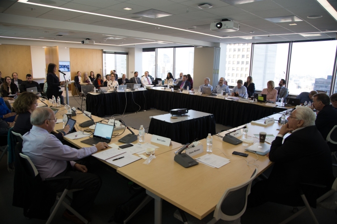
The CIRM Board discusses the future of the Stem Cell Agency

Budgets are very rarely exciting things; but they are important. For example, it’s useful for a family to know when they go shopping exactly how much money they have so they know how much they can afford to spend. Stem cell agencies face the same constraints; you can’t spend more than you have. Last week the CIRM Board looked at what we have in the bank, and set us on a course to be able to do as many of the things we want to, with the money we have left.

First some context. Last year CIRM spent a shade over $306 million on a wide range of research from Discovery, the earliest stage, through Translational and into Clinical trials. We estimate that is going to leave us with approximately $335 million to spend in the coming years.

A couple of years ago our Board approved a 5 year Strategic Plan that laid out some pretty ambitious goals for us to achieve – such as funding 50 new clinical trials. At the time, that many clinical trials definitely felt like a stretch and we questioned if it would be possible. We’re proving that it is. In just two years we have funded 26 new clinical trials, so we are halfway to our goal, which is terrific. But it also means we are in danger of using up all our money faster than anticipated, and not having the time to meet all our goals.

Doing the math

So, for the last couple of months our Leadership Team has been crunching the numbers and looking for ways to use the money in the most effective and efficient way. Last week they presented their plan to the Board.

It boiled down to a few options.

  • Keep funding at the current rate and run out of money by 2019
  • Limit funding just to clinical trials, which would mean we could hit our 50 clinical trial goal by 2020 but would not have enough to fund Discovery and Translational level research
  • Place caps on how much we fund each clinical trial, enabling us to fund more clinical trials while having enough left over for Discovery and Translational awards

The Board went for the third option for some good reasons. The plan is consistent with the goals laid out in our Strategic Plan and it supports Discovery and Translational research, which are important elements in our drive to develop new therapies for patients.

Finding the right size cap

Here’s a look at the size of the caps on clinical trial funding. You’ll see that in the case of late stage pre-clinical work and Phase 1 clinical trials, the caps are still larger than the average amount we funded those stages last year. For Phase 2 the cap is almost the same as the average. For Phase 3 the cap is half the amount from last year, but we think at this stage Phase 3 trials should be better able to attract funding from other sources, such as industry or private investors.

cap awards

Another important reason why the Board chose option three – and here you’ll have to forgive me for being rather selfish – is that it means the Administration Budget (which pays the salaries of the CIRM team, including yours truly) will be enough to cover the cost of running this research plan until 2020.

The bottom line is that for 2018 we’ll be able to spend $130 million on clinical stage research, $30 million for Translational stage, and $10 million for Discovery. The impact the new funding caps will have on clinical stage projects is likely to be small (you can see the whole presentation and details of our plan here) but the freedom it gives us to support the broad range of our work is huge.

And here is where to go if you are interested in seeing the different funding opportunities at CIRM.

UCLA scientists on track to develop a stem cell replacement therapy for Duchenne Muscular Dystrophy

Muscle cells generated by April Pyle’s Lab at UCLA.

Last year, we wrote about a CIRM-funded team at UCLA that’s on a mission to develop a stem cell treatment for patients with Duchenne muscular dystrophy (DMD). Today, we bring you an exciting update on this research just in time for the holidays (Merry Christmas and Happy Hanukkah and Kwanza to our readers!).

DMD is a deadly muscle wasting disease that primarily affects young boys and young men. The UCLA team is trying to generate better methods for making skeletal muscle cells from pluripotent stem cells to regenerate the muscle tissue that is lost in patients with the condition. DMD is caused by genetic mutations in the dystrophin gene, which codes for a protein that is essential for skeletal muscle function. Without dystrophin protein, skeletal muscles become weak and waste away.

In their previous study, the UCLA team used CRISPR gene editing technology to remove dystrophin mutations in induced pluripotent stem cells (iPSCs) made from the skin cells of DMD patients. These corrected iPSCs were then matured into skeletal muscle cells that were transplanted into mice. The transplanted muscle cells successfully produced dystrophin protein – proving for the first time that DMD mutations can be corrected using human iPSCs.

A Step Forward

The team has advanced their research a step forward and published a method for making skeletal muscle cells, from DMD patient iPSCs, that look and function like real skeletal muscle tissue. Their findings, which were published today in the journal Nature Cell Biology, address a longstanding problem in the field: not being able to make stem cell-derived muscle cells that are mature enough to model DMD or to be used for cell replacement therapies.

Dr. April Pyle, senior author on the study and Associate Professor at the Eli and Edythe Broad Center of Regenerative Medicine and Stem Cell Research at UCLA explained in a news release:

April Pyle, UCLA.

“We have found that just because a skeletal muscle cell produced in the lab expresses muscle markers, doesn’t mean it is fully functional. For a stem cell therapy for Duchenne to move forward, we must have a better understanding of the cells we are generating from human pluripotent stem cells compared to the muscle stem cells found naturally in the human body and during the development process.”

By comparing the proteins expressed on the cell surface of human fetal and adult muscle cells, the team identified two proteins, ERBB3 and NGFR, that represented a regenerative population of skeletal muscle cells. They used these two markers to isolate these regenerative muscle cells, but found that the muscle fibers they created in a lab dish were smaller than those found in human muscle.

First author, Michael Hicks, discovered that using a drug to block a human developmental signaling pathway called TGF Beta pushed these ERBB3/NGFR cells past this intermediate stage and allowed them to mature into functional skeletal muscle cells similar to those found in human muscle.

Putting It All Together

In their final experiments, the team combined the new stem cell techniques developed in the current study with their previous work using CRISPR gene editing technology. First, they removed the dystrophin mutations in DMD patient iPSCs using CRISPR. Then, they coaxed the iPSCs into skeletal muscle cells in a dish and isolated the regenerative cells that expressed ERBB3 and NGFR. Mice that lacked the dystrophin protein were then transplanted with these cells and were simultaneously given an injection of a TGF Beta blocking drug.

The results were exciting. The transplanted cells were able to produce human dystrophin and restore the expression of this protein in the Duchenne mice.

Skeletal muscle cells isolated using the ERBB3 and NGFR surface markers (right) restore human dystrophin (green) after transplantation significantly greater than previous methods (left). (Image courtesy of UCLA)

Dr. Pyle concluded,

“The results were exactly what we’d hoped. This is the first study to demonstrate that functional muscle cells can be created in a laboratory and restore dystrophin in animal models of Duchenne using the human development process as a guide.”

In the long term, the UCLA team hopes to translate this research into a patient-specific stem cell therapy for DMD patients. In the meantime, the team will use funding from a recent CIRM Quest award to make skeletal muscle cells that can regenerate long-term in response to chronic injury in hopes of developing a more permanent treatment for DMD.

The UCLA study discussed in this blog received funding from Discovery stage CIRM awards, which you can read more about here and here.

Stem Cell Stories that Caught Our Eye: GPS for Skin & Different Therapies for Aging vs. Injured Muscles?

Skin stem cells specialize into new skin by sensing neighborhood crowding
When embarking on a road trip, the GPS technology inside our smartphones helps us know where we are and how to get where we’re going. The stem cells buried in the deepest layers of our skin don’t have a GPS and yet, they do just fine determining their location, finding their correct destination and becoming the appropriate type of skin cell. And as a single organ, all the skin covering your body maintains the right density and just the right balance of skin stem cells versus mature skin cells as we grow from a newborn into adult.

crowdinginth

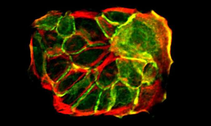
Skin cells growing in a petri dish (green: cytoskeleton, red: cell-cell junction protein).
Credit: MPI for Biology of Aging

This easily overlooked but amazing feat is accomplished as skin cells are continually born and die about every 30 days over your lifetime. How does this happen? It’s an important question to answer considering the skin is our first line of defense against germs, toxins and other harmful substances.

This week, researchers at the Max Planck Institute for Biology of Aging in Cologne, Germany reported a new insight into this poorly understood topic. The team showed that it all comes down to the skin cells sensing the level of crowding in their local environment. As skin stem cells divide, it puts the squeeze on neighboring stem cells. This physical change in tension on these cells “next door” triggers signals that cause them to move upward toward the skin surface and to begin maturing into skin cells.

Lead author Yekaterina Miroshnikova explained in a press release the beauty of this mechanism:

“The fact that cells sense what their neighbors are doing and do the exact opposite provides a very efficient and simple way to maintain tissue size, architecture and function.”

The research was picked up by Phys.Org on Tuesday and was published in Nature Cell Biology.

Stem cells respond differently to aging vs. injured muscle
From aging skin, we now move on to our aging and injured muscles, two topics I know oh too well as a late-to-the-game runner. Researchers at the Sanford Burnham Prebys Medical Discovery Institute (SBP) in La Jolla report a surprising discovery that muscle stem cells respond differently to aging versus injury. This important new insight could help guide future therapeutic strategies for repairing muscle injuries or disorders.

muscle stem cell

Muscle stem cell (pink with green outline) sits along a muscle fiber.
Image: Michael Rudnicki/OIRM

Muscle stem cells, also called satellite cells, make a small, dormant population of cells in muscle tissue that springs to life when muscle is in need of repair. It turns out that these stem cells are not identical clones of each other but instead are a diverse pool of cells.  To understand how the assortment of muscle stem cells might respond differently to the normal wear and tear of aging, versus damage due to injury or disease, the research team used a technology that tracks the fate of individual muscle stem cells within living mice.

The analysis showed a clear but unexpected result. In aging muscle, the muscle stem cells maintained their diversity but their ability to divide and grow declined. However, the opposite result was observed in injured muscle: the muscle stem cell diversity became limited but the capacity to divide was not affected. In a press release, team leader Alessandra Sacco explains the implications of these findings for therapy development:

sacco

Alessandra Sacco, PhD

“This study has shown clear-cut differences in the dynamics of muscle stem cell pools during the aging process compared to a sudden injury. This means that there probably isn’t a ‘one size fits all’ approach to prevent the decline of muscle stem cells. Therapeutic strategies to maintain muscle mass and strength in seniors will most likely need to differ from those for patients with degenerative diseases.”

This report was picked up yesterday by Eureka Alert and published in Cell Stem Cell.

jCyte Shares Encouraging Update on Clinical Trial for Retinitis Pigmentosa

Stepping out of the darkness into light. That’s how patients are describing their experience after participating in a CIRM-funded clinical trial targeting a rare form of vision loss called retinitis pigmentosa (RP). jCyte, the company conducting the trial, announced 12 month results for its candidate stem cell-based treatment for RP.

RP is a genetic disorder that affects approximately 1 in 40,000 individuals and 1.5 million people globally. It causes the destruction of the light-sensing cells at the back of the eye called photoreceptors. Patients experience symptoms of vision loss starting in their teenage years and eventually become legally blind by middle age. While there is no cure for RP, there is hope that stem cell-based therapies could slow its progression in patients.

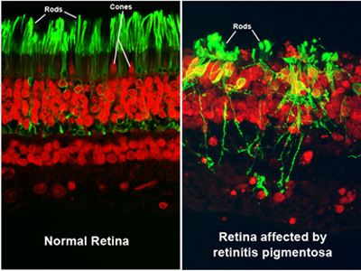
Photoreceptors look healthy in a normal retina (left). Cells are damaged in the retina of an RP patient (right). (Source National Eye Institute)

jCyte is one of the leaders in developing cell-based therapies for RP. The company, which was founded by UC Irvine scientists led by Dr. Henry Klassen, is testing a product called jCell, which is composed of pluripotent stem cell-derived progenitor cells that develop into photoreceptors. When transplanted into the back of the eye, they are believed to release growth factors that prevent further damage to the surviving cells in the retina. They also can integrate into the patient’s retina and develop into new photoreceptor cells to improve a patient’s vision.

Positive Results

At the Annual Ophthalmology Innovation Summit in November, jCyte announced results from its Phase 1/2a trial, which was a 12-month study testing two different doses of transplanted cells in 28 patients. The company reported a “favorable safety profile and indications of potential benefit” to patient vision.

The patients received a single injection of cells in their worst eye and their visual acuity (how well they can see) was then compared between the treated and untreated eye. Patients who received the lower dose of 0.5 million cells were able to see one extra letter on an eye chart with their treated eye compared to their untreated eye while patients that received the larger dose of 3 million cells were able to read 9 more letters. Importantly, none of the patients experienced any significant side effects from the treatment.

According to the company’s news release, “patient feedback was particularly encouraging. Many reported improved vision, including increased sensitivity to light, improved color discrimination and reading ability and better mobility. In addition, 22 of the 28 patients have been treated in their other eye as part of a follow-on extension study.”

One of these patients is Rosie Barrero. She spoke to us earlier this year about how the jCyte trial has not only improved her vision but has also given her hope. You can watch her video below.

Next Steps

These results suggest that the jCell therapy is safe (at least at the one year mark) to use in patients and that larger doses of jCell are more effective at improving vision in patients. jCyte CEO, Paul Bresge commented on the trial’s positive results:

Paul Bresge

“We are very encouraged by these results. Currently, there are no effective therapies to offer patients with RP. We are moving forward as quickly as possible to remedy that. The feedback we’ve received from trial participants has been remarkable. We look forward to moving through the regulatory process and bringing this easily-administered potential therapy to patients worldwide.”

Bresge and his company will be able to navigate jCell through the regulatory process more smoothly with the product’s recent Regenerative Medicine Advanced Therapy (RMAT) designation from the US Food and Drug Administration (FDA). The FDA grants RMAT to regenerative medicine therapies for serious diseases that have shown promise in early-stage clinical trials. The designation allows therapies to receive expedited review as they navigate their way towards commercialization.

jCyte is now evaluating the safety and efficacy of jCell in a Phase2b trial in a larger group of up to 85 patients. CIRM is also funding this trial and you can read more about it on our website.


Related Links:


A new study suggests CRISPR gene editing therapies should be customized for each patient

You know a scientific advance is a big deal when it becomes the main premise and title of a Jennifer Lopez-produced TV drama. That’s the case for CRISPR, a revolutionary gene-editing technology that promises to yield treatments for a wide range of genetic diseases.

In fact, clinical trials using the CRISPR method are already underway with more on the horizon. And at CIRM, we’re funding several CRISPR projects including a candidate gene and stem cell therapy that applies CRISPR to repair a genetic mutation found in sickle cell anemia patients.

geneeditingclip2

Animation by Todd Dubnicoff/CIRM

While these projects are moving full steam ahead, a study published this week in PNAS suggests a note of caution. They report that the natural genetic variability that is found when comparing  the DNA sequences of individuals has the potential to negatively impact the effectiveness of a CRISPR-based treatment and in some cases, could lead to dangerous side effects. As a result, the research team – a collaboration between Boston Children’s Hospital and the University of Montreal – recommends that therapy products using CRISPR should be customized to take into account the genetic variation between patients.

CRISPR 101
While other gene-editing methods pre-date CRISPR, the gene-editing technique has taken the research community by storm because of its ease of use. Pretty much any lab can incorporate it into their studies. CRISPR protein can cut specific DNA sequence within a person’s cells with the help of an attached piece of RNA. It’s pretty straight-forward to customize this “guide” RNA molecule so that it recognizes a desired DNA sequence that is in need of repair or modification.

https://player.vimeo.com/video/112757040

Because CRISPR activity heavily relies on the guide RNA molecule’s binding to a specific DNA sequence, there have been on-going concerns that a patient’s genetic variability could hamper the effectiveness of a given CRISPR therapy if it didn’t bind well. Even worse, if the genetic variability caused the CRISPR product to bind and inactivate a different region of DNA, say a gene responsible for suppressing cancer growth, it could lead to dangerous, so-called off target effects.

Although, studies have been carried out to measure the frequency of these potential CRISPR mismatches, many of the analyses depend on a reference DNA sequence from one individual. But as senior author Stuart Orkin, of Dana-Farber Boston Children’s Cancer and Blood Disorders Center, points out in a press release, this is not an ideal way to gauge CRISPR effectiveness and safety:

orkin

Stuart Orkin

“Humans vary in their DNA sequences, and what is taken as the ‘normal’ DNA sequence for reference cannot account for all these differences.”


One DNA sequence is not like the other
So, in this study, the research team analyzed previously published DNA sequence data from 7,444 people. And they focused on 30 disease genes that various researchers were targeting with CRISPR gene-editing. The team also generated 3,000 different guide RNAs with which to target those 30 disease genes.

The analysis showed that, in fact, about 50 percent of the guide RNAs could potentially have mismatches due to genetic variability found in these patients’ DNA sequences. These mismatches could lead to less effective binding of CRISPR to the disease gene target, which would reduce the effectiveness of the gene editing. And, though rare, the team also found cases in which an individual’s genetic variability could cause the CRISPR guide RNA to bind and cut in the wrong spot.

Matthew Canver, an MD-PhD student at Harvard Medical School who is also an author in the study, points out these less-than-ideal activities could also impact other gene editing techniques. Canver gives an overall recommendation how to best move forward with CRISPR-based therapy development:

canver, matthew

Matthew Canver

“The unifying theme is that all these technologies rely on identifying stretches of DNA bases very specifically. As these gene-editing therapies continue to develop and start to approach the clinic, it’s important to make sure each therapy is going to be tailored to the patient that’s going to be treated.”


Using the AIDS virus to help children battling a deadly immune disorder

Ronnie Kashyap, patient in SCID clinical trial: Photo Pawash Priyank

More than 35 million people around the world have been killed by HIV, the virus that causes AIDS. So, it’s hard to think that the same approach the virus uses to infect cells could also be used to help children battling a deadly immune system disorder. But that’s precisely what researchers at UC San Francisco and St. Jude Children’s Research Hospital are doing.

The disease the researchers are tackling is a form of severe combined immunodeficiency (SCID). It’s also known as ‘bubble baby’ disease because children are born without a functioning immune system and in the past were protected from germs within the sterile environment of a plastic bubble. Children with this disease often die of infections, even from a common cold, in the first two years of life.

The therapy involves taking the patient’s own blood stem cells from their bone marrow, then genetically modifying them to correct the genetic mutation that causes SCID. The patient is then given low-doses of chemotherapy to create space in their bone marrow for the news cells. The gene-corrected stem cells are then transplanted back into the infant, creating a new blood supply and a repaired immune system.

Unique delivery system

The novel part of this approach is that the researchers are using an inactivated form of HIV as a means to deliver the correct gene into the patient’s cells. It’s well known that HIV is perfectly equipped to infiltrate cells, so by taking an inactivated form – meaning it cannot infect the individual with HIV – they are able to use that infiltrating ability for good.

The results were announced at the American Society of Hematology (ASH) Annual Meeting and Exposition in Atlanta.

The researchers say seven infants treated and followed for up to 12 months, have all produced the three major immune system cell types affected by SCID. In a news release, lead author Ewelina Mamcarz, said all the babies appear to be doing very well:

“It is very exciting that we observed restoration of all three very important cell types in the immune system. This is something that’s never been done in infants and a huge advantage over prior trials. The initial results also suggest our approach is fundamentally safer than previous attempts.”

One of the infants taking part in the trial is Ronnie Kashyap. We posted a video of his story on our blog, The Stem Cellar.

If the stem cell-gene therapy combination continues to show it is both safe and effective it would be a big step forward in treating SCID. Right now, the best treatment is a bone marrow transplant, but only around 20 percent of infants with SCID have a sibling or other donor who is a good match. The other 80 percent have to rely on a less well-matched bone marrow transplant – usually from a parent – that can still leave the child prone to life-threatening infections or potentially fatal complications such as graft-versus-host disease.

CIRM is funding two other clinical trials targeting SCID. You can read about them here and here.