Stem cell stories that caught our eye: potential glaucoma therapy, Parkinson’s model, clinical trial list, cancer immune therapy

Here are some stem cell stories that caught our eye this past week. Some are groundbreaking science, others are of personal interest to us, and still others are just fun.

Stem cells may be option in glaucoma.  A few (potentially) blind mice did not run fast enough in an Iowa lab. But lucky for them they did not run into a farmer’s wife wielding a knife. Instead they had their eye sight saved by a team at the University of Iowa that corrected the plumbing in the back of their eyes with stem cells. They had a rodent version of glaucoma, which allows fluid to build up in the eye causing pressure that eventually damages the optic nerve and leads to blindness.

The fluid buildup results from a breakdown of the trabecular meshwork, a patch of cells that drains fluid from the eye. The Iowa researchers repaired that highly valuable patch with cells grown from iPS type stem cells created by reprogramming adult cells into an embryonic-like state. The trick with any early stage stem cell is getting it to mature into the desired tissue. This team pulled that off by growing the cells in a culture dish that had previously housed trabecular meshwork cells, which must have left behind some chemical signals that directed the growth of the stem cells.

The cells restored proper drainage in the mice. Also notable, the cells not only acted to replace damaged tissue directly, but they also seem to have summoned the eye’s own healing powers to do more repair. The research team also worked at the university affiliated Veterans Affairs Hospital, and the VA system issued a press release on the work published in the Proceedings of the National Academy of sciences, which was posted by Science Codex.

A “mini-brain” from a key area.   The brain is far from a uniform organ. Its many distinct divisions have very different functions. A few research teams have succeeded in coaxing stem cells into forming multi-layered clumps of cells referred to as “brain organoids” that mimic some brain activity, but those have generally been parts of the brain near the surface responsible for speech, learning and memory. Now a team in Singapore has created an organoid that shows activity of the mid-brain, that deep central highway for signals key to vision, hearing and movement.

The midbrain houses the dopamine nerves damaged or lost in Parkinson’s disease, so the mini-brains in lab dishes become immediate candidates for studying potential therapies and they are likely to provide more accurate results than current animal models.

 “Considering one of the biggest challenges we face in PD research is the lack of accessibility to the human brains, we have achieved a significant step forward. The midbrain organoids display great potential in replacing animals’ brains which are currently used in research,” said Ng Huck Hui of A*Star’s Genome Institute of Singapore where the research was conducted in a press release posted by Nanowerk.

The website Mashable had a reporter at the press conference in Singapore when the institute announce the publication of the research in Cell Stem Cell. They have some nice photos of the organoids as well as a microscopic image showing the cells containing a black pigment typical of midbrain cells, one of the bits of proof the team needed to show they created what they wanted.


Stem cell clinical trials listings.  Not a day goes by that I, or one of my colleagues, do not refer a desperate patient or family member—often several per day—to the web site clinicaltrials.gov. We do it with a bit of unease and usually some caveats but it is the only resource out there providing any kind of searchable listing of clinical trials. Not everything listed at this site maintained by the National Institutes of Health (NIH) is a great clinical trial. NIH maintains the site, and sets certain baseline criteria to be listed, but the agency does not vet postings.

Over the past year a new controversy has cropped up at the site. A number of for profit clinics have registered trials that require patients to pay many thousands of dollars for the experimental stem cell procedure.  Generally, in clinical trials, participation is free for patients. Kaiser Health News, an independent news wire supported by the Kaiser Family Foundation distributed a story this week on the phenomenon that was picked up by a few outlets including the Washington Post. But the version with the best links to added information ran in Stat, an online health industry portal developed by The Boston Globe, which has become one of my favorite morning reads.

The story leads with an anecdote about Linda Smith who went to the trials site to look for stem cell therapies for her arthritic knees. She found a listing from StemGenex and called the listed contact only to find out she would first have to pay $14,000 for the experimental treatment. The company told the author that they are not charging for participation in the posted clinical trial because it only covers the observation phase after the therapy, not the procedure itself. The reporter found multiple critics who suggested the company was splitting hairs a bit too finely with that explanation.

But the NIH came in for just as much criticism for allowing those trials to be listed at all. The web site already requires organizations listing trials to disclose information about the committees that oversee the safety of the patients in the trial, and critics said they should also demand disclosure of payment requirements, or outright ban such trials from the site.

Paul-Knoepfler-2013 “The average patient and even people in health care … kind of let their guard down when they’re in that database. It’s like, ‘If a trial is listed here, it must be OK,’” said Paul Knoepfler, a CIRM grantee and fellow blogger at the University of California, Davis. “Most people don’t realize that creeping into that database are some trials whose main goal is to generate profit.”

The NIH representative quoted in the article made it sound like the agency was open to making some changes. But no promises were made.

Added note 7/30. While this post factually describes an article that appeared in the mainstream media, the role of this column, I should add that while I did not take a position on paid trials, I am thrilled Stemgenex is collecting data and look forward to them sharing that data in a timely, peer-reviewed fashion.

Off the shelf T cells.  We at CIRM got some good news this week. We always like it when we see an announcement that technology from a researcher we have supported gets licensed to a company. That commercialization moves it a giant step closer to helping patients.

This week, Kite Pharma licensed a system developed in the lab of Gay Crooks at the University of California, Los Angeles, that creates an artificial thymus “organoid” in a dish capable of mass producing the immune system’s T cells from pluripotent stem cells. Just growing stem cells in the lab yields tiny amounts of T cells. They naturally mature in our bodies in the thymus gland, and seem to need that nurturing to thrive.

T-cell based immune therapy is all the rage now in cancer therapy because early trials are producing some pretty amazing results, and Kite is a leader in the field. But up until now those therapies have all been autologous—they used the patient’s own cells and manipulate them individually in the lab. That makes for a very expensive therapy. Kite sees the Crooks technology as a way to turn the procedure into an allogeneic one—using donor cells that could be pre-made for an “off-the-shelf” therapy. Their press release also envisioned adding some genetic manipulation to make the cells less likely to cause immune complications.

FierceBiotech published a bit more analysis of the deal, but we are not going to go into more detail on the actual science now. Crooks is finalizing publication of the work in a scientific journal, and when she does you can get the details here. Stay tuned.

How the Ice Bucket Challenge changed the fight against ALS

Ice Bucket2

200 people in Boston take the Ice Bucket Challenge: Photo courtesy Forbes

A couple of years ago millions of people did something they probably never thought they would: they dumped a bucket of ice cold water on their head to raise awareness about a disease most of them had probably never heard of, and almost certainly knew very little about.

The disease was ALS, also known as Lou Gehrig’s disease, and the Ice Bucket Challenge was something that went from a fun idea by a supporter of the ALS Association, to a blockbuster $220 million fundraiser. Like any good idea it sparked a backlash with critics accusing it of being a lazy way for people to feel good without actually doing anything, of diverting money from other charities, and even of just wasting water at a time of drought (at least here in California.)

But two years later we can now look back and see if those critics were correct, and if the money raised did make a difference. And the answer, I’m happy to say, is no and yes. In that order.

An article in the New Yorker magazine, by James Surowiecki, takes a look at what has happened since the Ice Bucket Challenge exploded on the scene and it has some good news:

  • Contributions to the ALS Association remain higher than before the Challenge
  • The average age of donors dropped from 50+ to 35
  • The Challenge may have helped spur an increase in overall donations to charity

All this is, of course, excellent news. But there’s an even more important point, which is that the money raised by the Challenge has helped advance ALS research further and faster than ever before.

Barbara Newhouse, the CEO of the ALS Association told Surowiecki:

“The research environment is dramatically different from what it was. We’re seeing research that’s really moving the needle not just on the causes of the disease but also on treatments and therapies.”

As an example Newhouse cites a study, published in Science  last summer, by researchers at Johns Hopkins that helped explain protein clumps found in the brains of people with ALS. Philip Wong, one of the lead authors of the study, says money raised by the Challenge helped make their work possible;

“Without it, we wouldn’t have been able to come out with the studies as quickly as we did. The funding from the ice bucket is just a component of the whole—in part, it facilitated our effort.”

And just this week the ALS Association said funding from the Challenge helped identify a gene connected to the disease.

Having been one of those who took a dunk for science – and we did ours early on, when the Challenge had only raised $4m – it’s nice to know something as silly and simple can have such a profound impact on developing treatments for a deadly disorder.


Cloning breakthrough: Dolly the sheep has sister clones and they’re healthy

On the topic of famous farm animals, a few come to mind: Babe the pig, Old Yeller, Mr. Ed, and the cast of Charlotte’s Web. Many of us grew up with these fictional characters and hold them near and dear to our heart, but what about real, living farm animals? The first that comes to my mind is Dolly the sheep.

Back in 1996, scientists made a major breakthrough when they cloned a sheep which they named after the famous singer and actress Dolly Parton. This famous sheep was born in a test tube – a product of a scientific process called somatic cell nuclear transfer (SCNT). It involves transferring the nucleus (which contains a cell’s genetic material) from an adult cell – a mammary gland cell in the case of Dolly – into an unfertilized egg cell that has had its own nucleus removed. Much like jumping a car, scientists use an electric shock to trigger the egg cell to divide and develop into an embryo that has the exact genetic makeup as the original organism it was derived from.

Are cloned animals healthy?

SCNT is a very inefficient process with a high failure rate during embryonic and fetal development. Dolly was a huge achievement for scientists as she was the first mammal to be successfully cloned using SCNT. Unfortunately, even though Dolly lived to the age of six and a half years, she wasn’t the healthiest of sheep. She suffered from a severe form of arthritis and tumors in her lungs and was eventually put down to relieve her from pain. Scientists hypothesized that the lung cancer was likely caused by a common virus that infects sheep, but they questioned whether some of Dolly’s other symptoms were caused by accelerated aging resulting from the cloning process.

Whether cloned animals are physically healthy and age normally are questions that have spurred much debate amongst scientists since Dolly’s inception. Further experiments have shown that cloned mammals that survive past their infancy are typically healthy, but some experiments in mice showed that cloned mice tended to be more obese, have diabetic symptoms, and live shorter lives. Concerns about the safety of cloning prompted many countries to ban reproductive cloning in mammals until more was known about the process.

Good news for Dolly’s sisters

Dolly’s 20th anniversary since her birth was earlier this year, and in celebration, many journals and news outlets wrote about the progress of SCNT and cloning over the past two decades. This week, a new study added an exciting new chapter to these recent stories about Dolly.

Published in Nature Communications, scientists from the University of Nottingham in Britain reported that cloned sheep are healthy and live normal lives. They studied 13 cloned sheep, four of which were Dolly’s sisters cloned from the same mammary gland cell line as Dolly. These sheep were between 7-9 years of age which is near the end of a healthy sheep’s average lifespan of 10 years.

Cloned sheep, sisters to the famous Dolly the Sheep. (University of Nottingham)

Cloned sheep, sisters to the famous Dolly the Sheep. (University of Nottingham)

The scientists wanted to know whether cloning had any negative impact on the health and lifespan of these sheep. Lead author on the study, Dr. Kevin Sinclair, explained to the Washington Post:

“When we did the study, these clones were already 2½ years older than Dolly was when she died. And they appeared to be perfectly healthy, but we wanted to see if they might be harboring subtle defects.”

They conducted studies that assessed symptoms typically caused by aging in both humans and sheep. These included tests for blood pressure, insulin sensitivity, arthritis, and heart disease. They also conducted MRI scans and X-rays to look at the integrity of their bones, joints, and muscles.

On the whole, the sheep were healthy and their tests yielded normal results. A few of the cloned sheep had early signs of arthritis, but their conditions were similar to normal non-cloned sheep of the same age. The scientists concluded that there were no obvious signs of premature aging in this group of cloned sheep and that the cloning process did not have negative effects on the health and lifespan of these animals.

“It was quite obvious that the concerns of Dolly just didn’t relate,” Sinclair said. “So you can’t extend beyond the Dolly experience and say this premature aging applies to all clones.”

Cloning breakthrough but questions remain about safety

This study, which many scientists are considering as a “breakthrough in cloning”, has received a lot of attention in the media from major news outlets like the New York Times, Washington Post, Statnews, and NPR.

The New York Times piece does a great job of discussing how the advancements in cloning could have positive impacts on reproductive technology, the farming industry (raising cloned farm animals as a food source), therapeutic development, and saving endangered species. But the article also balances this optimism with caution over the safety and ethics behind reproductive cloning. They posed the cloning safety question to Dr. Sinclair, the lead author on the study, whose response was positive but referenced the remaining issue of cloning being an inefficient process:

“If they [cloned sheep] could speak, they would say ‘yes; it’s perfectly safe. They’re perfectly healthy, and they’re old ladies now, and for them, their whole process worked perfectly. But there are others who struggled to adapt after birth.”

The STATNews piece also made a good point that further scientific studies on the cloned sheep need to be done to test for molecular signs of aging such as shortened telomeres, before the scientists can truly claim that these sheep are living normal healthy lives. The cloned sheep probably will live for another year at which point the scientists said they will conduct further experiments to look for other signs of aging at the cellular level.

Embryonic gene reverses old age in adult stem cells, in the lab

Getting old is an inevitable fact of life but what exactly causes it? One major hallmark of the aging process is cell senescence, in which cells gradually lose the ability to divide, leading to a breakdown in proper organ function. Adult stem cells that reside in our tissues usually spring into action to replenish cells lost to senescence (as well as injury and disease). But, unfortunately, senescence also affects stem cells, causing their natural regenerative capacity to diminish as we age.

gettingold

During the aging process, our stem cells gradually lose their regenerative potential (image source)

But what if we could tinker with senescence in these elderly stem cells? Could we slow down the aging process? A recent study by University of Buffalo scientists says yes, at least in a petri dish. Reporting in Stem Cells, the team shows that artificially activating the NANOG gene alone can reverse aging in adult mesenchymal stem cells (MSCs) and restore their full potential to form functional muscle tissue.

If you’re up on your induced pluripotent stem (iPS) cell knowledge, then you probably know that NANOG is a member of the “famous four”: the group of genes that can reprogram, say, a skin or blood cell, back into an embryonic stem cell-like state.  In this study, the research team derived human MSCs and mimicked senescence by allowing the cells to divide 12 to 16 times (Late Passage, or LP) in petri dishes and compared them to cells allowed to divide only a few times (Early Passage, or EP). The cells were genetically engineered to produce high levels of NANOG when the drug tetracycline was added to the cell culture.

First, the team looked at the impact of NANOG activation on various genes. They found that the activation level of several genes that had been suppressed in the senescent LP cells was restored to the levels seen in the pre-senescent EP cells. A closer look at the identity of those genes showed they were genes important for the capacity of a cell to develop into muscle and blood vessel which corresponds well with the MSCs potential to specialize into muscle and vascular tissue. Based on that genetic analysis, follow up experiments showed that NANOG indeed restored the senescent LP cells’ potential to develop into muscle and restore the muscle tissue’s contractile function.

Premature senescence is observed in diseases such as Hutchinson–Gilford Progeria Syndrome (HGPS), a fatal genetic disorder that causes rapid aging in childhood. NANOG was artificially activated in human MSCs, derived from a HGPS patient in this study, and also showed a restoration of the MSCs’ potential, as seen in the other donor cells.

In a university press release, lead author Stelios Andreadis, summarized the findings this way:

“Our research into Nanog is helping us to better understand the process of aging and ultimately how to reverse it.”


This work is very early days for this research especially given that these studies were performed in lab dishes and not animals. And because NANOG is a powerful gene that promotes embryonic and stem cell identity, the scientists will need to look into potential negative long term side effects for activating NANOG in adult stem cells. Ultimately, this path of research could uncover methods to treat aging-related diseases.

Out of the mouths, or in this case hearts, of babes comes a hopeful therapy for heart attack patients

Pediatric-Congenital-Heart-Disease-patient-300x200

Lessons learned from babies with heart failure could now help adults

Inspiration can sometimes come from the most unexpected of places. For English researcher Stephen Westaby it came from seeing babies who had heart attacks bounce back and recover. It led Westaby to a new line of research that could offer hope to people who have had a heart attack.

Westaby, a researcher at the John Radcliffe hospital in Oxford, England, found that implanting a novel kind of stem cell in the hearts of people undergoing surgery following a heart attack had a surprisingly significant impact on their recovery.

Westaby got his inspiration from studies showing babies who had a heart attack and experienced scarring on their heart, were able to bounce back and, by the time they reached adolescence, had no scarring. He wondered if it was because the babies’ own heart stem cells were able to repair the damage.

Scarring is a common side effect of a heart attack and affects the ability of the heart to be able to pump blood efficiently around the body. As a result of that diminished pumping ability people have less energy, and are at increased risk of further heart problems. For years it was believed this scarring was irreversible. This study, published in the Journal of Cardiovascular Translational Research, suggests it may not be.

Westaby and his team implanted what they describe as a “novel mesenchymal precursor (iMP)” type of stem cell in the hearts of patients who were undergoing heart bypass surgery following a heart attack. The cells were placed in parts of the heart that showed sizeable scarring and poor blood flow.

Two years later the patients showed a 30 percent improvement in heart function, a 40 percent reduction in scar size, and a 70 percent improvement in quality of life.

In an interview with the UK Guardian newspaper, Westaby admitted he was not expecting such a clear cut benefit:

“Quite frankly it was a big surprise to find the area of scar in the damaged heart got smaller,”

Of course it has to be noted that the trial was small, only involving 11 patients. Nonetheless the findings are important and impressive. Westaby and his team now hope to do a much larger study.

CIRM is funding a clinical trial with Capricor that is taking a similar approach, using stem cells to rejuvenate the hearts of patients who have had heart attacks.

Fred Lesikar, one of the patient’s in the first phase of that trial, experienced a similar benefit to those in the English trial and told us about it in our Stories of Hope.

Stem cell stories that caught our eye: turning on T cells; fixing our brains; progress and trends in stem cells; and one young man’s journey to recover from a devastating injury

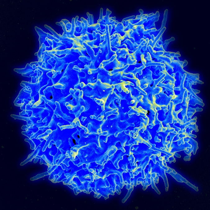
Healthy_Human_T_Cell

A healthy T cell

Here are some stem cell stories that caught our eye this past week. Some are groundbreaking science, others are of personal interest to us, and still others are just fun.

Directing the creation of T cells. To paraphrase the GOP Presidential nominee, any sane person LOVES, LOVES LOVES their T cells, in a HUGE way, so HUGE. They scamper around the body getting rid of viruses and the tiny cancers we all have in us all the time. A CIRM-funded team at CalTech has worked out the steps our genetic machinery must take to make more of them, a first step in letting physicians turn up the action of our immune systems.

We have known for some time the identity of the genetic switch that is the last, critical step in turning blood stem cells into T cells, but nothing in our body is as simple as a single on-off event. The Caltech team isolated four genetic factors in the path leading to that main switch and, somewhat unsuspected, they found out those four steps had to be activated sequentially, not all at the same time. They discovered the path by engineering mouse cells so that the main T cell switch, Bcl11b, glows under a microscope when it is turned on.

“We identify the contributions of four regulators of Bcl11b, which are all needed for its activation but carry out surprisingly different functions in enabling the gene to be turned on,” said Ellen Rothenberg, the senior author in a university press release picked up by Innovations Report. “It’s interesting–the gene still needs the full quorum of transcription factors, but we now find that it also needs them to work in the right order.”

Video primer on stem cells in the brain.  In conjunction with an article in its August issue, Scientific American posted a video from the Brain Forum in Switzerland of Elena Cattaneo of the University of Milan explaining the basics of adult versus pluripotent stem cells, and in particular how we are thinking about using them to repair diseases in the brain.

The 20-minute talk gives a brief review of pioneers who “stood alone in unmarked territory.” She asks how can stem cells be so powerful; and answers by saying they have lots of secrets and those secrets are what stem cell scientist like her are working to unravel.  She notes stem cells have never seen a brain, but if you show them a few factors they can become specialized nerves. After discussing collaborations in Europe to grow replacement dopamine neurons for Parkinson’s disease, she went on to describe her own effort to do the same thing in Huntington’s disease, but in this case create the striatal nerves lost in that disease.

The video closes with a discussion of how basic stem cell research can answer evolutionary questions, in particular how genetic changes allowed higher organisms to develop more complex nervous systems.

kelley and kent

CIRM Science Officers Kelly Shepard and Kent Fitzgerald

A stem cell review that hits close to home.  IEEE Pulse, a publication for scientists who mix engineering and medicine and biology, had one of their reporters interview two of our colleagues on CIRM’s science team. They asked senior science officers Kelly Shepard and Kent Fitzgerald to reflect on how the stem cell field has progressed based on their experience working to attract top researchers to apply for our grants and watching our panel of outside reviewers select the top 20 to 30 percent of each set of applicants.

One of the biggest changes has been a move from animal stem cell models to work with human stem cells, and because of CIRM’s dedicated and sustained funding through the voter initiative Proposition 71, California scientists have led the way in this change. Kelly described examples of how mouse and human systems are different and having data on human cells has been critical to moving toward therapies.

Kelly and Kent address several technology trends. They note how quickly stem cell scientists have wrapped their arms around the new trendy gene editing technology CRISPR and discuss ways it is being used in the field. They also discuss the important role of our recently developed ability to perform single cell analysis and other technologies like using vessels called exosomes that carry some of the same factors as stem cells without having to go through all the issues around transplanting whole cells.

“We’re really looking to move things from discovery to the clinic. CIRM has laid the foundation by establishing a good understanding of mechanistic biology and how stem cells work and is now taking the knowledge and applying it for the benefit of patients,” Kent said toward the end of the interview.

jake and family

Jake Javier and his family

Jake’s story: one young man’s journey to and through a stem cell transplant; As a former TV writer and producer I tend to be quite critical about the way TV news typically covers medical stories. But a recent story on KTVU, the Fox News affiliate here in the San Francisco Bay Area, showed how these stories can be done in a way that balances hope, and accuracy.

Reporter Julie Haener followed the story of Jake Javier – we have blogged about Jake before – a young man who broke his spine and was then given a stem cell transplant as part of the Asterias Biotherapeutics clinical trial that CIRM is funding.

It’s a touching story that highlights the difficulty treating these injuries, but also the hope that stem cell therapies holds out for people like Jake, and of course for his family too.

If you want to see how a TV story can be done well, this is a great example.

CIRM Board targets diabetes and kidney disease with big stem cell research awards

diabetes2

A recent study  estimated there may be more than 500 million people worldwide who have diabetes. That’s an astounding figure and makes diabetes one of the largest chronic disease epidemics in human history.

One of the most serious consequences of untreated or uncontrolled diabetes is kidney damage. That can lead to fatigue, weakness, confusion, kidney failure and even death. So two decisions taken by the CIRM Board today were good news for anyone already suffering from either diabetes or kidney disease. Or both.

The Board awarded almost $10 million to Humacyte to run a Phase 3 clinical trial of an artificial vein needed by people undergoing hemodialysis – that’s the most common form of dialysis for people with kidney damage. Hemodialysis helps clean out impurities and toxins from the blood. Without it waste will build up in the kidneys with devastating consequences.

The artificial vein is a kind of bioengineered blood vessel. It is implanted in the individual’s arm and, during dialysis, is connected to a machine to move the blood out of the body, through a filter, and then back into the body. The current synthetic version of the vein is effective but is prone to clotting and infections, and has to be removed regularly. All this puts the patient at risk.

Humacyte’s version – called a human acellular vessel or HAV – uses human cells from donated aortas that are then seeded onto a biodegradable scaffold and grown in the lab to form the artificial vein. When fully developed the structure is then “washed” to remove all the cellular tissue, leaving just a collagen tube. That is then implanted in the patient, and their own stem cells grow onto it, essentially turning it into their own tissue.

In earlier studies Humacyte’s HAV was shown to be safer and last longer than current versions. As our President and CEO, Randy Mills, said in a news release, that’s clearly good news for patients:

“This approach has the potential to dramatically improve our ability to care for people with kidney disease. Being able to reduce infections and clotting, and increase the quality of care the hemodialysis patients get could have a significant impact on not just the quality of their life but also the length of it.”

There are currently almost half a million Americans with kidney disease who are on dialysis. Having something that makes life easier, and hopefully safer, for them is a big plus.

The Humacyte trial is looking to enroll around 350 patients at three sites in California; Sacramento, Long Beach and Irvine.

While not all people with diabetes are on dialysis, they all need help maintaining healthy blood sugar levels, particularly people with type 1 diabetes. That’s where the $3.9 million awarded to ViaCyte comes in.

We’re already funding a clinical trial with ViaCyte  using an implantable delivery system containing stem cell-derived cells that is designed to measure blood flow, detect when blood sugar is low, then secrete insulin to restore it to a healthy level.

This new program uses a similar device, called a PEC-Direct. Unlike the current clinical trial version, the PEC-Direct allows the patient’s blood vessels to directly connect, or vasularize, with the cells inside it. ViaCyte believes this will allow for a more robust engraftment of the stem cell-derived cells inside it and that those cells will be better able to produce the insulin the body needs.

Because it allows direct vascularization it means that people who get the delivery system  will also need to get chronic immune suppression to stop their body’s immune system attacking it. For that reason it will be used to treat patients with type 1 diabetes that are at high risk for acute complications such as severe hypoglycemic (low blood sugar) events associated with hypoglycemia unawareness syndrome.

In a news release Paul Laikind, Ph.D., President and CEO of ViaCyte, said this approach could help patients most at risk.

“This high-risk patient population is the same population that would be eligible for cadaver islet transplants, a procedure that can be highly effective but suffers from a severe lack of donor material. We believe PEC-Direct could overcome the limitations of islet transplant by providing an unlimited supply of cells, manufactured under cGMP conditions, and a safer, more optimal route of administration.”

The Board also approved more than $13.6 million in awards under our Discovery program. You can see the winners here.


Researchers Identify Potential New Cell Source for Spinal Cord Injury Treatments

Now that Asterias Biotherapeutics’ CIRM-funded, stem cell-based clinical trial for spinal cord injury (SCI) has safely treated its first group of patients and begun recruiting the second, should other SCI researchers close up shop? Of course not. Since it’s a first-in-human trial, there certainly will be room for improvement even if the therapy proves successful. And it may not work for every SCI victim. So the development of other therapeutic approaches is critical to ensure effective treatments for all patients with this unmet medical need.

spinalcordinjury_bbcgraphic

Graphic of spinal cord injury site
(BBC via JACOPIN/BSIP/SCIENCE PHOTO LIBRARY)

Enter the lab of Michael Fehlings at the University of Toronto. Their recent Stem Cells Translational Medicine study describes a potential, minimally invasive therapeutic strategy which involves a type of brain cell not previously studied in the context of SCI.

In the case of the Asterias trial, embryonic stem cell-derived cells called oligodendrocytes are being transplanted directly into the injured spinal cord to help restore the disrupted nerve signals that cause a whole range of debilitating symptoms, including painful tingling and loss of movement in arms and legs, loss of bladder control and difficulty breathing.

Instead of trying to directly repair the disconnected nerve signals, Fehlings’ team looked at reducing the damaging effects of inflammation that occur at the site of injury in the days and weeks following the spinal cord trauma. This sounds like a perfect job for mesenchymal stem cells (MSCs) whose anti-inflammatory effects are well established. But previous animal studies using MSCs for spinal cord injury have had mixed results. Different sources of MSCs are known to have different anti-inflammatory actions so perhaps this is the culprit behind the variability. On top of that, the exact mechanism of action isn’t well understood which presents a barrier to getting FDA approval for clinical trials.

So the current study performed a careful comparative analysis of the healing effects of human cord blood MSCs and human brain vascular pericytes (HBVPs) – MSC-like cells found near blood vessels in the brain – in a rat model of spinal cord injury. Shortly after the SCI injury, the cells were delivered into the rats through the blood. The blood levels of various cytokines – proteins that modulate the inflammation response – were measured for several days. The only cytokine that increased in the days after the cell delivery of either cell type was IL-10 which is known for its anti-inflammatory effects.

Examining the spinal cord one to seven days after injury, the researchers found that both MSCs and HBVPs were better than controls at reducing hemorrhaging, with the HBVPs showing better improvement. In terms of long-term effects on functional behaviors, the researchers showed that after three weeks, grip strength, body coordination, and hind limb movement were most improved in the HBVPs.

In a university press release, Fehlings described these promising results:

fehlings

Michael Fehlings

“Our study demonstrates that these cells not only display a MSC phenotype in a dish, but also have similar immunomodulatory effects in animals after spinal cord injury that are more potent than those of non-central nervous system tissue-derived cells. Therefore, these cells are of interest for therapeutic use in acute spinal cord injury.”

A lot more work will be needed to translate these findings into clinical trials but for the sake of those suffering from spinal cord injury it’s encouraging that alternative approaches to treating this devastating, life-changing condition are in development.

Advancing Stem Cell Research at the CIRM Bridges Conference

Where will stem cell research be in 10 years?

What would you say to patients who wanted stem cell therapies now?

What are the most promising applications for stem cell research?

Why is it important for the government to fund regenerative medicine?

These challenging and thought-provoking questions were posed to a vibrant group of undergraduate and masters-level students at this year’s CIRM Bridges to Stem Cell Research and Therapy conference.

Educating the next generation of stem cell scientists

The Bridges program is one of CIRM’s educational programs that offers students the opportunity to take coursework at California state schools and community colleges and conduct stem cell research at top universities and industry labs. Its goal is to train the next generation of stem cell scientists by giving them access to the training and skills necessary to succeed in this career path.

The Bridges conference is the highlight of the program and the culmination of the students’ achievements. It’s a chance for students to showcase the research projects they’ve been working on for the past year, and also for them to network with other students and scientists.

Bridges students participated in a networking pitch event about stem cell research.

Bridges students participated in a networking pitch event about stem cell research.

CIRM kicked off the conference with a quick and dirty “Stem Cell Pitch” networking event. Students were divided into groups, given one of the four questions above and tasked with developing a thirty second pitch that answered their question. They were only given ten minutes to introduce themselves, discuss the question, and pick a spokesperson, yet when each team’s speaker took the stage, it seemed like they were practiced veterans. Every team had a unique, thoughtful answer that was inspiring to both the students and to the other scientists in the crowd.

Getting to the clinic and into patients

The bulk of the Bridges conference featured student poster presentations and scientific talks by leading academic and industry scientists. The theme of the talks was getting stem cell research into the clinic and into patients with unmet medical needs.

Here are a few highlights and photos from the talks:

On the clinical track for Huntington’s disease

Leslie Thompson, Professor at UC Irvine, spoke about her latest research in Huntington’s disease (HD). She described her work as a “race against time.” HD is a progressive neurodegenerative disorder that’s associated with multiple social and physical problems and currently has no cure. Leslie described how her lab is heading towards the clinic with human embryonic stem cell-derived neural (brain) stem cells that they are transplanting into mouse models of HD. So far, they’ve observed positive effects in HD mice that received human neural stem cell transplants including an improvement in the behavioral and motor defects and a reduction in the accumulation of toxic mutant Huntington protein in their nerve cells.

Leslie Thompson

Leslie Thompson

Leslie noted that because the transplanted stem cells are GMP-grade (meaning their quality is suitable for use in humans), they have a clear path forward to testing their potential disease modifying activity in human clinical trials. But before her team gets to humans, they must take the proper regulatory steps with the US Food and Drug Administration and conduct further experiments to test the safety and proper dosage of their stem cells in other mouse models as well as test other potential GMP-grade stem cell lines.

Gene therapy for SCID babies

Morton Cowan, a pediatric immunologist from UC San Francisco, followed Leslie with a talk about his efforts to get gene therapy for SCID (severe combined immunodeficiency disease) off the bench into the clinic. SCID is also known as bubble-baby disease and put simply, is caused by a lack of a functioning immune system. SCID babies don’t have normal T and B immune cell function and as a result, they generally die of infection or other conditions within their first year of life.

Morton Cowan

Morton Cowan, UCSF

Morton described how the gold standard treatment for SCID, which is hematopoietic or blood stem cell transplantation, is only safe and effective when the patient has an HLA matched sibling donor. Unfortunately, many patients don’t have this option and face life-threatening challenges of transplant rejection (graft-versus host disease). To combat this issue, Morton and his team are using gene therapy to genetically correct the blood stem cells of SCID patients and transplant those cells back into these patients so that they can generate healthy immune cells.

They are currently developing a gene therapy for a particularly hard-to-treat form of SCID that involves deficiency in a protein called Artemis, which is essential for the development of the immune system and for repairing DNA damage in cells. Currently his group is conducting the necessary preclinical work to start a gene therapy clinical trial for children with Artemis-SCID.

Treating spinal cord injury in the clinic

Casey Case, Asterias Biotherapeutics

Casey Case, Asterias Biotherapeutics

Casey Case, Senior VP of Research and Nonclinical Development at Asterias Biotherapeutics, gave an update on the CIRM-funded clinical trial for cervical (neck) spinal cord injury (SCI). They are currently testing the safety of transplanting different doses of their oligodendrocyte progenitor cells (AST-OPC1) in a group of SCI patients. The endpoint for this trial is an improvement in movement greater than two motor levels, which would offer a significant improvement in a patient’s ability to do some things on their own and reduce the cost of their healthcare. You can read more about these results and the ongoing study in our recent blogs (here, here).

Opinion: Scientists should be patient advocates

David Higgins gave the most moving speech of the day. He is a Parkinson’s patient and the Patient Advocate on the CIRM board and he spoke about what patient advocates are and how to become one. David explained how, these days, drug development and patient advocacy is more patient oriented and patients are involved at the center of every decision whether it be questions related to how a drug is developed, what side effects should be tolerated, or what risks are worth taking. He also encouraged the Bridges students to become patient advocates and understand what their needs are by asking them.

David Higgins, Parkinson's advocate and CIRM Board member

David Higgins

“As a scientist or clinician, you need to be an ambassador. You have a job of translating science, which is a foreign language to most people, and you can all effectively communicate to a lay audience without being condescending. It’s important to understand what patients’ needs are, and you’ll only know that if you ask them. Patients have amazing insights into what needs to be done to develop new treatments.”

Bridging the gap between research and patients

The Bridges conference is still ongoing with more poster presentations, a career panel, and scientific talks on discovery and translational stem cell research and commercializing stem cell therapies to all patients in need. It truly is a once in a lifetime opportunity for the Bridges students, many of whom are considering careers in science and regenerative medicine and are taking advantage of the opportunity to talk and network with prominent scientists.

If you’re interested in hearing more about the Bridges conference, follow us on twitter (@CIRMnews, @DrKarenRing, #CIRMBridges2016) and on Instagram (@CIRM_Stemcells).

CIRM-funded stem cell clinical trial for retinitis pigmentosa focuses on next stage

rp1

How retinitis pigmentosa erodes normal vision

The failure rate for clinical trials is depressingly high. A study from Tufts University in 2010  found that for small molecules – the substances that make up more than 90 percent of the drugs on the market today – the odds of getting from a Phase 1 trial to approval by the Food and Drug Administration are just 13 percent. For stem cell therapies the odds are even lower.

That’s why, whenever a stem cell therapy shows good results it’s an encouraging sign, particularly when that therapy is one that we at CIRM are funding. So we were more than a little happy to hear that Dr. Henry Klassen and his team at jCyte and the University of California, Irvine have apparently cleared the first hurdle with their treatment for retinitis pigmentosa (RP).

jCyte has announced that the first nine patients treated for RP have shown no serious side effects, and they are now planning the next phase of their Phase 1/2a safety trial.

In a news release Klassen, the co-founder of jCyte, said:

“We are pleased with the results. Retinitis pigmentosa is an incurable retinal disease that first impacts people’s night vision and then progressively robs them of sight altogether. This is an important milestone in our effort to treat these patients.”

The therapy involves injecting human retinal progenitor cells into one eye to help save the light sensing cells that are destroyed by the disease. This enables the researchers to compare the treated eye with the untreated eye to see if there are any changes or improvements in vision.

So far, the trial has undergone four separate reviews by the Data Safety Monitoring Board (DSMB), an independent group of experts that examines data from trials to ensure they meet all safety standards and that results show patients are not in jeopardy. Results from the first nine people treated are encouraging.

The approach this RP trial is taking has a couple of advantages. Often when transplanting organs or cells from one person into another, the recipient has to undergo some kind of immunosuppression, to stop their body rejecting the transplant. But earlier studies show that transplanting these kinds of progenitor cells into the eye doesn’t appear to cause any immunological response. That means patients in the study don’t have to undergo any immunosuppression. Because of that, the procedure is relatively simple to perform and can be done in a doctor’s office rather than a hospital. For the estimated 1.5 million people worldwide who have RP that could make getting treatment relatively easy.

Of course the big question now is not only was it safe – it appears to be – but does it work? Did any of those people treated experience improvements in their vision? We will share those results with you as soon as the researchers make them available.

Next step for the clinical trial is to recruit more patients, and treat them with a higher number of cells. There’s still a long way to go before we will know if this treatment works, if it either slows down, stops, or better still helps reverse some of the effects of RP. But this is a really encouraging first step.


Related links: