Here are some stem cell stories that caught our eye this past week. Some are groundbreaking science, others are of personal interest to us, and still others are just fun.
Video shows tumors growing. A team at the University of Iowa used video to capture breast cancer cells recruiting normal cells to the dark side where they help tumors grow.
Led by David Soll, the team reports that cancer cells secrete a cable that can reach out and actively grab other cells. Once the cable reaches another cell, it pulls it in forming a larger tumor.
“There’s nothing but tumorigenic cells in the bridge (between cells),” Soll said in a story in SciCasts, “and that’s the discovery. The tumorigenic cells know what they’re doing. They make tumors.”
They published their work in the American Journal of Cancer Research, and in a press release they suggested the results could provide an alternative to the theory that cancer stem cells are the engine of tumor growth. I would guess that before too long, someone will find a way to merge the two theories into one, more cohesive story of how cancer grows.
3-D home creates stem cells quicker. Using a 3-D gel to grow the cells, a Swiss team reprogrammed skin cells into iPS-type stem cells in half the time that it takes in a flat petri dish. Since these induced Pluripotent Stem cells have tremendous value now in research and potentially in the future treating of patients, this major improvement in a process that has been notoriously slow and inefficient is great news.
The senior researcher Matthias Lutoff from Polytechnique Federale explained that the 3-D environment gave the cells a home closer to the environment where they would grow in someone’s body. In an article in Healthline, he described the common method used today:
“What we currently have available is this two dimensional plastic surface that many, many stem cells really don’t like at all.”
At CIRM our goal is to get this research done as quickly as possible and to find ways to scale up any therapy so that it becomes practical to make it available to all patients who need it. Healthline quoted our CIRM scientist colleague Kevin Whittlesey on how the work would be a boon for stem cells scientists with its ability to shave months off the process of creating iPS cells.
Help for recent spinal cord injury. A team at Case Western Reserve University in Cleveland used the offspring of stem cells that they are calling multi-potent adult progenitor cells (MAPCs) to modulate the immune response after spinal cord injury. They wanted to preserve some of the role of the immune system in clearing debris after an injury but prevent any overly rambunctious activity that would result in additional damage to healthy tissue and scarring.

They published their work in Scientific Reports and at the web portal MD the senior researcher Jerry Silver described the project as targeting a specific immune cell, the macrophage, in the early days following stroke in mice:
“These were kinder, gentler macrophages. They do the job, but they pick and choose what they consume. The end result is spared tissue.”
The team injected the MAPCs into the mice one day after injury. Those cells were observed to go mostly to the spleen, which is know to be a reservoir for macrophages, and from their the MAPCs seemed to modulate the immune response.
“There was this remarkable neuroprotection with the friendlier macrophages,” Silver explained. “The spinal cord was just bigger, healthier, with much less tissue damage.”
Rundown on all the mini-organs. Regular readers of The Stem Cellar know researchers have made tremendous strides toward growing replacement organs from stem cells. You also know that with a few exceptions, like bladders and the esophagus, these are not ready for transplant into people.
Live Science web site does a fun rundown of progress with 11 different organs. They hit the more advanced esophagus and cover the early work on the reproductive tract, with items on fallopian tubes, vaginas and the penis. But most of the piece covers the early stage research that results in mini-organs, or as some have dubbed them, organoids. The author includes brain, heart, kidney, lung, stomach and liver. They also throw it the recent full ear grown on a scaffold.
Each short item comes with a photograph, mostly beautiful fluorescent microscopic images of cells forming the complex structures that become rudimentary organs.
3D printed human ear.
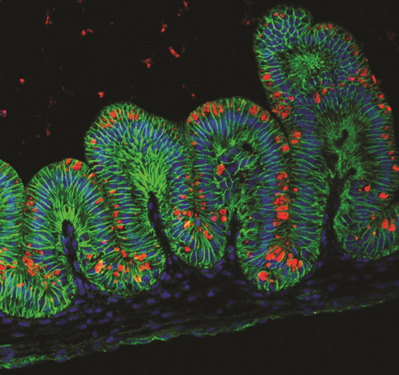
Mini-stomachs.
This past summer we wrote about an article on work at the University of Wisconsin on the many hurdles that have to be leapt to get actual replacement organs. Progress is happening faster that most of us expected, but we still have a quite a way to go.
