At birth, the human brain holds about 100 billion neurons. These nerve cells already form a complex network of connections essential for brain development and function.
During development, neural stem cells create neurons that migrate to their proper locations. Once there, neurons extend axons and dendrites, forming synaptic connections that let them communicate through electrical and chemical signals.
Early in development, neurons create far more synaptic connections than they need. From birth through early adulthood, the brain prunes weak or unnecessary connections. By your mid‑twenties, it has removed nearly half the synapses you started with.
This pruning process helps the brain refine its neural network and strengthen essential connections. It works much like a gardener trimming excess branches so the remaining ones grow stronger and produce better fruit.
The brain can make new neurons
Scientists once believed that synaptic pruning stopped by adulthood. But a new Salk Institute study, published in Nature Neuroscience and partly funded by CIRM, shows visual evidence that pruning continues in adults much like it does during development.

Salk Professor Rusty Gage led the study, known for his discovery in the late 1990s that the adult brain can generate new neurons. His work overturned the long‑held belief that the brain lacks stem cells and that we are born with all the neurons we will ever have.
In adults, two regions of the brain contain neural stem cells capable of producing new neurons. One of these regions is the dentate gyrus, part of the hippocampus, which is critical for forming memories. Gage and his team wanted to know whether new neurons in the dentate gyrus also undergo the same pruning process seen during early development.
Pruning the Adult Brain
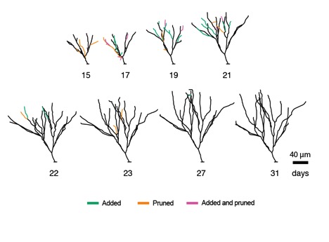
They developed a special microscope technique that allowed them to visually image the development of new neurons from stem cells in the dentate gyrus of the mouse brain. Every day, they imaged the growing neurons and monitored how many dendritic branches they sent out.

After observing the neurons for a few weeks, they were amazed to discover that these new neurons behaved similarly to neurons in the developing brain. They sent out dozens of dendritic branches and formed synaptic connections with other neurons, some of which were eventually pruned away over time.
This phenomenon was observed more readily when they made the mice exercise, which stimulated the stem cells in the dentate gyrus to divide and produce more neurons. These exercise-induced neurons robustly sent out dendritic branches only to have them pruned back later.
First author on the paper, Tiago Gonçalves commented on their observations:
“What was really surprising was that the cells that initially grew faster and became bigger were pruned back so that, in the end, they resembled all the other cells.”
Rusty Gage was also surprised by the findings but explained that developing neurons, whether in the developing or adult brain, have evolved this process to establish the best connections.
“We were surprised by the extent of the pruning we saw. The results suggest that there is significant biological pressure to maintain or retain the dendrite tree of these neurons.”
Potential new insights into brain disorders
This study is important because it increases our understanding of how neurons develop in the adult brain. Such knowledge can help scientists gain a better understanding of what goes wrong in brain disorders such as autism, schizophrenia, and epilepsy, where defects in how neurons form synaptic connections or how these connections are pruned are to blame.
Gonçalves also noted that this study raises another important question about regenerative medicine applications of stem cells for neurological disease.
“This also has big repercussions for regenerative medicine. Could we replace cells in this area of the brain with new stem cells and would they develop in the same way? We don’t know yet.”