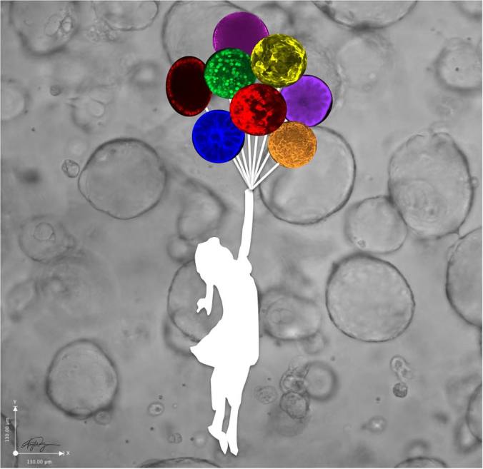
Creating a platform to help transplanted stem cells survive after a heart attack

heart

Developing new tools to repair damaged hearts

Repairing, even reversing, the damage caused by a heart attack is the Holy Grail of stem cell researchers. For years the Grail seemed out of reach because the cells that researchers transplanted into heart attack patients didn’t stick around long enough to do much good. Now researchers at Stanford may have found a way around that problem.

In a heart attack, a blockage cuts off the oxygen supply to muscle cells. Like any part of our body starved off oxygen the muscle cells start to die, and as they do the body responds by creating a layer of scars, effectively walling off the dead tissue from the surviving healthy tissue.  But that scar tissue makes it harder for the heart to effectively and efficiently pump blood around the body. That reduced blood flow has a big impact on a person’s ability to return to a normal life.

In the past, efforts to transplant stem cells into the heart had limited success. Researchers tried pairing the cells with factors called peptides to help boost their odds of surviving. That worked a little better but most of the peptides were also short-lived and weren’t able to make a big difference in the ability of transplanted cells to stick around long enough to help the heart heal.

Slow and steady approach

Now, in a CIRM-funded study published in the journal Nature Biomedical Engineering, a team at Stanford – led by Dr. Joseph Wu – believe they have managed to create a new way of delivering these cells, one that combines them with a slow-release delivery mechanism to increase their chances of success.

The team began by working with a subset of bone marrow cells that had been shown in previous studies to have what are called “pro-survival factors.” Then, working in mice, they identified three peptides that lived longer than other peptides. That was step one.

Step two involved creating a matrix, a kind of supporting scaffold, that would enable the researchers to link the three peptides and combine them with a delivery system they hoped would produce a slow release of pro-survival factors.

Step three was seeing if it worked. Using fluorescent markers, they were able to show, in laboratory tests, that unlinked peptides were rapidly released over two or three days. However, the linked peptides had a much slower release, lasting more than 15 days.

Out of the lab and into animals

While these petri dish experiments looked promising the big question was could this approach work in an animal model and, ultimately, in people. So, the team focused on cardiac progenitor cells (CPCs) which have shown potential to help repair damaged hearts, but which also have a low survival rate when transplanted into hearts that have experienced a heart attack.

The team delivered CPCs to the hearts of mice and found the cells without the pro-survival matrix didn’t last long – 80 percent of the cells were gone four days after they were injected, 90 percent were gone by day ten. In contrast the cells on the peptide-infused matrix were found in large numbers up to eight weeks after injection. And the cells didn’t just survive, they also engrafted and activated the heart’s own survival pathways.

Impact on heart

The team then tested to see if the treatment was helping improve heart function. They did echocardiograms and magnetic resonance imaging up to 8 weeks after the transplant surgery and found that the mice treated with the matrix combination had a statistically improved left ventricular function compared to the other mice.

Jayakumar Rajadas, one of the authors on the paper told CIRM that, because the matrix was partly made out of collagen, a substance the FDA has already approved for use in people, this could help in applying for approval to test it in people in the future:

“This paper is the first comprehensive report to demonstrate an FDA-compliant biomaterial to improve stem cell engraftment in the ischemic heart. Importantly, the biomaterial is collagen-based and can be readily tested in humans once regulatory approval is obtained.”


Researchers, beware: humanized mice not human enough to study stem cell transplants

A researcher’s data is only as good as the experimental techniques used to obtain those results. And a Stanford University study published yesterday in Cell Reports, calls into question the accuracy of a widely used method in mice that helps scientists gauge the human immune system’s response to stem cell-based therapies. The findings, funded in part by CIRM, urge a healthy dose of caution before using promising results from these mouse experiments as a green light to move on to human clinical trials.

Humanized mice aren’t quite human. Illustration: Pascal Gerard

Immune rejection of stem cell-based products is a major obstacle to translating these therapies from cutting-edge research into everyday treatments for the general population for people. If the genetic composition between the transplanted cells and the patient are mismatched, the patient’s immune system will see that cell therapy as foreign and will attack it. Unlike therapies derived from embryonic stem cells or from another person, induced pluripotent stem cells (iPSC) are exciting because scientists can potentially develop stem cell-based therapies from a patient’s own cells which relieves most of the immune rejection fears.

But manufacturing iPSC-derived therapies for each patient can take months, not to mention a lot of money, to complete. Some patients with life-threatening conditions like a heart attack or stroke don’t have the luxury of waiting that long. So even with these therapies, many researchers are working towards developing non-matched cell products which would be available “off-the-shelf. In all of these cases, immune-suppressing drugs would be needed which have their own set of concerns due to dangerous side effects, like serious infection or cancer. So, before testing in humans begins, it’s important to be able to test various immune-suppressing drugs and doses in animals to understand how well a stem cell-based therapy will survive once transplanted.

But how do you test a human immune response to a human cell product in an animal? Believe it or not, researchers – some of whom are authors in this Cell Reports publication – developed “humanized mice” back in the 1980’s. These mice were engineered to lack their own immune system to allow the engraftment of a human immune system. Over the years, advances in this mouse experimental system has gotten it closer and closer to imitating a human immune system response to transplantation of mismatched cell product.

Close but no cigar, it seems.

The team in the current study performed a detailed analysis of the immune response in two different strains of humanized mice. Both groups of animals did not mount a normal, healthy immune response and so they could not completely reject transplants of various human stem cells or stem cell-based products. Now, if you didn’t know about the abnormally weak immune response in these humanized mice, you might conclude that very little immunosuppression would be needed for a given cell therapy to keep a patient’s immune system in check. But conclusively making that interpretation is not possible, according to team lead Dr. Joseph Wu, director of Stanford’s Cardiovascular Institute:

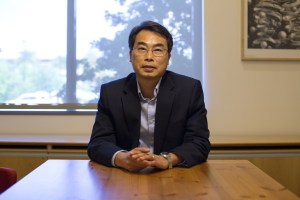
Joseph Wu. Photo: Steve Fisch/Stanford University

“In an ideal situation, these humanized mice would reject foreign stem cells just as a human patient would”, he said in a press release. “We could then test a variety of immunosuppressive drugs to learn which might work best in patients, or to screen for new drugs that could inhibit this rejection. We can’t do that with these animals.”

To uncover what was happening, the team took a step back and, rather than engrafting a human immune system into the mice, they engrafted immune cells from an unrelated mouse strain. Think of it as a mouse-ified mouse, if you will. When mouse iPSCs or human embryonic stem cells were transplanted into these mouse, the engrafted mouse immune system effectively rejected the stem cells. So, compared to these mice, some elements of the immune system in the humanized mouse strains are not quite capturing the necessary complexity to truly reproduce a human immune response.

More work will be needed to understand the underlying mechanisms of this difference. Other experiments in this study suggest that signals that inhibit the immune response may be elevated in the humanized mouse models. Dr. Leonard Shultz, a pioneer in the development of humanized mice at Jackson Laboratory and an author of this study, is optimistic about building a better model:

“The immune system is highly complex and there still remains much we need to learn. Each roadblock we identify will only serve as a landmark as we navigate the future. Already, we’ve seen recent improvements in humanized mouse models that foster enhancement of human immune function.”

Until then, the team urges other scientists to tread carefully when drawing conclusions from the humanized mice in use today.

Stem cell stories that caught our eye: drug safety for heart cells, worms hijack plant stem cells & battling esophageal cancer

Devising a drug safety measuring stick in stem cell-derived heart muscle cells
One of the mantras in the drug development business is “fail early”. That’s because most of the costs of getting a therapy to market occur at the later stages when an experimental treatment is tested in clinical trials in people. So, it’s best for a company’s bottom line and, more importantly, for patient safety to figure out sooner rather than later if a therapy has dangerous toxic side effects.

Researchers at Stanford reported this week in Science Translational Medicine on a method they devised that could help weed out cancer drugs with toxic effects on the heart before the treatment is tested in people.

In the lab, the team grew beating heart muscle cells, or cardiomyocytes, from induced pluripotent stem cells derived from both healthy volunteers and kidney cancer patients. A set of cancer drugs called tyrosine kinase inhibitors which are known to have a range of serious side effects on the heart, were added to the cells. The effect of the drugs on the heart cell function were measured with several different tests which the scientists combined into a single “safety index”.

roundup_wu

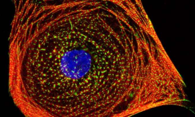
A single human induced pluripotent stem cell-derived cardiomyocyte. Cells such as these were used to assess tyrosine kinase inhibitors for cardiotoxicity in a high-throughput fashion. Credit: Dr. Arun Sharma at Dr. Joseph Wu’s laboratory at Stanford University

They found that the drugs previously shown to have toxic effects on patients’ hearts had the worst safety index values in the current study. And because these cells were in a lab dish and not in a person’s heart, the team was able to carefully examine cell activity and discovered that the toxic effects of three drugs could be alleviated by also adding insulin to the cells.

As lead author Joseph Wu, director of the Stanford Cardiovascular Institute, mentions in a press release, the development of this drug safety index could provide a powerful means to streamline the drug development process and make the drugs safer:

“This type of study represents a critical step forward from the usual process running from initial drug discovery and clinical trials in human patients. It will help pharmaceutical companies better focus their efforts on developing safer drugs, and it will provide patients more effective drugs with fewer side effects”

Worm feeds off of plants by taking control of their stem cells
In what sounds like a bizarre mashup of a vampire movie with a gardening show, a study reported this week pinpoints how worms infiltrate plants by commandeering the plants’ own stem cells. Cyst nematodes are microscopic roundworms that invade and kill soybean plants by sucking out their nutrients. This problem isn’t a trivial matter since nematodes wreak billions of dollars of damage to the world’s soybean crops each year. So, it’s not surprising that researchers want to understand how exactly these critters attack the plants.

nematode-feeding-site

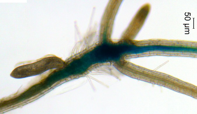
A nematode, the oblong object on the left, activates the vascular stem cell pathway in the developing nematode feeding site on a plant root. Credit: Xiaoli Guo, University of Missouri

Previous studies by Melissa Goellner Mitchum, a professor at the University of Missouri, had shown that the nematodes release protein fragments, called peptides, near a plant’s roots that help divert the flow of plant nutrients to the worm.

“These parasites damage root systems by creating a unique feeding cell within the roots of their hosts and leeching nutrients out of the soybean plant. This can lead to stunting, wilting and yield loss for the plant,” Mitchum explained in a press release.

In the current PLOS Pathogens study, Mitchum’s team identified another peptide produced by the nematode that is identical to a plant peptide that instructs stem cells to form the plant equivalent of blood vessels. This devious mimicking of the plant peptides is what allows the nematode to trick the plant stem cells into building vessels that reroute the plants’ nutrients directly to the worm.

Mitchum described the big picture implications of this fascinating discovery:

“Understanding how plant-parasitic nematodes modulate host plants to their own benefit is a crucial step in helping to create pest-resistant plants. If we can block those peptides and the pathways nematodes use to overtake the soybean plant, then we can enhance resistance for this very valuable global food source.”

Finding vulnerabilities in treatment-resistant esophageal cancer stem cells

diagram_showing_internal_radiotherapy_for_cancer_of_the_oesophagus_cruk_162-svg

Illustration of radiation therapy for esophageal cancer.
Credit: Cancer Research UK

The incidence of esophageal cancer has increased more than any other disease over the past 30 years. And while some patients respond well to chemotherapy and radiation treatment, most do not because the cancer becomes resistant to these treatments.

Focusing on cancer stem cells, researchers at Trinity College Dublin have identified an approach that may overcome treatment resistance.

Within tumors are thought to lie cancer stem cells that, just like stem cells, have the ability to multiply indefinitely. Even though they make up a small portion of a tumor, in some patients the cancer stem cells evade the initial rounds of treatment and are responsible for the return of the cancer which is often more aggressive. Currently, there’s no effective way to figure out how well a patient with esophageal cancer will response to treatment.

In the current study published in Oncotarget, the researchers found that a genetic molecule called miR-17 was much less abundant in the esophageal cancer stem cells. In fact, the cancer stem cells with the lowest levels of miR-17, were the most resistant to radiation therapy. The researchers went on to show that adding back miR-17 to the highly resistant cells made them sensitive again to the radiation. Niamh Lynam-Lennon, the study’s first author, explained in a press release that these results could have direct clinical applications:

“Going forward, we could use synthetic miR-17 as an addition to radiotherapy to enhance its effectiveness in patients. This is a real possibility as a number of other synthetic miR-molecules are currently in clinical trials for treating other diseases.”

Curing the Incurable through Definitive Medicine

“Curing the Incurable”. That was the theme for the first annual Center for Definitive and Curative Medicine (CDCM) Symposium held last week at Stanford University, in Palo Alto, California.

The CDCM is a joint initiative amongst Stanford Healthcare, Stanford Children’s Health and the Stanford School of Medicine. Its mission is to foster an environment that accelerates the development and translation of cell and gene therapies into clinical trials.

The research symposium focused on “the exciting first-in-human cell and gene therapies currently under development at Stanford in bone marrow, skin, cardiac, neural, pancreatic and neoplastic diseases.” These talks were organized into four different sessions: cell therapies for neurological disorders, stem cell-derived tissue replacement therapies, genome-edited cell therapies and anti-cancer cell-based therapies.

A few of the symposium speakers are CIRM-funded grantees, and we’ll briefly touch on their talks below.

Targeting cancer

The keynote speaker was Irv Weissman, who talked about hematopoietic or blood-forming stem cells and their value as a cell therapy for patients with blood disorders and cancer. One of the projects he discussed is a molecule called CD47 that is found on the surface of cancer cells. He explained that CD47 appears on all types of cancer cells more abundantly than on normal cells and is a promising therapeutic target for cancer.

Irv Weissman

Irv Weissman

“CD47 is the first gene whose overexpression is common to all cancer. We know it’s molecular mechanism from which we can develop targeted therapies. This would be impossible without collaborations between clinicians and scientists.”


At the end of his talk, Weissman acknowledged the importance of CIRM’s funding for advancing an antibody therapeutic targeting CD47 into a clinical trial for solid cancer tumors. He said CIRM’s existence is essential because it “funds [stem cell-based] research through the [financial] valley of death.” He further explained that CIRM is the only funding entity that takes basic stem cell research all the way through the clinical pipeline into a therapy.

Improving bone marrow transplants

judith shizuru

Judith Shizuru

Next, we heard a talk from Judith Shizuru on ways to improve current bone-marrow transplantation techniques. She explained how this form of stem cell transplant is “the most powerful form of cell therapy out there, for cancers or deficiencies in blood formation.” Inducing immune system tolerance, improving organ transplant outcomes in patients, and treating autoimmune diseases are all applications of bone marrow transplants. But this technique also carries with it toxic and potentially deadly side effects, including weakening of the immune system and graft vs host disease.

Shizuru talked about her team’s goal of improving the engraftment, or survival and integration, of bone marrow stem cells after transplantation. They are using an antibody against a molecule called CD117 which sits on the surface of blood stem cells and acts as an elimination signal. By blocking CD117 with an antibody, they improved the engraftment of bone marrow stem cells in mice and also removed the need for chemotherapy treatment, which is used to kill off bone marrow stem cells in the host. Shizuru is now testing her antibody therapy in a CIRM-funded clinical trial in humans and mentioned that this therapy has the potential to treat a wide variety of diseases such as sickle cell anemia, leukemias, and multiple sclerosis.

Tackling stroke and heart disease

img_1327We also heard from two CIRM-funded professors working on cell-based therapies for stroke and heart disease. Gary Steinberg’s team is using human neural progenitor cells, which develop into cells of the brain and spinal cord, to treat patients who’ve suffered from stroke. A stroke cuts off the blood supply to the brain, causing the death of brain cells and consequently the loss of function of different parts of the body.  He showed emotional videos of stroke patients whose function and speech dramatically improved following the stem cell transplant. One of these patients was Sonia Olea, a young woman in her 30’s who lost the ability to use most of her right side following her stroke. You can read about her inspiring recover post stem cell transplant in our Stories of Hope.

Dr. Joe Wu. (Image Source: Sean Culligan/OZY)

Dr. Joe Wu. (Image Source: Sean Culligan/OZY)

Joe Wu followed with a talk on adult stem cell therapies for heart disease. His work, which is funded by a CIRM disease team grant, involves making heart cells called cardiomyocytes from human embryonic stem cells and transplanting these cells into patient with end stage heart failure to improve heart function. His team’s work has advanced to the point where Wu said they are planning to file for an investigational new drug (IND) application with the US Food and Drug Administration (FDA) in six months. This is the crucial next step before a treatment can be tested in clinical trials. Joe ended his talk by making an important statement about expectations on how long it will take before stem cell treatments are available to patients.

He said, “Time changes everything. It [stem cell research] takes time. There is a lot of promise for the future of stem cell therapy.”

Stem cell stories that caught our eye: two studies of the heart and cool stem cell art

Here are some stem cell stories that caught our eye this past week. Some are groundbreaking science, others are of personal interest to us, and still others are just fun.

Image from Scope Blog.

Image from Scope Blog.

Understanding Heart Defects. Healthy heart tissue is made up of smooth, solid muscle, which is essential for normal heart function. Patients with a heart defect called left ventricular non-compaction (LVNC), lack normal heart tissue in their left ventricle – the largest, strongest blood-pumping chamber – and instead have spongy-looking tissue.

LVNC occurs during early heart development where pieces of heart muscle fail to condense (compact) and instead form an airy, sponge-like network that can leave patients at risk for heart failure and other complications.

A team at Stanford is interested in learning how LVNC occurs in humans, and they’re using human stem cells for the answer. Led by CIRM grantee Joe Wu, the scientists generated induced pluripotent stem cells (iPSCs) from four patients with LVNC. iPSCs are cells that can be turned into any other cell in the body, so Wu turned these cells into iPSC-derived heart muscle in a dish.

Wu’s team was particularly interested in determining why some LVNC patients have symptoms of disease while others seem perfectly normal. After studying the heart muscle cells derived from the four LVNC patients, they identified a genetic mutation in a gene called TBX20. This gene produces a type of protein called a cardiac transcription factor, which controls the expression of other heart related genes.

Upon further exploration, the scientists found that the genetic mutation in TBX20 prevented LVNC heart muscle cells from dividing at their normal rate. If they blocked the signal of mutant TBX20, the heart cells went back to their normal activity and created healthy looking heart tissue.

This study was published in Nature Cell Biology and covered by the Stanford Medicine Scope blog. In an interview with Scope, Joe Wu highlighted the big picture of their work:

Joseph Wu Stanford

Joseph Wu Stanford

“This study shows the feasibility of modeling such developmental defects using human tissue-specific cells, rather than relying on animal cells or animal models. It opens up an exciting new avenue for research into congenital heart disease that could help literally the youngest — in utero — patients.”

Stem Cell Heart Patch. Scientists from the University of Wisconsin, Madison are creating stem cell-based heart patches that they hope one day could be used to treat heart disease.

In a collaboration with Duke and the University of Alabama at Birmingham, they’re developing 3D stem cell-derived patches that contain the three main cell types found in the heart: cardiomyocytes (heart muscle cells), fibroblasts (support cells), and endothelial cells (cells that line the insides of blood vessels). These patches would be transplanted into heart disease patients to replace damaged heart tissue and improve heart function.

As with all research that has the potential for reaching human patients, the scientists must first determine whether the heart patches are safe in animal models. They plan to transplant the heart patches into a pig model – chosen because pigs have similar sized hearts compared to humans.

In a UW-Madison News release, the director of the UW-Madison Stem Cell and Regenerative Medicine Center Timothy Kamp, hinted at the potential for this technology to reach the clinic.

“The excitement here is we’re moving closer to patient applications. We’re at a stage when we need to see how these cells do in a large animal heart attack model. We’ll be making patches of heart muscle that can be applied to these injured areas.”

Kamp and his team still have a lot of work to do to perfect their heart patch technology, but they are thinking ahead. Two issues that they are trying to address are how to prevent a patient’s immune system from rejecting the heart patch transplant, and how to make sure the heart patches beat in sync with the heart they are transplanted into.

Check out the heart patches in action in this video:

(Video courtesy of Xiaojun Lian)

Cool Stem Cell Art! When I was a scientist, I worked with stem cells all the time. I grew them in cell culture dishes, coaxed them to differentiate into brain cells, and used a technique called immunostaining to take really beautiful, colorful pictures of my final cell products. I took probably thousands of pictures over my PhD and postdoc, but sadly, only a handful of these photos ever made it into journal publications. The rest collected dust either on my hard drive or in my lab notebook.

It’s really too bad that at the time I didn’t know about this awesome stem cell art contest called Cells I See run by the Centre for Commercialization of Regenerative Medicine (CCRM) in Ontario Canada and sponsored by the Stem Cell Network.

The contest “is about the beauty of stem cells and biomaterials, seen directly through the microscope or through the interpretive lens of the artist.” Scientists can submit their most prized stem cell images or art, and the winner receives a cash prize and major science-art street cred.

The submission deadline for this year’s contest was earlier this month, and you can check out the contenders on CCRM’s Facebook page. Even better, you can vote for your favorite image or art by liking the photo. The last date to vote is October 15th and the scientist whose image has the most likes will be the People’s Choice winner. CCRM will also crown a Grand Prize winner at the Till & McCulloch Stem Cell Meeting in October.

I’ll leave you with a few of my favorite photos, but please don’t let this bias your vote =)!

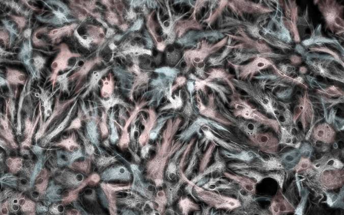
"Icy Astrocytes" by Samantha Yammine

“Icy Astrocytes” by Samantha Yammine (Vote here!)

"Reaching for organoids" by Amy Wong

“Reaching for organoids” by Amy Wong (Vote here!)

"Iris" by Sabiha Hacibekiroglu

“Iris” by Sabiha Hacibekiroglu (Vote here!)

CIRM Grantees Reflect on Ten Years of iPS Cells

For the fourth entry for our “Ten Years of Induced Pluripotent Stem (iPS) Cells” series, which we’ve been posting all month, I reached out to three of our CIRM grantees to get their perspectives on the impact of iPSC technology on their research and the regenerative medicine field as a whole:
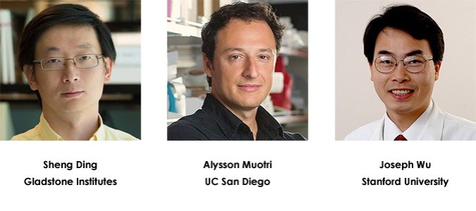
granteesStep back in time for us to August 2006 when the landmark Takahashi/Yamanaka Cell paper was published which described the successful reprogramming of adult skin cells into an embryonic stem cell-like state, a.k.a. induced pluripotent stem (iPS) cells. What do you remember about your initial reactions to the study?

Sheng Ding, MD, PhD
Senior Investigator, Gladstone Institute of Cardiovascular Disease
Shinya had talked about the (incomplete) iPS cell work well before his 2006 publication in several occasions, so seeing the paper was not a total surprise.

Alysson Muotri, PhD
Associate Professor, UCSD Dept. of Pediatrics/Cellular & Molecular Medicine
At that time, I was a postdoc. I was in a meeting when Shinya first presented his findings. I think he did not give the identity of the 4 factors at that time. I was very excited but remember hearing rumors in the corridors saying the data was too good to be true. Soon after, the publication come out and it was a lot of fun reading it.

Joseph Wu, MD, PhD
Director, Stanford Cardiovascular Institute
I remember walking to the parking lot after work. One of my colleagues called me on my cell phone and he asked if I had seen “the Cell paper” published earlier that day. I said I haven’t and I would look it up when I get back home. I read it that night and found it quite interesting because the concept was simple but yet powerful.

How soon after the publication did you start using the iPSC technique in your own research? At that time, what research questions were you able to start exploring that weren’t possible in the “pre-iPS” era?

Ding:
I think many of us in the (pluripotent stem cell) field quickly jumped on this seminal discovery and started working on the iPSC technology itself as, at the time, there were many aspects of the discovery that would need to be better understood and further improved for its applications.

Muotri:
Immediately after the first mouse Cell paper, but I started with human cells. There were some concerns if the 4 factors will also work in humans. Nonetheless, I start using the mouse cDNA factors in human cells and it worked! I was amazed to witness the transformation and see the iPSC colonies in my dish – I showed the results to everyone in the lab.

Soon after, the papers showing that the procedure worked in human cells were published but I already knew that. Thus, I started to apply this to model disease, my main focus. In 2010, we published the modeling of the first neurodevelopmental disease using the iPSC technology. It is still a landmark publication, and I am very happy to be among the pioneers who believed in the Yamanaka technology.

Wu:
We started working on iPS cells about a couple of months after the initial publication. To our surprise, it was incredibly easy to reproduce, and we were able to get successful clones after a few initial attempts, in part because we had already been working on human embryonic stem (ES) cells for several years.

I think the biggest advantage of iPS cells is that we can know the medical record of the donor. So we can study the correlation between the donor’s underlying genetic makeup and their resulting cellular and whole-body characteristics using iPS cells as a platform for integrating these analyses. Examining these correlations is simply not possible with ES cells since no adult donor exists.

Dr. Ding, what do you think made you and your research team especially skilled at pioneering the use of small molecules to replace the “Yamanaka” reprogramming factors?

Ding:
We had been working on identifying and using small molecules to modulate stem cell fate (including cell proliferation, differentiation, and reprogramming) before iPS cell technology was reported. So when the iPS cell work was reported, it was obvious to us that we could apply our expertise in small molecule discovery to better understand and improve iPS cell reprogramming and replace the genetic factors by pharmacological approaches.

Now, come back to the present and reflect on how the paper has impacted your research over the past 10 years. Describe some of the key findings your lab has made over the past 10 years through iPSC studies

Ding:
We’ve worked on three aspects that are related to iPS cell research: one is to identify small molecule drugs that can functionally replace the genetic reprogramming factors, and enhance reprogramming efficiency and iPS cell quality (to mitigate risks associated with genetic manipulation, to make the iPS cell generation process more robust and efficient, and reduce the cost etc).

Second is to better understand the reprogramming mechanisms, that would allow us to improve reprogramming and better utilize cellular reprogramming technology. For example, we had uncovered and characterized several fundamental mechanisms underlying the reprogramming process.

The third is to “repurpose/re-direct” the iPS cell reprogramming into directly generating tissue/organ-specific precursor cells without generating iPS cell (itself, which is tumorigenic and needs to be differentiated for most of its applications). This so-called “Cell-Activation and Signaling-Directed/CASD” reprogramming approach allowed us to directly generate cells in the brain, heart, pancreas, liver, and blood vessels.

Muotri:
My lab has focused on the use of iPS cells to model autism spectrum disorder, a condition that is very heterogeneous both clinically and genetically. Previous models for autism, such as animals and postmortem tissues, were limited because we could not have access to live neurons to test experimentally several hypotheses. Thus, the attractiveness of the iPS cell model, by capturing the genome of patients in pluripotent stem cells and then guide them to become neural networks.

While the modeling in a dish was a great potential, there were some clear limitations too: the variability in the system was too high for example. My lab has worked hard to develop a chemically-defined culture media (iDEAL) to grow iPS cells and reduce the variability in the system. Moreover, we have developed robust protocols to analyze the morphology and electrophysiological properties of cortical neurons derived from iPS cells. We have used these methods to learn more about how genes impact neuronal networks and to screen drugs for several diseases.

We also used these methods to create cerebral organoids or “mini-brains” in a dish and have applied this technology to test the impact of several genetic and environmental factors. For example, we recently showed that the Zika virus could target neural progenitor cells in these organoids, leading to defects in the human developing cortex. Without this technology, we would be limited to mouse models that do not recapitulate the microcephaly of the babies born in Brazil.

Wu:
Our lab has taken advantage of the iPS cell platform to better understand cardiovascular diseases and to advance the precision medicine initiative. For example, we have used iPS cells to elucidate the molecular mechanisms of diseases related to an enlarged heart, cardiac arrhythmias, viral- and chemotherapy-induced heart disease, the genetics of coronary artery disease, among other diseases. We have also used iPS cells for testing the safety and efficacy of various cardiovascular drugs (i.e., “clinical trial in a dish”).

How are your findings important in terms of accelerating stem cell treatments to patients with unmet medical needs?

Ding:
Better understanding the reprogramming process and developing small molecule drugs for enhancing reprogramming would allow more effective generation of safe stem cells with reduced cost for treating diseases or doing research.

Muotri:
We work with two concepts. First, we screen drugs that could repair the disorder at a cellular level in a dish, hoping these drugs will be useful for a large fraction of autistic individuals. This approach can also be used to stratify the autistic population, finding subgroups that are more responsive to a particular drug. This strategy should help future clinical trials.

In parallel, we also work with the idea of personalized medicine by using patient-derived cells to create “disease in a dish” models in the lab. We then examine the genomic information of these cells to help us find drugs that are more specific to that individual. This approach should allow us to better design the treatment, testing ideal drugs and dosage, before prescribing it to the patient.

Wu:
The iPS cell technology provides us with an unprecedented glimpse into cardiovascular developmental biology. With this knowledge, we should be able to better understand how cardiac and vascular cells regenerate in the heart during different phases of human life and also during times of stress such as in the case of a heart attack. However, to be able to translate this knowledge into clinical care for patients will take a significant amount of time. This is because we still need to tackle the issues of immunogenicity, tumorigenicity, and safety for products that are derived from ES and iPS cells. Equally importantly, we need to understand how transplanted cells integrate into the patient because based on our experience so far, most of the injected cells die upon transplant into the heart. Finally, the economics of this type of personalized regenerative medicine is a daunting challenge.

Finally, it’s foolhardy to predict the future but, just for fun, imagine that I revisit you in August 2026. What key iPSC-related accomplishments do you think your lab will achieve by then?

Ding:
We are hoping to have cell-based therapy and small molecule drugs developed based on iPS cell-related research for treating human diseases. Particularly, we are also hoping our cellular reprogramming research would lead us to identify and develop small molecule drugs that control tissue/organ regeneration in vivo [in an animal].

Muotri:
We hope to have improved several steps on the neural differentiation, dramatically reducing costs and increasing efficiency.

Wu:
We would like to use the iPS cell platform to discover several new drugs (or repurpose existing drugs) for our cardiovascular patients; to replace the current industry standard of drug toxicity testing using the hERG assay (which I believe is outdated); to predict what medications patients should be taking (i.e., precision cardiovascular medicine); and to elucidate risk index of genetic variants (in combination with genome editing approach).
Chemo-Induced Heart Failure: Using Stem Cells to Identify Those at Risk

The good news is you’re cancer free, the bad news is you need a heart transplant.

It almost sounds like the punchline to a joke, but it’s no laugher matter because the scenario is real for some cancer patients.  Chemotherapy is a life saver for many but certain doses can be so toxic that it’s often hard to tell which symptoms are due to the cancer and which are due to the drug. Doxorubicin, used to treat around 50% of people diagnosed with breast cancer, is particularly awful. It’s been estimated that about 8% of those treated with doxorubicin experience side effects to the heart with symptoms ranging from arrhythmias to congestive heart failure severe enough to require heart transplantation.

800px-Doxorubicin_3D_ball

doxorubicin, a chemotherapy drug that carries a risk of serious heart damage

Avoiding the fire after jumping out of the frying pan
To avoid this predicament, doctors need a way to screen for an increased risk of heart damage due to doxorubicin before a patient even sets foot in a chemotherapy clinic. A CIRM-funded Stanford research team has made a big step toward that goal. Reporting yesterday in Nature Medicine, the scientists describe a non-invasive laboratory method that could help pinpoint which breast cancer patients are most likely to experience so-called doxorubicin-induced cardiotoxicity, or DIC.

Eight woman with breast cancer who had received doxorubicin treatment were recruited for the study. Four suffered from DIC while the other four did not. Skin samples were obtained from each person as well as four healthy volunteers. In the lab, the skin fibroblasts were reprogrammed into embryonic-like induced pluripotent stem cells (iPS) and then specialized into beating heart muscle cells or cardiomyocytes.

Chemo-induced heart damage in a dish
To find out if these patient-derived heart cells in the lab reflect what happened inside the patients’ hearts, the team compared the effects of doxorubicin on the different groups of cells. Looking at cell survival and the rhythmic beating of the heart cells, differences emerged. Lead author Paul Burridge summarized the results in a university press release:

“We found that cells from the patients who had experienced doxorubicin toxicity responded more negatively to the presence of the drug. They beat more irregularly in response to increased levels of doxorubicin, and we saw a significant increase in cell death after 72 hours of exposure to the drug when we compared those cells to cells from healthy controls or patients who didn’t have heart damage.”

Screen Shot 2016-04-19 at 9.27.23 AM

iPS-derived heart muscle cells from patients without (DOX, first row) and with (DOXTOX, bottom row) doxorubicin toxicity were treated with increasing amounts of the doxorubicin. The regular green stripe patterns indicate normal, intact muscle structures. By 0.1 µM of drug (second column), the DOXTOX structures become disarrayed while the DOX cells remain intact. Image: Burridge et al. Nat Med. 2016 Apr 18.

So how exactly does doxorubicin wreak havoc on the heart and why are some patients more sensitive to the drug?

Feeling the burn of reactive oxygen species (ROS)
The answers, in part, lie inside cellular structures called mitochondria where calories, stored in the form of sugar and fat, are “burned” to generate the body’s energy needs.  A harmful byproduct of this energy metabolism is reactive oxygen species (ROS), a chemically reactive form of oxygen that damages the mitochondria and other cell components. This damage is especially bad for heart cells which are 35% mitochondria by volume due to their intense energy needs as they busily beat for a lifetime.

Now, earlier research studies had pointed to ROS production in mitochondria as a key deliverer of doxorubicin’s destructive effects on the hearts of chemotherapy patients. So the Stanford team investigated the drug’s effects on ROS production and on mitochondria function in context of their patient derived heart cells. In response to doxorubicin, the scientists found that the cells from patients with doxorubicin induced heart damage generated more ROS compared to the cells from patients who had no heart damage. Along with the higher ROS production, mitochondria function was more compromised in the doxorubicin-sensitive heart cells.

And even in the absence of treatment, there was a lower baseline function and quantity of mitochondria in the doxorubicin-sensitive cells. These results suggest some underlying genetic differences in the heart muscle cells of patients with DIC. The team plans to perform DNA comparisons to pinpoint the genes involved and ultimately help patients survive cancer without the fear of swapping it for another life threatening illness.

iPS cells: opening new paths for helping cancers patients
Compared to tools he had previously relied on, Joseph Wu, the team leader and director of the Stanford’s Cardiovascular Institute, is very excited about his lab’s future research possibilities:

“In the past, we’ve tried to model this doxorubicin toxicity in mice by exposing them to the drug and then removing the heart for study. Now we can continue our studies in human cells with iPS-derived heart muscle cells from real patients. One day we may even be able to predict who is likely to get into trouble.”

Stem Cells become Tool to Screen for Drugs; Fight Dangerous Heart Infections.

A Stanford study adds a powerful example to our growing list of diseases that have yielded their secrets to iPS-type stem cells grown in a dish. These “disease-in-a-dish” models have become one of the most rapidly growing areas of stem cell science. But this time they did not start with skin from a patient with a genetic disease and see how that genetic defect manifests in cells in a dish. Instead they started with normal tissue and looked at how the resulting cells reacted to viral infection.

They were looking at a nasty heart infection called viral myocarditis, which can begin to cause damage to heart muscle within hours and often leads to death. Existing antiviral drugs have only a modest impact on reducing these infections. So even though there is an urgent need to find better drugs, animal models have not proven very useful and there is no ready supply of human heart tissue for lab study.

To create a ready supply of human heart tissue Joseph Wu’s CIRM-funded team at Stanford started with skin samples from three healthy donors, reprogrammed them into iPS cells and then matured those into heart muscle tissue. Then they took one of the main culprits of this infection, coxsackievirus, and labeled it with a fluorescent marker so they could track its activity in the heart cells.

They were able to verify that the virus infected the cells in a dish just as they do in normal heart tissue. And when they tried treating the cells with four existing antiviral drugs they saw the same modest decrease in the rate of infected cells seen in patients. For one of the drugs that had been shown to cause some heart toxicity, they also saw some damage to the cells in the dish.

They propose that their model can now be used to screen thousands of compounds for potentially more effective and safer drugs. They published their results in Circulation Research July 15.

What a Difference Differentiation can Make: a Little Change can Reduce the Risk of Rejection

No one likes to be rejected. It hurts. But while rejection is something most of us experience at least a couple of times in our life, researchers at Stanford have found a way to reduce the risk of rejection, at least when it comes to one form of stem cells. Reporting in the latest issue of Nature Communications they have found that turning iPS or induced pluripotent stem cells into other, more specialized kinds of cells, can reduce the risk the immune system will attack them.

Our immune systems are things of beauty. They hunt out invaders like viruses and when they spot something that shouldn’t be there, they attack it. It’s a critical part of our body’s way of fighting off disease and staying healthy.

However, the immune system is not perfect. Researchers have known for some time that when you take skin from an individual and turn it into an iPS cell – one that is capable of turning into any other cell in the body – and then transplant that cell back into the same individual their immune system often attacks it. So far, the only individuals this has been done with are mice, but we assume the same would happen in people.

So Joseph Wu and his CIRM-funded Stanford team decided to see what would happen if they took those same iPS cells and, before transplanting them back into the individual they came from, turned them into a more specialized form of cell. The results were encouraging: the immune system didn’t wage an attack.

Dr. Joseph Wu, Stanford University School of Medicine

Dr. Joseph Wu, Stanford University School of Medicine


Researchers were not sure why the body would attack something that was created from its own tissue, but speculated that turning ordinary skin cells into iPS cells created a kind of cell that the immune system hadn’t seen before, or at least hadn’t seen since it since it was an embryo.

In the Stanford news release, Wu said this finding could be really important in helping avoid rejection in organ or other tissue transplants:

“Induced pluripotent stem cells have tremendous potential as a source for personalized cellular therapeutics for organ repair. This study shows that undifferentiated iPS cells are rejected by the immune system upon transplantation in the same recipient, but that fully differentiating these cells allows for acceptance and tolerance by the immune system without the need for immunosuppression.” 

The team first transplanted some iPS cells into genetically identical recipient mice. The transplants were rejected and within 42 days there were no signs that any cells had survived.

Then they took the same kind of iPS cell and differentiated, or ‘re-programmed,’ them so that they turned into endothelial cells, the kind found in the inner lining of blood vessels. Then they transplanted those cells into the mice. At the same time they took some of the mice’s own endothelial cells out, and transplanted them back into genetically identical mice to see how they would compare. Both sets of cells, the iPS-turned-into endothelial and the endothelial cells, survived for at least 63 days after transplantation.

When the researchers repeated the experiment and examined the areas where the cells had been transplanted, they found much greater signs of immune system activity in the mice that were given iPS cells compared to the mice who got iPS cells that had been turned into endothelial cells, and the mice that just got endothelial cells.

For Wu, the bottom line was simple:

“This study certainly makes us optimistic that differentiation — into any nonpluripotent cell type — will render iPS cells less recognizable to the immune system. We have more confidence that we can move toward clinical use of these cells in humans with less concern than we’ve previously had.” 

 We work closely with Joseph Wu and his team on a number of other different projects, most focusing on heart disease.

kevin mccormack