Stem Cell Agency Board Invests in Therapy Targeting Deadly Blood Cancers

THIS BLOG IS ALSO AVAILABLE AS AN AUDIO CAST

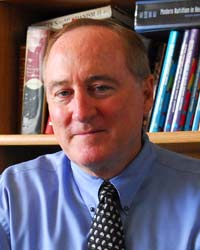
Dr. Ezra Cohen, photo courtesy UCSD

Hematologic malignancies are cancers that affect the blood, bone marrow and lymph nodes and include different forms of leukemia and lymphoma. Current treatments can be effective, but in those patients that do not respond, there are few treatment options. Today, the governing Board of the California Institute for Regenerative Medicine (CIRM) approved investing $4.1 million in a therapy aimed at helping patients who have failed standard therapy.

Dr. Ezra Cohen, at the University of California San Diego, and Oncternal Therapeutics are targeting a protein called ROR1 that is found in B cell malignancies, such as leukemias and lymphomas, and solid tumors such as breast, lung and colon. They are using a molecule called a chimeric antigen receptor (CAR) that can enable a patient’s own T cells, an important part of the immune system, to target and kill their cancer cells. These cells are derived from a related approach with an antibody therapy that targets ROR1-binding medication called Cirmtuzumab, also created with CIRM support. This CAR-T product is designed to recognize and kill cancer stem cells that express ROR1.

This is a late-stage preclinical project so the goal is to show they can produce enough high-quality cells to treat patients, as well as complete other regulatory measures needed for them to apply to the US Food and Drug Administration (FDA) for permission to test the therapy in a clinical trial in people.

If given the go-ahead by the FDA the therapy will target patients with chronic lymphocytic leukemia (CLL), mantle cell lymphoma (MCL) and acute lymphoblastic leukemia (ALL).  

“CAR-T cell therapies represent a transformational advance in the treatment of hematologic malignancies,” says Dr. Maria T. Millan, CIRM’s President and CEO. “This approach addresses the need to develop new therapies for patients whose cancers are resistant to standard chemotherapies, who have few therapeutic options and a very poor chance or recovery.”

Blocking pancreatic cancer stem cells

John Cashman

Cancer stem cells are one of the main reasons why cancers are able to survive surgery, chemotherapy and radiation. They are able to hide from those therapies and, at a future date, emerge and spread the cancer in the body once again.

Jionglia Cheng, PhD.

Jionglia Cheng, PhD., the lead author of a new CIRM-funded study, says that’s one of the reasons why pancreatic cancer has proved so difficult to treat.

“Pancreatic cancer remains a major health problem in the United States and soon will be the second most common cause of mortality due to cancer. A majority of pancreatic cancer patients are often resistant to clinical therapies. Thus, it remains a challenge to develop an efficacious clinically useful pancreatic cancer therapy.”

Dr. Cheng, a researcher with ChemRegen Inc., teamed up with John Cashman at the Human BioMolecular Research Institute and identified a compound, that seems to be effective in blocking the cancer stem cells.

In earlier studies the compound, called PAWI-2, demonstrated effectiveness in blocking breast, prostate and colon cancer. When tested in the laboratory PAWI-2 showed it was able to kill pancreatic cancer stem cells, and also was effective in targeting drug-resistant pancreatic cancer stem cells.

In addition, when PAWI-2 was used with a drug called erlotinib (brand name Tarceva) which is commonly prescribed for pancreatic cancer, the combination proved more effective against the cancer stem cells than erlotinib alone.

In a news release Dr. Cheng said: “In the future, this molecule could be used alone or with other chemotherapy albeit at lower doses, as a new therapeutic drug to combat pancreatic cancer. This may lead to much less toxicity to the patient,”

The study is published in the journal Scientific Reports.

First patient treated for colon cancer using reprogrammed adult cells

Dr. Sandip Patel (left) and Dr. Dan Kaufman (center) of UC San Diego School of Medicine enjoy a light-hearted moment before Derek Ruff (right) receives the first treatment for cancer using human-induced pluripotent stem cells (hiPSCs). Photo courtesy of UC San Diego Health.

For patients battling cancer for the first time, it can be quite a draining and grueling process. Many treatments are successful and patients go into remission. However, there are instances where the cancer returns in a much more aggressive form. Unfortunately, this was the case for Derek Ruff.

After being in remission for ten years, Derek’s cancer returned as Stage IV colon cancer, meaning that the cancer has spread from the colon to distant organs and tissues. According to statistics from Fight Colorectal Cancer, colorectal cancer is the 2nd leading cause of cancer death among men and women combined in the United States. 1 in 20 people will be diagnosed with colorectal cancer in their lifetime and it is estimated that there will be 140,250 new cases in 2019 alone. Fortunately, Derek was able to enroll in a groundbreaking clinical trial to combat his cancer.

In February 2019, as part of a clinical trial at the Moores Cancer Center at UC San Diego Health in collaboration with Fate Therapeutics, Derek became the first patient in the world to be treated for cancer with human-induced pluripotent stem cells (hiPSCs). hiPSCs are human adult cells, such as those found on the skin, that are reprogrammed into stem cells with the ability to turn into virtually any kind of cell. In this trial, hiPSCs were reprogrammed into natural killer (NK) cells, which are specialized immune cells that are very effective at killing cancer cells, and are aimed at treating Derek’s colon cancer.

A video clip from ABC 10 News San Diego features an interview with Derek and the groundbreaking work being done.

In a public release, Dr. Dan Kaufman, one of the lead investigators of this trial at UC San Diego School of Medicine, was quoted as saying,

“This is a landmark accomplishment for the field of stem cell-based medicine and cancer immunotherapy. This clinical trial represents the first use of cells produced from human induced pluripotent stem cells to better treat and fight cancer.”

In the past, CIRM has given Dr. Kaufman funding related to the development of NK cells. One was a $1.9 million grant for developing a different type of NK cell from hiPSCs, which could also potentially treat patients with lethal cancers. The second grant was a $4.7 million grant for developing NK cells from human embryonic stem cells (hESCs) to potentially treat patients with acute myelogenous leukemia (AML).

In the public release, Dr. Kaufman is also quoted as saying,

“This is a culmination of 15 years of work. My lab was the first to produce natural killer cells from human pluripotent stem cells. Together with Fate Therapeutics, we’ve been able to show in preclinical research that this new strategy to produce pluripotent stem cell-derived natural killer cells can effectively kill cancer cells in cell culture and in mouse models.”

Second “Don’t Eat Me” Signal Identified in Cancer Cells, Points to New Immunotherapies

When the immune system comes up as a topic in everyday conversation, it’s usually related to fighting off a cold or flu. While our immune cells certainly do detect and neutralize invading bacteria and viruses, they also play a critical role in killing abnormal, cancerous cells from within our bodies.

“Don’t Eat Me” Signal 101
A white blood cell called a macrophage (macro = “big”; phage = “eater”) is part of the so-called innate immune system and acts as a first line of defense by patrolling our organs and gobbling up infected as well as cancerous cells (see macrophages in action in the cool video below).

Unfortunately, cancer cells possess the ability to cloak themselves and escape a macrophage’s engulfing grasp. Nearly all cancer cells carry a protein called CD47 on their surface. When CD47 binds to a protein called SIRPalpha on the surface of macrophages, a “don’t eat me” signal is triggered and the macrophage ignores the cancer cell.

Stanford researcher Irv Weissman and his team discovered this “don’t eat me” signal several years ago and showed that adding an antibody protein that binds tightly to CD47 interferes with the CD47/SIRPalpha signal. As a result, the anti-CD47 antibody deactivates the cancer cell’s “don’t eat me” signal and restores the macrophage’s ability to detect and kill the cancer cells.

cd47-gene3

CD47 protein on surface of cancer cells triggers “don’t eat me signal” which can be blocked with anti-CD47 antibody. Image: Acrobiosystems

Because CD47 is found on the surface of most cancer cells, this anti-CD47 antibody represents an exciting new strategy for targeting cancer stem cells – the cells thought to maintain cancer growth and cause tumor relapse – in a wide variety of cancers. In fact, CIRM has provided funding for three clinical trials, one sponsored by Stanford University and two by Forty-Seven Inc. (a company that was spun out of Stanford), that are testing anti-CD47 therapy for the treatment of the blood cancer acute myeloid leukemia (AML), as well as colon cancer and other solid tumors.

“Reaching Clinical Trials” does not equal “The Research is Done”
Although these clinical trials are underway, the Weissman team continues to seek new insights related to blocking the CD47 “don’t eat me” signal. They observed that although anti-CD47 led to increased macrophage-induced killing of most cancer cell samples tested, some were resistant to anti-CD47 and remained cloaked from macrophages. And even the cancer cells that did respond to the antibody varied widely in the amount of increased killing by macrophages.

These results suggested that alternate processes may exist that allow some cancers to evade macrophages even when the CD47 “don’t eat me” signal is blocked. In a report published this week in Nature Immunology, the researchers report the identification of a second, independent “don’t eat me” signal, which may lead to more precise methods to disarm a cancer’s evasiveness.

To track down this alternate “don’t eat me” signal, they looked for, but didn’t find, correlations between specific types of cancer cells and the cancer’s resistance to anti-CD47 treatment.  So instead they analyzed surface proteins found on the various cancer cell samples and found that cancer cells that had high levels of MHC (Major Histocompatibility Complex) class I proteins were more likely to be resistant to anti-CD47 antibodies.

A Second “Don’t Eat Me” Signal
MHC class I proteins help another arm of the immune system, the adaptive immune response, detect what’s going inside a cell. They are found on nearly all cells and display, at the cell surface, bits of proteins sampled from inside the cell. If cells of the adaptive immune response, such as T or B cells, recognize one of those protein bits as abnormal or foreign, efficient killing mechanisms are kicked into high gear to destroy those cells.

But in the case of cancers cells, the MHC class I protein are harnessed as a “don’t eat me” signal by binding to a protein called LILRB1 on macrophages. When either the MHC class I proteins or LILRB1 were blocked, the “don’t eat me” signal was lifted and restored the macrophages’ ability to kill the cancer cells both in petri dish samples as well as in mice that carried human cancers.

Graduate student and co-lead author Amira Barkal described in a press release the impact of blocking both “don’t eat me” signals at the same time:

barkalSm

Amira Barkal

“Simultaneously blocking both these pathways in mice resulted in the infiltration of the tumor with many types of immune cells and significantly promoted tumor clearance, resulting in smaller tumors overall. We are excited about the possibility of a double- or perhaps even triple-pronged therapy in humans in which we combine multiple blockades to cancer growth.”

The Big Picture for Cancer Immunotherapies
Because MHC protein class I proteins play an important role in stimulating immune cells called T cells to kill cancer cells as part of the adaptive immune response, the level of MHC protein on an individual patient’s cancer cells could serve as an indicator, or “biomarker”, for what type of cancer therapy to pursue.  The big picture implications of this idea are captured in the press release:

“Understanding the balance between adaptive and innate immunity is important in cancer immunotherapy. For example, it’s not uncommon for human cancer cells to reduce the levels of MHC class 1 on their surfaces to escape destruction by T cells. People with these types of tumors may be poor candidates for cancer immunotherapies meant to stimulate T cell activity against the cancer. But these cells may then be particularly vulnerable to anti-CD47 treatment, the researchers believe. Conversely, cancer cells with robust MHC class 1 on their surfaces may be less susceptible to anti-CD47.”

CIRM-Funded Clinical Trials Targeting Cancers

Welcome to the Month of CIRM!

As we mentioned in last Thursday’s blog, during the month of October we’ll be looking back at what CIRM has done since the agency was created by the people of California back in 2004. To start things off, we’ll be focusing on CIRM-funded clinical trials this week. Supporting clinical trials through our funding and partnership is a critical cornerstone to achieving our mission: to accelerate stem cell treatments to patients with unmet medical needs.

Over the next four days, we will post infographics that summarize CIRM-funded trials focused on therapies for cancer, neurologic disorders, heart and metabolic disease, and blood disorders. Today, we review the nine CIRM-funded clinical trial projects that target cancer. The therapeutic strategies are as varied as the types of cancers the researchers are trying to eradicate. But the common element is developing cutting edge methods to outsmart the cancer cell’s ability to evade standard treatment.

For more details about all CIRM-funded clinical trials, visit our clinical trials page and read our clinical trials brochure which provides brief overviews of each trial.

Protein that turns normal cells into cancer stem cells offers target to fight colon cancer

colon-cancer

Colon cancer: Photo courtesy WebMD

Colon cancer is a global killer. Each year more than one million people worldwide are diagnosed with it; more than half a million die from it. If diagnosed early enough the standard treatment involves surgery, chemotherapy, radiation or targeted drug therapy to destroy the tumors. In many cases this may work. But in some cases, while this approach helps put people in remission, eventually the cancer returns, spreads throughout the body, and ultimately proves fatal.

Now researchers may have identified a protein that causes normal cells to become cancerous, and turn into cancer stem cells (CSCs). This discovery could help provide a new target for anti-cancer therapies.

Cancer stem cells are devilishly tricky. While most cancer cells are killed by chemotherapy or other therapies, cancer stem cells are able to lie dormant and hide, then emerge later to grow and spread, causing the person to relapse and the cancer to return.

In a study published in Nature Research’s Scientific Reports, researchers at SU Health New Orleans School of Medicine and Stanley S. Scott Cancer Center identified a protein, called SATB2, that appears to act as an “on/off” switch for specific genes inside a cancer cell.

In normal, healthy colorectal tissue SATB2 is not active, but in colorectal cancer it is highly active, found in around 85 percent of tumors. So, working with mice, the researchers inserted extra copies of the SATB2 gene, which produced more SATB2 protein in normal colorectal tissue. They found that this produced profound changes in the cell, leading to uncontrolled cell growth. In effect it turned a normal cell into a cancer stem cell.

As the researchers state in their paper:

“These data suggest that SATB2 can transform normal colon epithelial cells to CSCs/progenitor-like cells which play significant roles in cancer initiation, promotion and metastasis.”

When the researchers took colorectal cancer cells and inhibited SATB2 they found that this not only suppressed the growth of the cancer and it’s ability to spread, it also prevented those cancer cells from becoming cancer stem cells.

In a news release about the study Dr. Rakesh Srivastava,  the senior author on the paper, said the findings are important:

“Since the SATB2 protein is highly expressed in the colorectal cell lines and tissues, it can be an attractive target for therapy, diagnosis and prognosis.”

Because SATB2 is found in other cancers too, such as breast cancer, these findings may hold significance for more than just colorectal cancer.

The next step is to repeat the study in mice that have been genetically modified to better reflect the way colon cancer shows up in people. The team hope this will not only confirm their findings, but also give them a deeper understanding of the role that SATB2 plays in cancer formation and spread.

New Cellular Tracking Device Tests Ability of Cell-Based Therapies to Reach Intended Destination

Therapies aimed at replacing damaged cells with a fresh, healthy batch hold immense promise—but there remains one major sticking point: once you have injected new, healthy cells into the patient, how do you track them and how do you ensure they do the job for which they were designed?

New tracking technique could improve researchers' ability to test potential cell therapies.

New tracking technique could improve researchers’ ability to test potential cell therapies.

Unfortunately, there’s no easy solution. The problem of tracking the movement of cells during cell therapy is that it’s hard to stay on their trail they enter the body. They can get mixed up with other, native cells, and in order to test whether the therapy is working, doctors often have to rely on taking tissue samples.

But now, scientists at the University of California, San Diego School of Medicine and the University of Pittsburgh have devised an ingenious way to keep tabs on where cells go post injection. Their findings, reported last week in the journal Magnetic Resonance in Medicine, stand to help researchers identify whether cells are arriving at the correct destination.

The research team, lead by UCSD Radiology Professor Dr. Eric Ahrens, developed something called a periflourocarbon (PFC) tracer in conjunction with MRI technology. Testing this new technology in patients receiving immune cell therapy for colorectal cancer, the team found that they were better able to track the movement of the cells than with traditional methods.

“This is the first human PFC cell tracking agent, which is a new way to do MRI cell tracking,” said Ahrens in a news release. “It’s the first example of a clinical MRI agent designed specifically for cell tracking.”

They tagged these cells with atoms of fluorine, a compound that normally occurs at extremely low levels. After tagging the immune cells, the researchers could then see where they went after being injected. Importantly, the team found that more than one-half of the implanted cells left the injection site and headed towards the colon. This finding marks the first time this process had been so readily visible.

Ahrens explained the technology’s potential implications:

“The imaging agent technology has been shown to be able to tag any cell type that is of interest. It is a platform imaging technology for a wide range of diseases and applications.”

A non-invasive cell tracking solution could serve as not only as an attractive alternative to the current method of tissue sampling, it could even help fast-track through regulatory hurdles new stem cell-based therapies. According to Ahrens:

“For example, new stem cell therapies can be slow to obtain regulatory approvals in part because it is difficult, if not impossible, with current approaches to verify survival and location of transplanted cells…. Tools that allow the investigator to gain a ‘richer’ data set from individual patients mean it may be possible to reduce patient numbers enrolled in a trial, thus reducing total trial cost.”

What are the ways scientists see stem cells in the body? Check out our Spotlight Video on Magnetic Particle Imaging.

Stem Cell Stories that Caught our Eye: “Let it Grow” Goes Viral, Stroke Pilot Study, The Bowels of Human Stem Cells, Tumor ‘Safety Lock.’

Here are some stem cell stories that caught our eye this past week. Some are groundbreaking science, others are of personal interest to us, and still others are just fun.

“Let it Grow” Goes Viral (and National!): Last week on The Stem Cellar we shared one of our favorite student videos from our annual Creativity Program. The video, a parody of the hit song from the movie Frozen, highlighted the outstanding creativity of a group of high school students from City of Hope in Los Angeles. And now, the song has made a splash nationwide—with coverage from ABC 7 Bay Area and even NBC New York!

Students from the City of Hope practice their routine for the group video

Students from the City of Hope practice their routine for the group video

Watch the full video on our YouTube page.

Stroke Pilot Study Shows Promise. Researchers at Imperial College London are currently testing whether stem cells extracted from a patient’s bone marrow can reverse the after effects of a stroke.

Reporting in this week’s Stem Cells Translational Medicine the team, lead by Dr. Soma Banjeree, describe their pilot study in which they collect a type of bone marrow stem cells called CD34+ cells. These cells can give rise to cells that make up the blood and the blood vessel lining. Earlier research suggested that treating stroke victims with these cells can improve recovery after a stroke—not because they replace the brain cells lost during a stroke, but because they release a chemical that triggers brain cells to grow. So the team decided to take the next step with a pilot study of five individuals.

As reported in a recent news release, this initial pilot study was only designed to test the safety of the procedure. But in a surprising twist, all patients in the study also showed significant improvement over a period of six months post-treatment. Even more astonishing, three of the patients (who had suffered one of the most severe forms of stroke) were living assistance-free. But since the first six months after injury is a time when many patients see improved function, these results need to be tested in a controlled trial where not all patients receive the cells

Immediate next steps include using advancing imaging techniques to more closely monitor what exactly happens in the brain after the patients are treated.

Want to learn more about using stem cells to treat stroke? Check out our Stroke Fact Sheet.

Deep in the Bowels of Stem Cell Behavior. Another research advance from UK scientists—this time at Queen Mary University of London researchers—announces important new insight into the behavior of adult stem cells that reside in the human gastro-intestinal tract (which includes the stomach and intestines). As described in a news release, this study, which examined the stem cells in the bowels of healthy individuals, as well as cells from early-stage tumors, points to key differences in their behaviors. The results, published this week in the journal Cell Reports, point to a potential link between stem cell behavior and the development of some forms of cancer.

By measuring the timing and frequency of mutations as they occur over time in aging stem cells, the research team, led by senior author Dr. Trevor Graham, found a key difference in stem cell behaviors between healthy individuals, and those with tumors.

In the healthy bowel, there is a relative stasis in the number of stem cells at any given time. But in cancer, that delicate balance—called a ‘stem cell niche’—appears to get thrown out of whack. There appears to be an increased number of cells, paired with more intense competition. And while these results are preliminary, they mark the first time this complex stem cell behavior has been studied in humans. According to Graham:

“Unearthing how stem cells behave within the human bowel is a big step forward for stem cell research. We now want to use the methods developed in this study to understand how stem cells behave inside bowel cancer, so we can increase our understanding of how bowel cancer grows. This will hopefully shed more light on how we can prevent bowel cancer—the fourth most common cancer in the UK.”

Finding the ‘Safety Lock’ Against Tumor Growth. It’s one of the greatest risks when transplanting stem cells: the possibility that the transplanted cells will grow out of control and form tumors.

But now, scientists from Keio University School of Medicine in Japan have devised an ingenious method that could negate this risk.

Reporting in the latest issue of Cell Transplantation and summarized in a news release, Dr. Masaya Nakamura and his team describe how they transplanted stem cells into the spinal columns of laboratory mice.

And here’s where they switched things up. During the transplantation itself, all mice were receiving immunosuppressant drugs. But then they halted the immunosuppressants in half the mice post-transplantation.

Withdrawing the drugs post-transplantation, according to the team’s findings, had the interesting effect of eliminating the tumor risk, as compared to the group who remained on the drugs. Confirmed with bioluminescent imaging that tracked the implanted cells in both sets of mice, these findings suggest that it in fact may be possible to finely tweak the body’s immune response after stem-cell transplantation.

Want to learn more about stem cells and tumor risk? Check out this recent video from CIRM Grantee Dr. Paul Knoepfler: Paul Knoepfler Talks About the Tendency of Embryonic Stem Cells to Form Tumors.

Immune System “Double Agent” Fuels Colon Cancer Stem Cells, New Study Finds

Researchers have discovered that a type of cell normally tasked with defending the body against foreign invaders actually spurs the development of colon cancer stem cells. This discovery, published in the journal Immunity, offers new insight into why some forms of cancer come back again and again—even in the face of aggressive treatment.

Led by University of Michigan Medical School’s Dr. Weiping Zou, the research team uncovered how a subset of the human immune system is actually working against itself—by turning run-of-the-mill colon cancer cells into the far more dangerous cancer stem cells.

Cancer stem cells are a subpopulation of cancer cells that can self-renew, and transform into the many types of cells that are found in a tumor. They are a relatively new concept and are not quite fully understood—or even accepted at all—by researchers. But as Zou explained in today’s news release, they may hold the key to controlling aggressive cancers:

“If you want to control cancer stem cells through new therapies, then you need to understand what controls the cancer stem cells.”

The human body’s defense mechanism—its immune system—is managed by several types of T cells. In some forms of cancer, such as colon cancer, the T cells’ job is to attack and destroy the tumor cells.

But in this study, Zou and his team found one type of T cell that was going rogue. The Th22 T cell, rather than working with other T cells to target the tumor for destruction, Th22 actually acts as a ‘tumor helper,’ keeping the cells alive and helping them replicate—causing the tumor to grow.

Upon closer examination, the researchers identified a specific molecule within the Th22 cell, called DOT1L, which was the underlying culprit. In tumor samples taken from patients, the team found that when the samples had higher levels of DOT1L the patients had a survival time. According to the research team, these results suggest first of all that DOT1L could be used as a marker for colon cancer progression.

And if DOT1L levels could be artificially manipulated, Zou and his team foresee a way to halt Th22’s effects on tumor cells—thereby curbing the cells’ rogue tendencies.

Want to learn more about cancer stem cells? Check out our Spotlight on Cancer Stem Cells from our video archive.

Anne Holden