The Ogawa-Yamanaka Prize Crowns Its First Stem Cell Champion

A world of dark

Imagine if you woke up one day and couldn’t see. Your life would change drastically, and you would have to painfully relearn how to function in a world that heavily relies on sight.

A retina of a patient with macular degeneration. (Photo credit: Paul Parker/SPL)

A retina of a patient with macular degeneration. (Photo credit: Paul Parker/SPL)

While most people don’t lose their sight overnight, many suffer from visual impairments that slowly happen over time. Glaucoma, cataracts, and macular degeneration are examples of debilitating eye diseases that eventually lead to blindness.

With almost 300 million people world wide with some form of visual impairment, there’s urgency in the scientific community to develop safe therapies for clinical applications. One of the most promising strategies is using human induced pluripotent stem (iPS) cells derived from patients to generate cell types suitable for transplantation into the human eye.

However, this task is more easily said than done. Safety, regulatory, and economical concerns make the process of translating iPS cell therapies from the bench into the clinic an enormous challenge worthy only of a true scientific champion.

A world of light

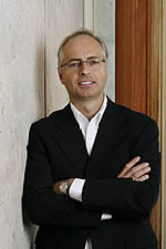
Dr. Masayo Takahashi

Dr. Masayo Takahashi

Meet Dr. Masayo Takahashi. She is a faculty member at the RIKEN Centre for Developmental Biology, a prominent female scientist in Japan, and a bona fide stem cell champion. Her mission is to cure diseases of blindness using iPS cell technology.

Since the Nobel Prize-winning discovery of iPS cells by Dr. Shinya Yamanaka eight years ago, Dr. Takahashi has made fast work using this technology to generate specific cells from human iPS cells that can be transplanted into patients to treat an eye disease called macular degeneration. This disease results in the degeneration of the retina, an area in the back of the eye that receives light and translates the information to your brain to produce sight.

Dr. Takahashi generates cells called retinal pigment epithelial (RPE) cells from human iPS cells that can replace lost or dying retinal cells when transplanted into patients with macular degeneration. What makes this therapy so exciting is that Dr. Takahashi’s iPS-derived RPE cells appear to be relatively safe and don’t cause an immune system reaction or cause tumors when transplanted into humans.

Because of the safety of her technology, and the unfulfilled needs of millions of patients with eye diseases, Dr. Takahashi made it her goal to take iPS cells into humans within five years of Dr. Yamanaka’s discovery.

Ogawa-Yamanaka Stem Cell Prize

It’s no surprise that Dr. Takahashi succeeded in her ambitious goal. Her cutting edge work has led to the first clinical trial using iPS cells in humans, specifically treating patients with macular degeneration. In September 2014, the first patient, a 70-year-old Japanese woman, received a transplant of her own iPS-derived RPE cells and no complications were reported.

Currently, the trial is on hold “as part of a safety validation step and in consideration of anticipated regulatory changes to iPS cell research in Japan” according to a Gladstone Institute news release. Nevertheless, this first iPS cell trial in humans has overcome significant regulatory hurdles, has set an important precedent for establishing the safety of stem cell therapies, and has given scientists hope that iPS cell therapies can become a reality.

Dr. Deepak Srivastava presents Dr. Takahashi with the Ogawa-Yamanaka Prize.

Dr. Deepak Srivastava presents Dr. Takahashi with the Ogawa-Yamanaka Prize.

For her accomplishments, Dr. Takahashi was recently awarded the first ever Ogawa-Yamanaka Stem Cell Prize and honored at a special event held at the Gladstone Institutes in San Francisco yesterday. This prize was established by a generous gift from Mr. Hiro Ogawa in collaboration with Dr. Shinya Yamanaka and Dr. Deepak Srivastava at the Gladstone Institutes. The award recognizes scientists who conduct translational iPS cell research that will eventually be applied to patients in the clinic.

In an interview with CIRM, Dr. Deepak Srivastava, the Director of the Gladstone Institute of Cardiovascular Disease and the Roddenberry Center for Stem Cell Biology and Medicine at Gladstone, described the prestigious prize and the ceremony held at the Gladstone to honor Dr. Takahashi:

Dr. Deepak Srivastava

The Ogawa-Yamanaka prize prize is meant to incentivize and honor those whose work is advancing the translational use of stem cells for regenerative medicine. Dr. Masayo Takahashi is a pioneer in pushing the technology of iPS cell-derived cell types and actually introducing them into people. She’s the very first person in the world to successfully overcome all the regulatory barriers and the scientific barriers to introduce this new type of stem cell into a patient. And she’s done so for a condition of blindness called macular degeneration, which affects millions of people world wide, and for which there are very few treatments currently. We are honoring her with this prize for her pioneering efforts at making this technology one that can be applied to patients.

The new world that iPS cells will bring

As part of the ceremony, Dr. Takahashi gave a scientific talk on the new world that iPS cells will bring for patients with diseases that lack cures, including those with visual impairments. The Stem Cellar team was lucky enough to interview Dr. Takahashi as well as attend her lecture during the Gladstone ceremony. We will cover both her talk and her interview with CIRM in an upcoming blog.

The Stem Cellar team at CIRM was excited to attend this momentous occasion, and to know that CIRM-funding has supported many researchers in the field of iPS cell therapy and regenerative medicine. We would like to congratulate Dr. Takahashi on her impressive and impactful accomplishments in this area and look forward to seeing progress in iPS cell trial for macular degeneration.


Related Links:

Patching up a Broken Heart with FSTL1

Get-Over-Heartbreak-Step-08How do you mend a broken heart? It’s a subject that songwriters have pondered for generations, without success. But if you pose the same question to a heart doctor, they would give you a number of practical options that focus on the prevention or management of the physical symptoms you are dealing with.

That’s because heart disease is complicated. There are many different types of diseases that affect the health and function of the heart. And once damage happens to your heart, say from a heart attack, it’s really hard to fix.

New regenerative factor for heart disease

Scientists from Stanford University, the University of California, San Diego, and the Sanford-Burnham-Prebys Medical Discovery Institute have teamed up to figure out how to fix a broken heart. In a CIRM-funded study published today in the journal Nature, the group reported that the gene follistatin-like 1 (FSTl1) has the ability to regenerate heart tissue when it’s delivered within a patch to the injured heart.

2004_Heart_Wall

The different layers of the heart.

The wall of the heart is made up of three different layers: the endocardium (inner), myocardium (middle), and epicardium (outer). The epicardium not only protects the inner two layers of the heart, but also supports the growth of the fetal heart.

The group decided to study epicardial cells to determine whether these cells produced specific factors that protect or even regenerate adult heart tissue. They took epicardial cells from rodents and cultured them with heart cells (called cardiomyocytes), and found that the heart cells divided and reproduced much more quickly when cultured with the epicardial cells. This suggested that the epicardial cells might secrete factors that promote the expansion of the heart cells.

Patching up a broken heart

They next asked whether factors secreted from epicardial cells could improve heart function in mice after heart injury. They designed and engineered tiny patches that contained a cocktail of special epicardial factors and sewed them onto the heart tissue of mice that had just experienced the equivalent of a human heart attack. When they monitored these mice two weeks later, they saw an improvement in heart function in mice with the patch compared to mice without.

When they analyzed the cocktail of epicardial factors in the patch, they identified one factor that had potential for regenerating heart tissue. It was FSTL1. To test its regenerative abilities, they cultured rodent heart cells in a dish and treated them with FSTL1 protein. This treatment caused the heart cells to divide like crazy, thus proving that FSTL1 had regenerative qualities.

Moving from the dish into animal models, the scientists explored which layers of the heart FSTL1 was expressed in after heart injury. In healthy hearts, FSTL1 is expressed in the epicardium. However, in injured hearts, they found that FSTL1 expression was missing in the epicardium and was instead present in the middle layer of the heart, the myocardium.

FSTL1 to the rescue

patch

Cross sections of a healthy (control) or injured mouse heart. Injured hearts treated with patches containing FSTL1 show the most recovery of healthy heart tissue (red). Image adapted from Wei et al. 2015)

In a eureka moment, the scientists decided to add a FSTL1 protein back to the epicardial layer of the heart, post heart injury, using the same patch system they used earlier in mice, to see whether this would promote heart tissue regeneration. Their guess was correct. FSTL1 delivery through the engineered epicardium patch system resulted in a number of beneficial effects to the heart including better function and survival, reduced scar tissue build up (a consequence of heart injury), and increased blood flow to the area of the patch.

Upon further inspection, they found that the FSTL1 epicardial patch caused heart cells to divide and proliferate. The same effect did not happen when FSTL1 is expressed in the myocardium layer of the injured heart.

To make sure their findings translated to other animal models, they studied the regenerative effects of FSTL1 in a pig model of heart injury. They applied patches infused with FSTL1 to the injured heart and as expected, observed that FSTL1 delivery improved symptoms and caused heart cells to divide.

No more heartbreak?

The authors concluded that heart injury turns off the activity of an important factor, FSTL1, in specific heart cells needed for heart regeneration. By turning on FSTL1 back on in the epicardium after injury, heart cells will receive the signal to divide and regenerate heart tissue.

Co-first author and CIRM postdoctoral scholar Ke Wei spoke to CIRM about the next steps for this study and its relevance:

Ke Wei

Co-first author, Ke Wei

In the future, we hope that our engineered epicardium patch technology can be used as a clinical platform to deliver drugs or cells to the injured heart. This strategy differs from conventional tools to treat heart attack, and may provide a novel approach in our repertoire battling heart diseases.

Thus it seems that scientists have found a potential way to patch-up a broken heart and to extend a lifeline for those suffering from heart disease. It’s comforting to know that the regenerative abilities of FSTl1 will be explored in human models and will hopefully reach clinical trials.

Ke Wei (UCSD, Sanford-Burnham-Prebys) and Vahid Serpooshan (Stanford) were co-first authors on this study. The senior authors were Daniel Bernstein (Stanford), Mark Mercola (UCSD, Sanford-Burnham-Prebys), and Pilar Ruiz-Lozano (Stanford). Both Ke Wei and Mark Mercola received CIRM funding for this study.


Related Links:

CIRM-funded team traces molecular basis for differences between human and chimp face

So similar yet so different
Whenever I go to the zoo, I could easily spend my entire visit hanging out with our not-so-distant relatives, the chimpanzees. To say we humans are similar to them is quite an understatement. Sharing 96% of our DNA, chimps are more closely related to us than they are to gorillas. And when you just compare our genes – that is, the segments of DNA that contain instructions for making proteins – we’re even more indistinguishable.

Chimps and Humans: So similar yet so different

Chimps and Humans: So similar yet so different

And yet you wouldn’t mistake a human for a chimp. I mean, I do have hairy arms, but they’re not that hairy. So what accounts for our very different appearance if our genes are so similar?

To seek out answers, a CIRM-funded team at Stanford University used both human and chimp induced pluripotent stem cells (iPSCs) to derive cranial neural crest cells (CNCCs). This cell type plays a key role in shaping the overall structure of the face during the early stages of embryo development. In a report published late last week in Cell, the team found differences, not in the genes themselves, but in gene activity between the human and chimp CNCCs.

Enhancers: Volume controls for your genes
Pinpointing the differences in gene activity relied on a comparative analysis of so-called enhancer regions of human and chimp DNA. Unlike genes, the enhancer regions of DNA do not provide instructions for making proteins. Instead they dictate how much protein to make by acting like volume control knobs for specific genes. A particular volume level, or gene activity, is determined by specific combinations of chemical tags and DNA-binding proteins on an enhancer region of DNA.

Enhancers: DNA segments that act like a volume control know for gene activity (Image source: xxxx)

Enhancers: DNA segments that act like a volume control knobs for gene activity (Image source: FANTOM Project, University of Copenhagen)

The researchers used several sophisticated lab techniques to capture a snapshot of this enhancer tagging and binding in the CNCCSs. They mostly saw similarities between human and chimp enhancers but, as senior author Joanna Wysocka explains in a Stanford University press release, they did uncover some differences:

“In particular, we found about 1,000 enhancer regions that are what we termed species-biased, meaning they are more active in one species or the other. Interestingly, many of the genes with species-biased enhancers and expression have been previously shown to be important in craniofacial development.”

PAX Humana: A genetic basis for our smaller jawline and snout?
For example, their analysis revealed that the genes PAX3 and PAX7 are associated with chimp-biased enhancer regions, and they had higher levels of activity in chimp CNCCs. These results get really intriguing once you learn a bit more about the PAX genes: other studies in mice have shown that mutations interfering with PAX function lead to mice with smaller, lower jawbones and snouts. So the lower level of PAX3/PAX7 gene activity in humans would appear to correlate with our smaller jaws and snout (mouth and nose) compared to chimps. Did that just blow your mind? How about this:

The researchers also found a variation in the enhancer region for the gene BMP4. But in this case, BMP4 was highly related to human-biased enhancer regions and had higher activity in humans compared to chimps. Previous mouse studies have shown that forcing higher levels of BMP4 specifically in CNCCs leads to shorter lower and upper jawbones, rounder skulls, and eyes positioned more to the front of the face. These changes caused by BMP4 sound an awful lot like the differences in human and chimp facial structures. It appears the Stanford group has established a terrific strategy for tracing the genetic basis for differences in humans and chimps.

So what’s next? According to Wysocka, the team is digging deeper into their data:

“We are now following up on some of these more interesting species-biased enhancers to better understand how they impact morphological differences. It’s becoming clear that these cellular pathways can be used in many ways to affect facial shape.”

And in the bigger picture, the researchers also suggest that this “cellular anthropology” approach could also be applied to a human to human search for DNA enhancer regions that play a role in the variation between healthy and disease states.

The best scientists always want to know more

Sir Isaac Newton

Sir Isaac Newton

Some years ago I was in the Wren Library at Trinity College, Cambridge in England when I noticed a display case with a cloth over it. Being a naturally curious person, downright nosy in fact, I lifted the cloth. In the display case was a first edition of Sir Isaac Newton’s Principia Mathematica and in the margins were notes, corrections put there by Newton for the second edition.

It highlighted for me how the best scientists never stop working, never stop learning, never stop trying to improve what they do.

That came back to me when I saw a news release from ViaCyte, a company we are funding in a Phase 1 clinical trial to treat type 1 diabetes.  The news release announced results of a study showing that insulin-producing cells, created in the lab from embryonic stem cells, can not only mature but also function properly after being implanted in a capsule-like device and placed under the skin of an animal model.

VC-01-cross-section-5

Now the clinical trial we are funding with ViaCyte uses a similar, but slightly different set of cells in people. The device in the trial contains what ViaCyte calls PEC-01™ pancreatic progenitor cells. These are essentially an earlier stage of the mature pancreatic cells that our body uses to produce insulin. The hope is that when implanted in the body, the cells will mature and then behave like adult pancreatic cells, secreting insulin and other hormones to keep blood glucose levels stable and healthy.

Those cells and that device are being tested in people with type 1 diabetes right now.

Learning more

But in this study ViaCyte wanted to know if beta cells, a more mature version of the cells they are using in our trial, would also work or have any advantages over their current approach.

The good news, published in the journal Stem Cells Translational Medicine,  is that these cells did work. As they say in their news release:

“The animal study also demonstrated for the first time that when encapsulated in a device and implanted into mice, these more mature cells are capable of producing functional pancreatic beta cells. ViaCyte is also the first to show that these further differentiated cells can function in vivo following cryopreservation, a valuable process step when contemplating clinical and commercial application.”

This does not mean ViaCyte wants to change the cells it uses in the clinical trial. As President and CEO Paul Laikind, PhD, makes clear:

“For a number of reasons we believe that the pancreatic progenitor cells that are the active component of the VC01 product candidate are better suited for cell replacement therapy. However, the current work has expanded our fundamental knowledge of beta cell maturation and could lead to further advances for the field.”

And that’s what I mean about the best scientists are the ones who keeping searching, keeping looking for answers. It may not help them today, but who knows how important that work will prove in the future.

Stem cell stories that caught our eye: diabetes drug hits cancer, video stem cell tracker and quick n’ easy stem cells for fatal lung disease


The chemical structure of Metformin (Image source: WikiMedia Commons)

The chemical structure of Metformin (Image source: WikiMedia Commons)

Teaching an old drug new tricks.
One the quickest way to get a drug to market is to find one that’s already been FDA approved for other diseases. Reporting this week in Cell Metabolism, researchers from London and Madrid identified the mechanisms that enable the anti-diabetic drug, metformin, to kill pancreatic cancer stem cells (PanCSCs).

Though they make up a tiny portion of a tumor, cancer stem cells (CSCs) are thought to lie dormant most of the time. As a result, they evade chemotherapy only to later revive the tumor and cause relapse. So, the hypothesis goes, target and kill the CSCs and you’ll eradicate the cancer.

Mitochondrion_mini.svg

Mitochondria – a cell’s power station (image source: WikiMedia Commons)

While most cancer cells produce their energy needs without the use of oxygen, the team found that PanCSCs use oxygen-dependent energy production that occurs in a cell structure called the mitochondria. Because metformin blocks key components of the mitochondria’s energy factory, the drug essentially shuts down power to the PanCSCs leading to cell death.

The PanCSCs still have another trick up their proverbial sleeves: some switch over to a mitochondria-independent form of energy production so the metformin becomes useless against the PanCSCs. However, by tweaking the levels of two proteins, the researchers forced the PanCSCs to only use the mitochondria for energy production, which restored metformin’s cancer-killing ways.

Pancreatic cancer has very poor survival rates with very limited treatment options. Let’s hope this work leads to alternatives for patients and their doctors.

It’s all about location, location, location. Or is it?
We’ve written numerous times at the Stem Cellar about the importance of a stem cell’s “neighborhood” for determining the cell type into which it will eventually specialize. But a study published this week in Stem Cell Reports put the role of a cell’s surroundings somewhat into question.

A research team at Drexel University in Philadelphia compared stem cells in the back of the brain – an area that interprets visual information – with stem cells in the front of the brain – an area responsible for controlling movement. A fundamental question about brain development is how these areas form very different structures. Are the stem cells in each part of the brain already programmed to take on different fates or are they blank slates which rely on protein signals in the local environment to determine the type of nerve cell they become?

To chip away at this question, the team isolated mouse stem cells from the back and the front on the brain. Each set was grown in the lab using the same nutrients and conditions. You might have guessed the stem cells would behave the same but that’s not what happened. Compared to the stem cells from the back of the brain, the front brain stem cells gave rise to smaller daughter cells that divided more slowly. This suggests these brain stem cells already have some built-in properties that set them apart.

The methods used in the study are as fascinating as the results themselves. The team developed a time-lapse cell-tracking system from scratch that, with minimal human intervention, tags individual daughter cells and analyzes their fate as they grow, move and specialize on the petri dish. In the movie below, Professor Andrew Cohen, one of the authors who helped design the web-based software, succinctly describes the work. Also this movie of the tracking system in action is stunning.

Kudos to the team for making the software and their data set open access. There’s no doubt this technology will lead to important new discoveries.

Quick and easy stem cells to fight deadly lung disease
Lung disease is the 3rd deadliest disease in the U.S. It afflicts 33 million people and accounts for one in six deaths. One of those diseases is Idiopathic Pulmonary Fibrosis (IPF), an incurable disease that causes scarring and thickening of the lungs and makes breathing more and more labored. People often succumb to the disease within 3 to 5 years of their diagnosis. Use of lung stem cells to replace and heal damaged tissue is a promising therapeutic strategy for IPF.

Red and green indicate lung stem cells within a spheroid. (Image credit: Henry et al. Stem Cells Trans Med September 2015-0062)

Red and green indicate lung stem cells within a spheroid. (Image credit: Henry et al. Stem Cells Trans Med September 2015-0062)

This week, a research team from North Carolina State University reported in Stem Cells Translational Medicine on a quick and easy method for growing large amounts of lung stem cells from healthy lung tissue. The typical process of harvesting the tissue, sorting the individual lung cells, and growing the cells on petri dishes can be costly and time-consuming.

Instead, the NCSU team grew the human lung stem cells in three dimensional spheres containing multiple cell types and allowed them to float in liquid nutrients. The lung stem cells are at the center of the sphere surrounded by support cells. This method better resembles the natural cellular environment of the stem cells compared to a flat homogenous lawn of cells in a petri dish.

When introduced intravenously into mice with IPF-like symptoms, these lung spheroids reduced lung scarring and inflammation, nearly matching the animals without IPF. And in a head-to-head comparison, the lung spheroids were more effective than fat-derived mesenchymal stem cells, another proposed cell source for treating lung disease. Alas, humans are not mice and more studies are necessary to reach the ultimate goal of treating IPF patients. But I’m excited about this team’s progress and look forward to hearing more from them.

Related Press Releases:

Using satellites to build bigger biceps

Arnold Schwarzenegger: Photo courtesy Awesome-Body.info

Arnold Schwarzenegger:
Photo courtesy Awesome-Body.info

There are several ways you can build bigger, stronger muscles. You can take the approach favored by our former Governor, Arnold Schwarzenegger, and pump iron till your biceps are as inflated as a birthday balloon. Or you could follow the lead of a research team we are funding and try to use stem cells to do the trick.

Our muscles contain a group of stem cells called satellite cells. These normally lie dormant until the muscle is damaged and then they spring into action to repair the injury. However, satellite cells are relatively rare and are hidden in the muscle itself, making them hard to find and notoriously difficult to study. In the past researchers have struggled to get these satellite cells to grow outside the body, which made it difficult to study muscle regeneration and develop new ways of treating muscle problems.

Finding a solution

Now a team at the University of California, San Francisco has found a solution to the problem. They started by analyzing samples of 7 different kinds of muscles (in the body, legs and head) from 43 patients. In all but two samples they found that the gene PAX7 was specifically turned on in satellite cells and the PAX7 protein was present with little variation from one muscle group to another.

Upon further sleuthing, they discovered that PAX7-positive satellite cells were the real deal because they also expressed two common cell surface markers of human satellite cells: CD29 and CD56.

The researchers then transplanted PAX7-positive cells into mice that had experienced muscle injuries. As they report in the journal Stem Cell Reports these cells not only engrafted in the mice but they also created hundreds of human-derived muscle fibers. This finding shows that satellite cells were regenerating and potentially helping to heal the damaged muscle.

What’s next

As always, anything done in mice is interesting but still needs to be replicated in people before we know for sure we are on to something.

In their conclusion the team freely admit this is just a first step but, compared to where we were before, it’s a very important step. As senior author Jason Pomerantz says:

“This is the first definitive experimental description of adult human endogenous muscle stem cell function.”

Harnessing the power of satellite cells would be of tremendous benefit to people suffering from facial paralysis, loss of hand function or muscle-wasting diseases such as sarcopenia, and could even be used as a way to deliver gene therapy to people with muscular dystrophies.

Using satellite cells to do all that, would be out of this world.

CIRM CAP Kickoff to New Clinical Trials

Alisha Bouge is the project manager for CIRM’s Clinical Advisory Panels (CAPs)

On the cusp of the official kickoff to football season, CIRM has had its own kickoff to celebrate.  The first Clinical Advisory Panel (CAP) meeting took place on August 18, 2015 in Irvine, CA with Caladrius Bioscience, Inc.  And just as every NFL team starts the season hopeful of a Super Bowl win, all our CAPs start out with equally lofty goals. That’s because under CIRM 2.0, the role of the CAP is to work with the clinical stage project teams we fund to help accelerate the development of therapies for patients with unmet medical needs and to give these projects the greatest likelihood of success.

In the case of Caladrius, the work is focused on treating metastatic melanoma, an aggressive and deadly form of skin cancer. You can read more about this clinical trial here.

Obstacles and challenges are inevitable in the lifecycle of research. CIRM hopes to help its grantees navigate through these hurdles as quickly and positively as possible by providing recommendations from expert advisors in the field.  The intention is for the CAP meeting process to be that navigating vessel throughout the lifetime of each clinical stage project.

The CAPs will include at least three members: one CIRM science officer, a patient representative, and an external scientific advisor.  The CAP will meet with the project team approximately four times a year, with the first meeting taking place in-person.  Consider the CAP as the grantee’s special team, doing all they can to get that two-point conversion at the end of an already successful outcome, giving the grantee and their team just a few more points in their pocket to reach the ultimate success.

CAP1

CIRM CAP on a tour of Caladrius’ facility in Irvine, CA.  The CIRM CAP can be seen in the far right of the photo (left to right) Randy Lomax (Patient Representative), Ingrid Caras (CIRM Sr. Science Officer), and Hassan Movahhed (External Scientific Advisor).

As the lead Science Officer on this first CAP, CIRM’s Ingrid Caras stated: “This is our opportunity to be good stewards of the taxpayers’ money.”

The mission and the message of the CAP was well received by Caladrius.  After the CAP meeting, Anna Crivici, VP of Operations & Program Management at Caladrius, had this to say about her experience:

anna crivici

Anna Crivici, Caladrius

I thought that the meeting was very productive.  Everyone on the Caladrius team appreciates the collaborative approach CIRM is taking on the program, as amply demonstrated during our successful first meeting.  The discussion on every agenda topic was helpful and insightful.  The opportunity to better understand the patient perspective will be especially beneficial and increasingly important as the Phase 3 program progresses.  We are confident that this and future CAP meetings will help us advance and refine our strategic planning and execution.

CAP2

CIRM CAP and members of Caladrius discussing operational strategies for success.

CIRM is looking forward to the 2015/2016 CAP season. And while there is no Super Bowl incentive at the end of our season, there is the hope that CIRM’s efforts, both financially and collaboratively, will contribute to successful treatments for so many out there in need. That’s something well worth cheering for.

Brain Stem Cells in a Dish to the Rescue

braindish

Image credit: CureCDKL5.org

The best way to impress your friends at the next party you attend might be to casually mention that scientists can grow miniature brain models in a dish using human stem cells. Sure, that might scare away some people, but when you explain how these tiny brain models can be used to study many different neurological diseases and could help identify new therapies to treat these diseases, your social status could sky rocket.

Recently, a group at UC San Diego used human stem cells to model a rare neurological disorder and identified a drug molecule that might be able to fix it. This work was funded in part by CIRM, and it was published today in the journal Molecular Psychiatry.

The disorder is called MECP2 duplication syndrome. It’s caused by a duplication of the MECP2 gene located in the X chromosome, and is genetically inherited as an X-linked disorder, meaning the disease is much more common in males. Having extra copies of this gene causes a number of unfortunate symptoms including reduced muscle tone (hypotonia), intellectual disabilities, impaired speech, seizures, and developmental delays, to name a few. So far, treatments for this disorder only help ease the symptoms and do not cure the disease.

The group from UCSD decided to model this disease using induced pluripotent stem cells (iPSCs) derived from patients with MECP2 duplication syndrome. iPSCs can form any cell type in the body, and the group used this to their advantage by coaxing the iPSCs into the specific type of nerve cell affected by the disorder. Their hard work was rewarded when they observed that the diseased nerve cells acted differently than normal nerve cells without the disease.

In fact, the diseased nerve cells generated more connections with other nearby nerve cells, and this altered their ability to talk to each other and perform their normal functions. The senior author Alysson Muotri described the difference as an “over-synchronization of the neuronal networks”, meaning that they were more active and tended to fire their signals in unison.

After establishing a relevant nerve cell model of MECP2 duplication disorder, the group tested out a library of drug molecules and identified a new drug candidate that was able to rescue the diseased nerve cells from their “over-synchronized” activity.

The senior author Alysson Muotri commented on the study in a press release:

Alysson Muotri (Photo by David Ahntholz)  

This work is encouraging for several reasons. First, this compound had never before been considered a therapeutic alternative for neurological disorders. Second, the speed in which we were able to do this. With mouse models, this work would likely have taken years and results would not necessarily be useful for humans.


The press release goes on to describe how Muotri and his team plan to push their preclinical studies using human stem-cell based models forward in hopes of entering clinical trials in the near future.


Related Links:
Stem cell stories that caught our eye: our earliest days, cell therapy without the cells and unproven therapies

Here are some stem cell stories that caught our eye this past week. Some are groundbreaking science, others are of personal interest to us, and still others are just fun.

2-cell embryoMapping our earliest days—as an embryo. We have some 23,000 genes in every cell of our body, but on day two after fertilization just 32 are switched on. A team at Sweden’s Karolinska Institute has for the first time mapped all the gene activity for the first few days of embryo development—information that is yielding clues for developing better fertility treatments.

One of their more interesting finding was that by day three, the number of active genes has grown to 129 even though the embryo has only grown from four to eight cells.

“These genes are the ‘ignition key’ that is needed to turn on human embryonic development. It is like dropping a stone into water and then watching the waves spread across the surface,” said the project leader Juha Kere in an article in Health News Digest.

As with much science today, the team took advantage of rapidly advancing technology in genetic analysis to map the gene activation.

Sending the message without the messenger. In many experimental stem cell therapies it’s not the stem cells themselves that are expected to make the repair but rather other cells in the body that respond to chemical messages released by the stem cells. Stem cells often send out those messages in packets called exosomes, which has led several teams to start using the exosomes alone for repair.

A stem cell releasing exosomes, sort of like little medicine bags.

A stem cell releasing exosomes, sort of like little medicine bags.

One group in Germany just published results in Stem Cells Translational Medicine suggesting that in at least one disease, a mouse model of stroke, exosomes trigger as much repair as whole stem cells. The researchers from the University of Duisburg-Essen claim to be the first to publish a side-by-side comparison of exosomes, which are also called extracellular vesicles (EVs) and whole stem cells.

“The fact that intravenous EV delivery alone was enough to protect the post-stroke brain and help it recover highlights the clinical potential of EVs in future stroke treatment,” said the two lead researchers in a press release distributed by the journal and picked up by BioSpace.

 They reported that the exosomes promoted brain recovery and protected the mice from post stroke inflammation that can cause ongoing damage.

Three hits on unproven stem cell therapy. Yesterday’s news feed brought in three welcomed pieces trying to explain why so many stem cell treatments being offered today are unproven, skirt regulations and may be a rip-off.  

 A group of neurologists from Ohio State University wrote a piece on the growing problem of “stem cell tourism” in the journal JAMA Neurology. The piece got picked up by many news outlets including Fox News. In particular, the authors noted internet advertisements suggesting stem cells are proven to help multiple sclerosis, ALS (Lou Gehrig’s disease) and other hard or impossible to treat neurological conditions. They called upon their fellow neurologists to do a better job of advising their patients on the issue.

“We must help educate our patients not only in the clinic setting, but also by working with patient advocacy groups such as the National Multiple Sclerosis Society and the ALS Association,” said one of the Ohio State authors, Jaime Imitola.

USA Today published a pair of pieces. One dealt with athletes making very visible trips to clinics offering unproven stem cell therapies and the power their fame has to attract other customers. The second piece discussed face creams that claim to have benefit due to stem cells and the lack of data and often lack of logic behind most of those claims.

stem cell drive through

From Paul Knoepfler’s Stem Cell Blog

Brent Schrotenboer wrote both pieces and he did the best job I’ ve seen in the consumer press of explaining how the US-based clinics skirt FDA regulation and fail to provide data showing their stem cells caused an improvement in the athletes beyond the other therapies they received at the same time. At almost 3,000 words the piece is unusually long for USA Today and at that length he has the opportunity to cover quite a bit of nuance between some of the various clinic offerings. He quotes a CIRM-grantee from the University of California, San Diego, Lawrence Goldstein a couple times, including in the concluding paragraph:

“It’s hard to write good law and regulation that allows legitimate work to proceed as rapidly as possible while prohibiting illegitimate work. Part of the problem is it is a new area of medicine where the regulations didn’t anticipate this sort of thing. The regulators on the ground in the field, they themselves don’t have adequate background to tell what’s legit and what’s not. It’s hard.”

Watch Spinal Cord Cells Take a Hike!

magic school busWhat exactly goes on inside the human body? If you asked this question to the children’s book character Ms. Frizzle, she would throw you into her Magic School Bus and take you on a wild ride “Inside the Human Body” to get you up close and personal with the different organs and structures within our bodies.

Ms. Frizzle had a wild imagination, but she was on to something with her crazy adventures. Recently, scientists took a page out of one of Ms. Frizzle children’s books and took their own wild ride to check out what’s going on with the human spinal cord.

In a paper published yesterday in Neuron, scientists from the Salk Institute in San Diego reported that they were able to watch spinal cord cells walk around the spine of mice in real-time. They used a special microscope that could track and record the movement of motor neurons, an important nerve cell that controls the movement of muscles in your body. What they found when they watched these cells was equivalent to a pot of gold at the end of the rainbow.

Check out their stunning movie here:

The scientists not only recorded the activity of these motor neurons, but they identified the other spinal cord cells that these neurons interact and make connections with. One of their most significant findings was a population of spinal cord cells that connected to a subtype of motor neurons to foster important muscle movements like walking.

Understanding how the different cells of the spinal cord work together is very important because it will allow scientists and doctors to figure out better ways to treat patients with spinal cord injuries or neurodegenerative diseases, like ALS, that affect motor neurons.

Senior author Samuel Pfaff commented in a press release on the importance of this study and how easy his team’s technology is to use:

Pfaff_S09

Samuel Pfaff

Using optical methods to be able to watch neuron activity has been a dream over the past decade. Now, it’s one of those rare times when the technology is actually coming together to show you things you hadn’t been able to see before. You don’t need to do any kind of post-image processing to interpret this. These are just raw signals you can see through the eyepiece of a microscope. It’s really a jaw-dropping kind of visualization for a neuroscientist.

While this study doesn’t provide a direct avenue for therapeutic development, it does pave the way for a better understanding of the normal, healthy processes that go on in the human spine. Having more knowledge of “what is right” will help scientists to develop better strategies to fix “what is wrong” in spinal cord injuries and diseases like ALS.


Related Links: