Each individual muscle in our body—no matter how large or how small—is controlled by several types of motor neurons. Damage to one or more types of these neurons can give rise to some of the most devastating motor neuron diseases, many of which have no cure. But now, stem cell scientists at UCLA have manufactured a way to efficiently generate motor neuron subtypes from stem cells, thus providing an improved system with which to study these crucial cells.
“Motor neuron diseases are complex and have no cure; currently we can only provide limited treatments that help patients with some symptoms,” said senior author Bennett Novitch, in a news release. “The results from our study present an effective approach for generating specific motor neuron populations from embryonic stem cells to enhance our understanding of motor neuron development and disease.”
Normally, motor neurons work by transmitting signals between the brain and the body’s muscles. When that connection is severed, the individual loses the ability to control individual muscle movement. This is what happens in the case of amyotrophic lateral sclerosis, or ALS, also known as Lou Gehrig’s disease.
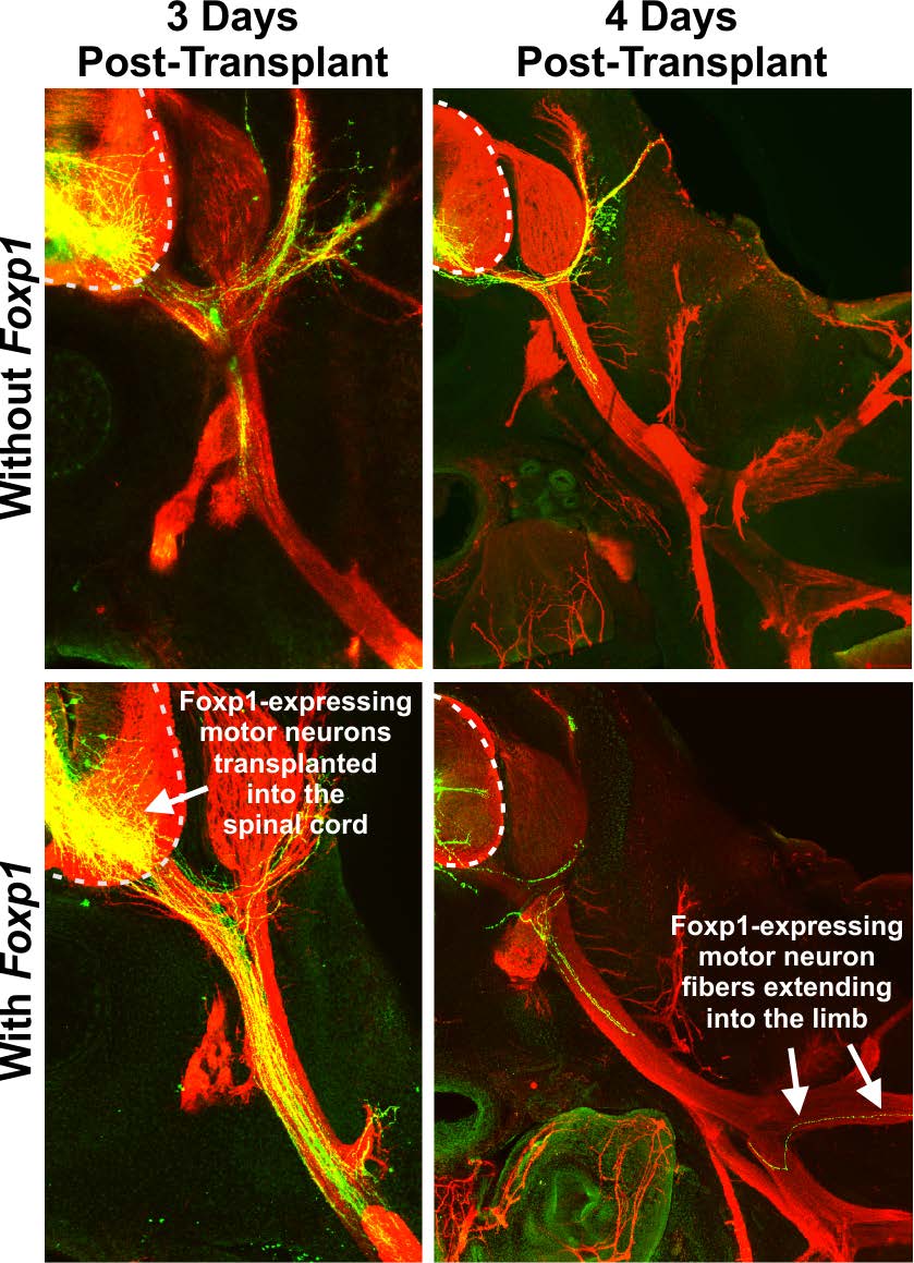
These images reveal the significance of the ‘Foxp1 effect.’ The Foxp1 transcription factor is crucial to the normal growth and function of motor neurons involved in limb-movement.
Recent efforts had focused on ways to use stem cell biology to grow motor neurons in the lab. However, such efforts to generate a specific type of motor neuron, called limb-innervating motor neurons, have not been successful. This motor-neuron subtype is particularly affected in ALS, as it supplies nerves to the arms and legs—the regions usually most affected by this deadly disease.
In this study, published this week in Nature Communications, Novitch and his team, including first author Katrina Adams, worked to develop a better method to produce limb-innervating motor neurons. Previous efforts had only had a success rate of about 3 percent. But Novitch and Adams were able to boost that number five-fold, to 20 percent.
Specifically, the UCLA team—using both mouse and human embryonic stem cells—used a technique called ‘transcriptional programming,’ in order to coax these stem cells into become fully functional, limb-innervating motor neurons.
In this approach, which was funded in part by a grant from CIRM, the team added a single transcription factor (a type of protein that regulates gene function), which would then guide the stem cell towards becoming the right type of motor neuron. Here, Novitch, Adams and the team used the Foxp1 transcription factor to do the job.
Normally, Foxp1 is found in healthy, functioning limb-innervating motor neurons. But in stem cell-derived motor neurons, Foxp1 was missing. So the team reasoned that Foxp1 might actually be the key factor to successfully growing these neurons.
Their initial tests of this theory, in which they inserted Foxp1 into a developing chicken spinal cord (a widely used model for neurological research), were far more successful. This result, which is not usually seen with most unmodified stem-cell-derived motor neurons, illustrates the important role played by Foxp1.
The most immediate implications of this research is that now scientists can now use this method to conduct more robust studies that enhance the understanding of how these cells work and, importantly, what happens when things go awry.
