A big focus of stem cell research is trying to figure how to make a stem cell specialize, or differentiate, into a desired cell type like muscle, liver or bone. When we write about these efforts in the Stem Cellar, it’s usually in terms of researchers identifying proteins that bind to a stem cell’s surface and trigger changes in gene activity inside the cell that ultimately leads to a specific cell fate.
But, that’s not the only game in town. As incredible as it sounds, affecting a cell’s shape through mechanical forces also plays a profound role in gene activity and determining a cell’s fate. In one study, mesenchymal stem cells would specialize into fat cells or bone-forming cells depending on how much the MSCs were stretched out on a petri dish.

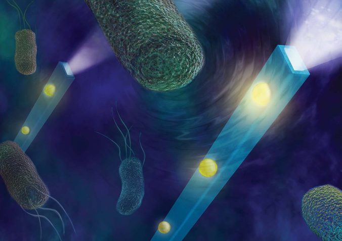
An artist’s illustration of nano optical fibers detecting the minuscule forces produced by swimming bacteria. Credit: Rhett S. Miller/UC Regents
Since we’re talking about individual cells, the strength of these mechanical forces is tiny, making measurements nearly impossible. But now, a research team at UC San Diego has engineered a device 100 times thinner than a human hair that can detect these miniscule forces. The study, funded in part by CIRM, was reported yesterday in Nature Photonics.
The device is made of a very thin optical fiber that’s coated with a resin which contains gold particles. The fiber is placed directly into the liquid that cells are grown in and then hit with a beam of light. The light is scattered by the gold particles and measured with a conventional light microscope. Forces and even sound waves caused by cells in the petri dish change the intensity of the light scattering which is detected by the microscope.

Donald Sirbuly,
team lead
In this study, the researchers measured astonishingly small forces (0.0000000000001 pound of force, to be exact!) in a culture of gut bacteria which swim around in the solution with the help of their whip-like flagella. The team also detected the sound of beating heart muscle cells at a level that’s a thousand times below the range of human hearing.
Dr. Donald Sirbuly, the team lead and a professor at UCSD’s Jacobs School of Engineering is excited about the research possibilities with this device:
“This work could open up new doors to track small interactions and changes that couldn’t be tracked before,” he said in a press release.
Bradley Fikes, the biotechnology reporter for the San Diego Union Tribune, reached out to others in the field to get their take on potential applications of this nanofiber device. Dr. John Marohn at Columbia University told Fikes in a news article (subscription is needed to access) that it could help stem cell scientists’ fully understand all of the intricacies of cell fate:
“So one of the cues that cells get, and they listen to these cues to decide how to change how to evolve, are just outside forces. This would give a way to kind of feel the outside forces that the cells feel, in a noninvasive way.”
And Eli Rothenberg at NYU School of Medicine, also not part of the study, summed up the device’s novelty, power and ease of use in an interview with Fikes:
“One of the main challenges in measuring things in biology is forces. We have no idea what’s going on in terms of forces in cells, in term of motion of molecules, the forces they interact with. But these sensors, you can put anywhere. They’re tiny, you can place them on the cells. If a cancer cell’s surface is moving, you can measure the forces…The fabrication of this device is quite straightforward. So, the simplicity of having this device and what you can measure with it, that’s kind of striking.”